

The Cellular Composition of the Uveal Immune Environment
- PMID: 34778287
- PMCID: PMC8586083
- DOI: 10.3389/fmed.2021.721953
The Cellular Composition of the Uveal Immune Environment
Abstract
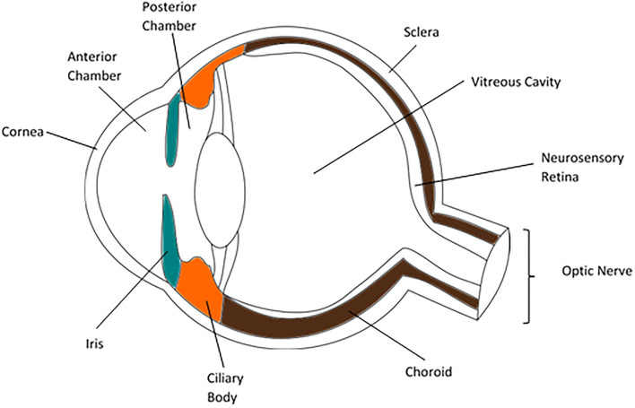
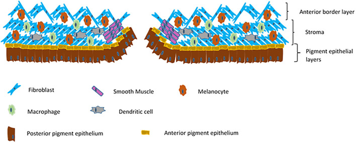
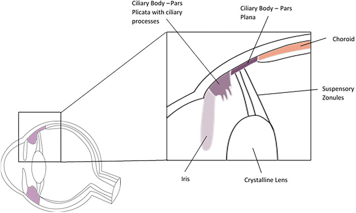
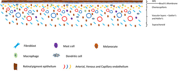
The uveal tract consists of the iris, the ciliary body and the choroid; these three distinct tissues form a continuous layer within the eye. Uveitis refers to inflammation of any region of the uveal tract. Despite being grouped together anatomically, the iris, ciliary body and choroid are distinct functionally, and inflammatory diseases may affect only one part and not the others. Cellular structure of tissues direct their function, and understanding the cellular basis of the immune environment of a tissue in health, the "steady state" on which the perturbations of disease are superimposed, is vital to understanding the pathogenesis of those diseases. A contemporary understanding of the immune system accepts that haematopoietic and yolk sac derived leukocytes, though vital, are not the only players of importance. An array of stromal cells, connective tissue cells such as fibroblasts and endothelial cells, may also have a role in the inflammatory reaction seen in several immune-mediated diseases. In this review we summarise what is known about the cellular composition of the uveal tract and the roles these disparate cell types have to play in immune homeostasis. We also discuss some unanswered questions surrounding the constituents of the resident leukocyte population of the different uveal tissues, and we look ahead to the new understanding that modern investigative techniques such as single cell transcriptomics, multi-omic data integration and highly-multiplexed imaging techniques may bring to the study of the uvea and uveitis, as they already have to other immune mediated inflammatory diseases.
Keywords: choroid; ciliary body; iris; uvea; uveitis.
Copyright © 2021 Reekie, Sharma, Foers, Sherlock, Coles, Dick, Denniston and Buckley.
Conflict of interest statement
JS is now an employee of Janssen. The remaining authors declare that the research was conducted in the absence of any commercial or financial relationships that could be construed as a potential conflict of interest.
Figures
References
Publication types
Grants and funding
LinkOut - more resources
Full Text Sources