Review
. 2021 Dec:25:101417.
doi: 10.1016/j.genrep.2021.101417. Epub 2021 Nov 5.

Immunopathology and Immunopathogenesis of COVID-19, what we know and what we should learn

Affiliations
Review

Immunopathology and Immunopathogenesis of COVID-19, what we know and what we should learn

Mehdi Shahgolzari et al. Gene Rep. 2021 Dec.

Abstract

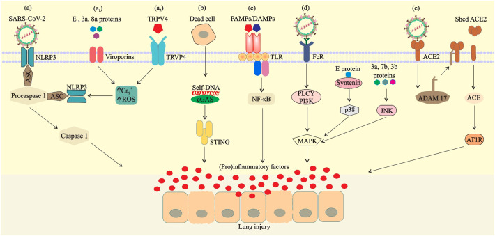
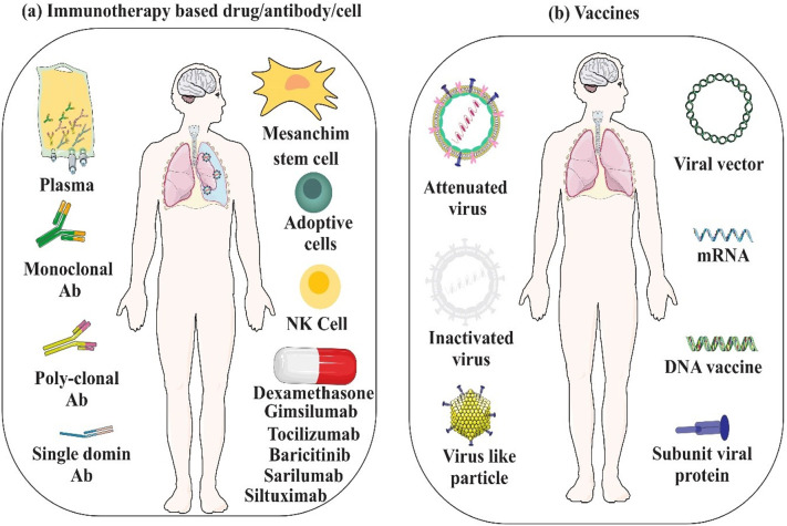
Severe Acute Respiratory Syndrome Coronavirus-2 (SARS-CoV-2) directly interacts with host's epithelial and immune cells, leading to inflammatory response induction, which is considered the hallmark of infection. The host immune system is programmed to facilitate the clearance of viral infection by establishing a modulated response. However, SARS-CoV-2 takes the initiative and its various structural and non-structural proteins directly or indirectly stimulate the uncontrolled activation of injurious inflammatory pathways through interaction with innate immune system mediators. Upregulation of cell-signaling pathways such as mitogen-activate protein kinase (MAPK) in response to recognition of SARS-CoV-2 antigens by innate immune system receptors mediates unbridled production of proinflammatory cytokines and cells causing cytokine storm, tissue damage, increased pulmonary edema, acute respiratory distress syndrome (ARDS), and mortality. Moreover, this acute inflammatory state hinders the immunomodulatory effect of T helper cells and timely response of CD4+ and CD8+ T cells against infection. Furthermore, inflammation-induced overproduction of Th17 cells can downregulate the antiviral response of Th1 and Th2 cells. In fact, the improperly severe response of the innate immune system is the key to conversion from a non-severe to severe disease state and needs to be investigated more deeply. The virus can also modulate the protective immune responses by developing immune evasion mechanisms, and thereby provide a more stable niche. Overall, combination of detrimental immunostimulatory and immunomodulatory properties of both the SARS-CoV-2 and immune cells does complicate the immune interplay. Thorough understanding of immunopathogenic basis of immune responses against SARS-CoV-2 has led to developing several advanced vaccines and immune-based therapeutics and should be expanded more rapidly. In this review, we tried to delineate the immunopathogenesis of SARS-CoV-2 in humans and to provide insight into more effective therapeutic and prophylactic strategies.

Keywords: Immune evasion; Immunotherapy; Inflammatory; SARS-CoV-2; Vaccine; Virus.


Conflict of interest statement

The authors declare that they have no competing interests.

Figures

Fig. 1
Fig. 1
Virus particles, complete genome sequences, spike structure, cleavage sites and fusion reaction of SARS-CoV and SARS-CoV-2. a) Virions contain a +ssRNA genome of 26–32 kb in size. The genome ORF1a/b encodes polyproteins, which form the viral replicase transcriptase complex. The other ORFs on the genome encode four main structural proteins: S, E, N and M proteins, as well as several accessory proteins. b) The spike protein structure is composed of an extracellular (EC) domain, a transmembrane anchor domain and a short intracellular tail. EC domain contains two functional subunits, a receptor-binding subunit (S1) and a membrane-fusion subunit (S2), S1 contains two independent domains, an N-terminal domain (S1-NTD) and receptor binding domain (RBD), the S1/S2 cleavage site is shown in its uncleaved, native state and resides in an unstructured region between S1 and S2, the S2’ cleavage site is exposed only after receptor binding. c) Fusion reaction; S1 attaches to a receptor on the target cells, induces a conformational change in the S, exposing cleavage sites between S1 and S2. In SARS-CoV-2, the trimeric S protein then cleaves into S1 and S2 subunits by cellular proteases (scissors), the fusion peptide (FP) latches onto the target membrane, anchoring the virus and cell together. The heptad repeat 2 (HR2) then folds to interact with the heptad repeat 1 (HR1), bringing the membranes together. The successful refolding of enough adjacent S2s leads to fusion of viral and cell membranes and release of the viral genome into the target cell cytoplasm.
Fig. 2
Fig. 2
The life cycle of SARS-CoV and SARS-CoV-2 in host cells. Both viruses enter target cells through fusion at the cell surface (early entry) or in the endocytic compartment (late entry). Entry route depends on which proteases activate the spike proteins. In early entry, the virus is cleaved at S protein by cell-surface transmembrane serine proteases (TTSPs). If an S protein is unable to be cleaved by transmembrane proteases, due to S protein sequence or lack of protease expression on target cell, the virus must undergo endocytosis and be activated by Cathepsin in the endosome/lysosome. The S proteins of SARS-CoV and SARS-CoV-2 bind to cellular receptor angiotensin-converting enzyme 2 (ACE2). Early and late entry lead to release of the viral +ssRNA genome to the cytoplasm. ORF1a and ORF1ab are translated and processed to form the RNA replicase–transcriptase complex for driving the production of negative-sense Full-length RNAs [(−) RNA]. Full-length (−) RNA is transcribed into four sub-genomic mRNAs which during translation encode viral structural proteins including S, E, M and N. Viral nucleocapsids are assembled from genomic RNA and N protein in the cytoplasm, followed by budding into the lumen of the endoplasmic reticulum (ER)–Golgi intermediate compartment (ERGIC). Virions are then released from the infected cell through exocytosis and unlike nonenveloped viruses, do not lyse cells.
Fig. 3
Fig. 3
Coronavirus immunity cycle. Following SARS-CoV-2 uptake in the endosome and virion degradation the viral antigens are presented in (1) the exogenous pathway, (2) cross-presentation pathway and (3) endogenous pathway; partial genome transcription may provide a source of antigen for priming T-cells by MHC class-1 antigen processing following endogenous pathway. Viral pathogen-associated molecular patterns (PAMPs), using endosomal or cytosolic PRRs, as well as the production of cytokines such as IFN-1, can promote potent cellular mediated immune responses. Structural or nonstructural proteins might be recognized by TLR-4 or inflammasome, leading to the activation of proinflammatory cytokines. Abbreviations: APC, antigen-presenting cells; IFN-1, interferon 1; ssRNA, single-stranded RNA; TBK1, TANK-binding kinase 1.
Fig. 4
Fig. 4
Likely mechanisms of suppression of the type 1 interferon response during SARS-CoV-2 infection. Structural and nonstructural proteins of virus or double membrane vesicles (DMVs) can shield PAMPs from immune sensors. Structural and nonstructural proteins also inactivate immune sensors or components of downstream type I IFN signaling. Abbreviations: DMV, double membrane vesicle; ISG, IFN-stimulated gene; MDA5, melanoma differentiation-associated protein 5.
Fig. 5
Fig. 5
Possible mechanisms of SARS-CoV-2-mediated inflammatory responses. a) NOD-like receptor protein 3 (NLRP3) is activated by virus, Ca2+ influx or ROS (induced via viroporins (a1) or TRPV4 (a2)) and binds to the precursor of caspase-1 (procaspase-1) through the adaptor protein ASC in the cell to form a multiprotein complex, thereby activating caspase-1. b) Entry of danger-associated molecular pattern (self- DNA) into the cytoplasm from the nucleus of mitochondria or dead cells activates the cGAS–STING pathway. c) Binding of viral PAMPs/DAMPs to the TLRs and activation of transcription factors for inducing proinflammatory factors. d) Viral structural proteins or binding of virus-Ab complex to FcR can also activate proinflammatory responses via MAPK signaling. e) Binding of the spike protein to ACE2 induces ADAM 17 activity, thereby reducing the number of ACE2 expressed on the cell surface.
Fig. 6
Fig. 6
Non-severe and severe COVID19. a) During non-severe stage, innate and specific adaptive immune responses can eliminate the virus and prevent disease progression to severe stages. b) Nevertheless, high viral load and propagation, followed by dysregulated immune response and massive destruction of the affected tissues can induce innate inflammation in the lungs that is largely mediated by inflammatory monocyte-macrophages (IMMs). Massive accumulation of pathogenic inflammatory IMMs and exuberant inflammation increase the severity of disease and lead to lung damage in the severe stages of the disease.
Fig. 7
Fig. 7
Immunotherapy and immunization against COVID-19. a) Immunotherapy based drugs/antibodies/cells can directly/indirectly suppress virus particles, and modulate immune system in severe COVID-19. b) Strategies for developing COVID-19 vaccines.

References

    1. Abdulamir A.S., Hafidh R.R. The possible immunological pathways for the variable Immunopathogenesis of COVID--19 infections among healthy adults, elderly and children. Electronic Journal of General Medicine. 2020;17
    1. Acuña-Castroviejo D., Escames G., Figueira J.C., de la Oliva P., Borobia A.M., Acuña-Fernández C. Clinical trial to test the efficacy of melatonin in COVID-19. J. Pineal Res. 2020;69:12683. - PMC - PubMed
    1. Alam I., Kamau A., Kulmanov M., Arold S.T., Pain A., Gojobori T., Duarte C.M. bioRxiv; 2020. Functional Pangenome Analysis Suggests Inhibition of the Protein E as a Readily Available Therapy for COVID-2019. (2020.2002.2017.952895)
    1. Alosaimi B., Hamed M.E., Naeem A., Alsharef A.A., AlQahtani S.Y., AlDosari K.M., Alamri A.A., Al-Eisa K., Khojah T., Assiri A.M., et al. MERS-CoV infection is associated with downregulation of genes encoding Th1 and Th2 cytokines/chemokines and elevated inflammatory innate immune response in the lower respiratory tract. Cytokine. 2020;126 - PMC - PubMed
    1. Alvarez D.F., King J.A., Weber D., Addison E., Liedtke W., Townsley M.I. Transient receptor potential vanilloid 4–mediated disruption of the alveolar septal barrier: a novel mechanism of acute lung injury. Circ. Res. 2006;99:988–995. - PMC - PubMed