Review
. 2021 Oct 27;3(4):fcab261.
doi: 10.1093/braincomms/fcab261. eCollection 2021.

VGF as a biomarker and therapeutic target in neurodegenerative and psychiatric diseases

Affiliations
Review

VGF as a biomarker and therapeutic target in neurodegenerative and psychiatric diseases

James P Quinn et al. Brain Commun. .

Abstract

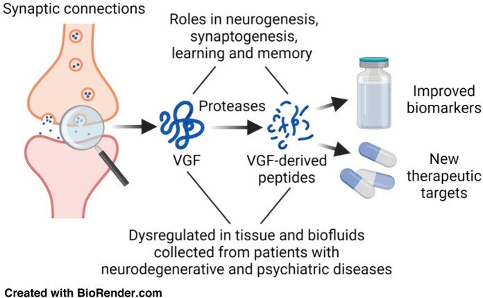
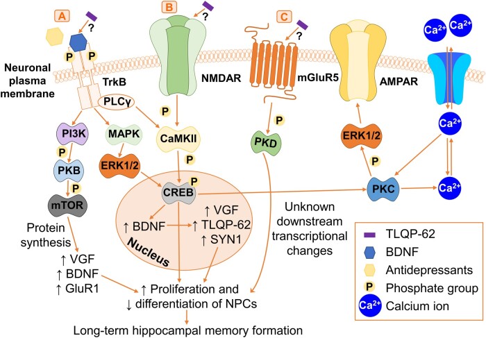
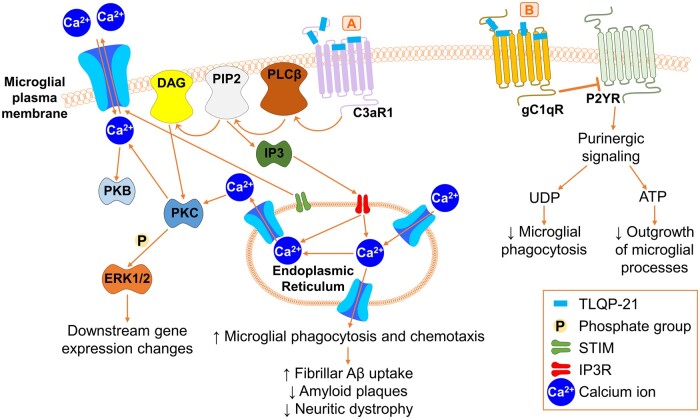
Neurosecretory protein VGF (non-acronymic) belongs to the granin family of neuropeptides. VGF and VGF-derived peptides have been repeatedly identified in well-powered and well-designed multi-omic studies as dysregulated in neurodegenerative and psychiatric diseases. New therapeutics is urgently needed for these devastating and costly diseases, as are new biomarkers to improve disease diagnosis and mechanistic understanding. From a list of 537 genes involved in Alzheimer's disease pathogenesis, VGF was highlighted by the Accelerating Medicines Partnership in Alzheimer's disease as the potential therapeutic target of greatest interest. VGF levels are consistently decreased in brain tissue and CSF samples from patients with Alzheimer's disease compared to controls, and its levels correlate with disease severity and Alzheimer's disease pathology. In the brain, VGF exists as multiple functional VGF-derived peptides. Full-length human VGF1-615 undergoes proteolytic processing by prohormone convertases and other proteases in the regulated secretory pathway to produce at least 12 active VGF-derived peptides. In cell and animal models, these VGF-derived peptides have been linked to energy balance regulation, neurogenesis, synaptogenesis, learning and memory, and depression-related behaviours throughout development and adulthood. The C-terminal VGF-derived peptides, TLQP-62 (VGF554-615) and TLQP-21 (VGF554-574) have differential effects on Alzheimer's disease pathogenesis, neuronal and microglial activity, and learning and memory. TLQP-62 activates neuronal cell-surface receptors and regulates long-term hippocampal memory formation. TLQP-62 also prevents immune-mediated memory impairment, depression-like and anxiety-like behaviours in mice. TLQP-21 binds to microglial cell-surface receptors, triggering microglial chemotaxis and phagocytosis. These actions were reported to reduce amyloid-β plaques and decrease neuritic dystrophy in a transgenic mouse model of familial Alzheimer's disease. Expression differences of VGF-derived peptides have also been associated with frontotemporal lobar dementias, amyotrophic lateral sclerosis, Lewy body diseases, Huntington's disease, pain, schizophrenia, bipolar disorder, depression and antidepressant response. This review summarizes current knowledge and highlights questions for future investigation regarding the roles of VGF and its dysregulation in neurodegenerative and psychiatric disease. Finally, the potential of VGF and VGF-derived peptides as biomarkers and novel therapeutic targets for neurodegenerative and psychiatric diseases is highlighted.

Keywords: VGF; biomarker; disease; neuropeptide; therapy.


Figures

Graphical Abstract
Graphical Abstract
Figure 1
Figure 1
Comparison of human and mouse VGF mRNA expression from the Allen Brain Atlas. (A) VGF mRNA expression data were downloaded from the Allen Mouse Brain Atlas; https://celltypes.brain-map.org/rnaseq/mouse (Accessed 03/30/2020). These data represent log2 VGF mRNA expression intensity in 265 different cell types across the whole mouse cortex and hippocampus obtained through single cell RNA-sequencing. Data were sorted by VGF expression and 27 representative cell types were included based on the following criteria: those with the highest VGF expression across a range of brain regions, different neuronal subtypes, glial cells and neurovascular unit cells to ensure a representative sample. (B) VGF mRNA expression data were downloaded from the Allen Human Brain Atlas; https://celltypes.brain-map.org/rnaseq/human/cortex (Accessed 03/30/2020). These data represent log2 VGF mRNA expression intensity in 120 different cell types across multiple human cortical regions (middle temporal gyrus, anterior cingulate gyrus, primary visual cortex, primary motor cortex, primary somatosensory cortex and primary auditory cortex) obtained through single nucleus RNA-sequencing. Data were sorted by VGF expression and 19 representative cell types were included based on the same criteria from the mouse atlas to ensure a representative sample. For each cell type examined, L refers to the layer(s) of the cortex where that cell type was identified along with their associated marker genes in italics. (C) Medial view and (D) lateral view of the predicted whole-brain mRNA expression of VGF using microarray data from the Allen Human Brain Atlas on the surface of the left cortical hemisphere. Colour scales represent log2 mRNA expression intensity and data were accessed from http://www.meduniwien.ac.at/neuroimaging/mRNA.html (Accessed 04/10/2020). Act = activated; Art = arterial; Astro = astrocyte; Cere = cerebellum; CC = cerebral cortex; Cho = cholinergic; Cor = cortex; DRG = dorsal root ganglion; Endo = endothelial cell; Exc = excitatory; Glut proj = glutamatergic projection; GL = granular layer; HB = hindbrain; HC = hippocampus; HT = hypothalamus; Inh = inhibitory; IN = interneuron; LRIN = long-range interneurons; Mat = mature; MG = microglia; MB, midbrain; NFL1 = neurofilament 1; Neu = neuron; Olf = olfactory; Olig = oligodendrocyte; OPC = oligodendrocyte progenitor cell; Ore = orexin-producing; Pep = peptidergic; Peri = pericyte; PV mac = perivascular macrophages; Sel = selective; Ser = serotonergic; SC = spinal cord; Tel = telencephalon; Thal = thalamus; VEC = vascular endothelial cells; VLMC = vascular and leptomeningeal cell.
Figure 2
Figure 2
Regulated processing and secretion of VGF and its resulting downstream effects on microglia and neurons. (A) Action potential-mediated VGF mRNA synthesis is triggered by different factors, including conventional cell depolarization, the antidepressant imipramine, exercise, EGF, IL-6, insulin, NGF, BDNF and NT3. (B) VGF is translated in the rough endoplasmic reticulum, where the signal peptide is removed to promote its sorting into the regulated secretory pathway. VGF promotes the biogenesis of immature LDCVs and targets itself into LDCVs in the Golgi apparatus. (C) Immature LDCVs containing VGF, PCs and other proteases are released from the Golgi apparatus and undergo homotypic fusion with other immature LDCVs. Inside the fused immature LDCVs, VGF is cleaved into a variety of active VGF-derived peptides by different proteases including PCs. (D) Anterograde axonal transport to the pre-synaptic terminal and local transport within the soma and to dendrites promotes condensation and maturation of LDCVs. (E) Mature LDCVs containing VGF and VGF-derived peptides undergo regulated secretion by exocytosis from the soma, dendrites, the axon and the pre-synaptic terminal. (F) VGF and VGF-derived peptides locally diffuse after regulated secretion where they bind to and activate different receptors on the soma, triggering action potentials and inducing VGF mRNA synthesis to rapidly replace VGF protein levels. (G) VGF and VGF-derived peptides, such as TLQP-62, diffuse across the synaptic cleft where they activate neuronal cell-surface receptors (Fig. 4). (H) VGF-derived peptides, such as TLQP-21 bind and activate microglial cell-surface receptors (Fig. 5). (I) Extracellular proteases cleave VGF and VGF-derived peptides, which reduces their active diffusion and is the only known mechanism for inhibiting their function. BDNF = brain-derived neurotrophic factor; EGF = epidermal growth factor; IL-6 = Interleukin-6; LDCV = large dense core vesicles; NGF = nerve growth factor; NT3 = neurotrophin-3; PC = prohormone convertase.
Figure 3
Figure 3
Proteolytic processing of VGF. Schematic of human VGF1–615 showing the known proteolytic cleavage sites from the literature as indicated by the P1–P1’ nomenclature of Schechter and Berger to indicate the amino acids N-terminal and C-terminal to the peptide bond that is cleaved. Putative PC cleavage sites are highlighted in bold while the other cleavage sites do not have a known protease responsible for their cleavage as determined by literature searches. Letters refer to the single amino acid code while the number refers to the position along the length of VGF. NERP = neuroendocrine regulatory peptide-1, -2, -3 and -4.
Figure 4
Figure 4
Activation of neuronal tyrosine kinase receptors, G-protein-coupled receptors and ionotropic glutamate receptors by TLQP-62. (A) Activation of the BDNF/TrkB pathway by TLQP-62 or antidepressants through TrkB phosphorylation initiates PI3K activation, which subsequently phosphorylates and activates PKB followed by mTOR. This induces VGF, BDNF and GluR1 protein synthesis, promoting NPC proliferation and decreasing NPC differentiation, which modulates long-term hippocampal memory formation. Activation of BDNF/TrkB sequentially activates MAPK followed by ERK1/2. This triggers CREB activation in the nucleus and leads to increased mRNA expression of BDNF followed by VGF and SYN1. This results in increased synapsin-1 and VGF protein synthesis, followed by TLQP-62 production, together promoting NPC-mediated long-term hippocampal memory formation. Activation of BDNF/TrkB triggers PLCγ on the plasma membrane to activate CaMKII, which converges with pathway B. (B) TLQP-62 binds via an unknown mechanism and activates the ionotropic glutamate receptor, NMDAR, which triggers the sequential phosphorylation and activation of CaMKII followed by CREB. CREB subsequently activates PKC, which regulates intracellular Ca2+ levels and phosphorylates and activates ERK1/2 to promote AMPAR formation on the plasma membrane. This contributes to long-term hippocampal memory formation. Activation of CREB also directly induces gene transcription to trigger NPC-mediated hippocampal memory formation. (C) TLQP-62 binds via an unknown mechanism and activates the GPCR, mGluR5, which induces the phosphorylation and activation of PKD. PKD induces downstream transcriptional changes that contribute to NPC-mediated long-term memory formation by an unknown mechanism. AMPAR = AMPA receptor; BDNF = brain-derived neurotrophic factor; CaMKII = calcium/calmodulin-dependent protein kinase II; CREB = cAMP response element-binding protein; ERK1/2 = extracellular-signal-regulated kinase 1/2; GluR1 = glutamate receptor 1; MAPK = mitogen-activated protein kinase; mGluR5 = metabotropic glutamate receptor 5; mTOR = mammalian target of rapamycin; NMDAR = NMDA receptor; NPCs = neural progenitor cells; PLCγ = phospholipase C-γ; PI3K = phosphoinositide 3-kinase; PKB = protein kinase B; PKC = protein kinase C; PKD = protein kinase D; TrkB = tropomyosin receptor kinase B.
Figure 5
Figure 5
Binding of TLQP-21 to microglial G-protein-coupled receptors. (A) TLQP-21 binds and activates microglial C3aR1, which sequentially activates PLCβ, PIP2 and DAG on the microglial plasma membrane. DAG activates PKC, which subsequently phosphorylates ERK1/2 and triggers downstream changes in gene expression. PIP2 activates IP3, which subsequently binds to IP3R on the surface of the endoplasmic reticulum. IP3R triggers an increase in the endoplasmic reticulum Ca2+ levels, which regulate intracellular and extracellular Ca2+ levels through STIM. Intracellular Ca2+ levels also regulate PKC and PKB activity. Ca2+ release from the endoplasmic reticulum increases microglial phagocytosis and chemotaxis. This triggered increased fibrillar Aβ uptake in vitro, while in 5xFAD mice, this decreased Aβ plaques and neuritic dystrophy. (B) TLQP-21 binds microglial gC1qR, which inhibits P2YR on the microglial plasma membrane. This inhibits UDP- and ATP-mediated purinergic signalling, which results in a reduction in microglial phagocytosis and a reduction in the outgrowth of microglial processes, respectively. C3aR1 = complement 3a receptor 1; DAG = diacylglycerol; ERK1/2 = extracellular-signal-regulated kinase 1/2; gC1qR = globular head of complement component 1q receptor; IP3 = inositol trisphosphate; IP3R = inositol trisphosphate receptor; PIP2 = phosphatidylinositol 4,5-bisphosphate; PLCβ = phospholipase C-β; PKB = protein kinase B; PKC = protein kinase C; P2YR = P2Y purinergic receptor; STIM = stromal interaction molecule; UDP = uridine diphosphate.

References

    1. Helle KB. The granin family of uniquely acidic proteins of the diffuse neuroendocrine system: Comparative and functional aspects. Biol Rev Camb Philos Soc. 2004;79(4):769–794. - PubMed
    1. Levi A, Eldridge JD, Paterson BM.. Molecular cloning of a gene sequence regulated by nerve growth factor. Science. 1985;229(4711):393–395. - PubMed
    1. Alder J, Thakker-Varia S, Bangasser DA, et al. Brain-derived neurotrophic factor-induced gene expression reveals novel actions of VGF in hippocampal synaptic plasticity. J Neurosci. 2003;23(34):10800–10808. - PMC - PubMed
    1. Bonni A, Ginty DD, Dudek H, Greenberg ME.. Serine 133-Phosphorylated CREB induces transcription via a cooperative mechanism that may confer specificity to neurotrophin signals. Mol Cell Neurosci. 1995;6(2):168–183. - PubMed
    1. Ferri GL, Possenti R.. vgf: A neurotrophin-inducible gene expressed in neuroendocrine tissues. Trends Endocrinol Metab. 1996;7(7):233–239. - PubMed

LinkOut - more resources