Review
. 2021 Oct 28;3(5):e200550.
doi: 10.1148/ryct.2021200550. eCollection 2021 Oct.

Coronary Artery Aneurysm in Kawasaki Disease: Coronary CT Angiography through the Lens of Pathophysiology and Differential Diagnosis

Affiliations
Review

Coronary Artery Aneurysm in Kawasaki Disease: Coronary CT Angiography through the Lens of Pathophysiology and Differential Diagnosis

Jenica Thangathurai et al. Radiol Cardiothorac Imaging. .

Abstract

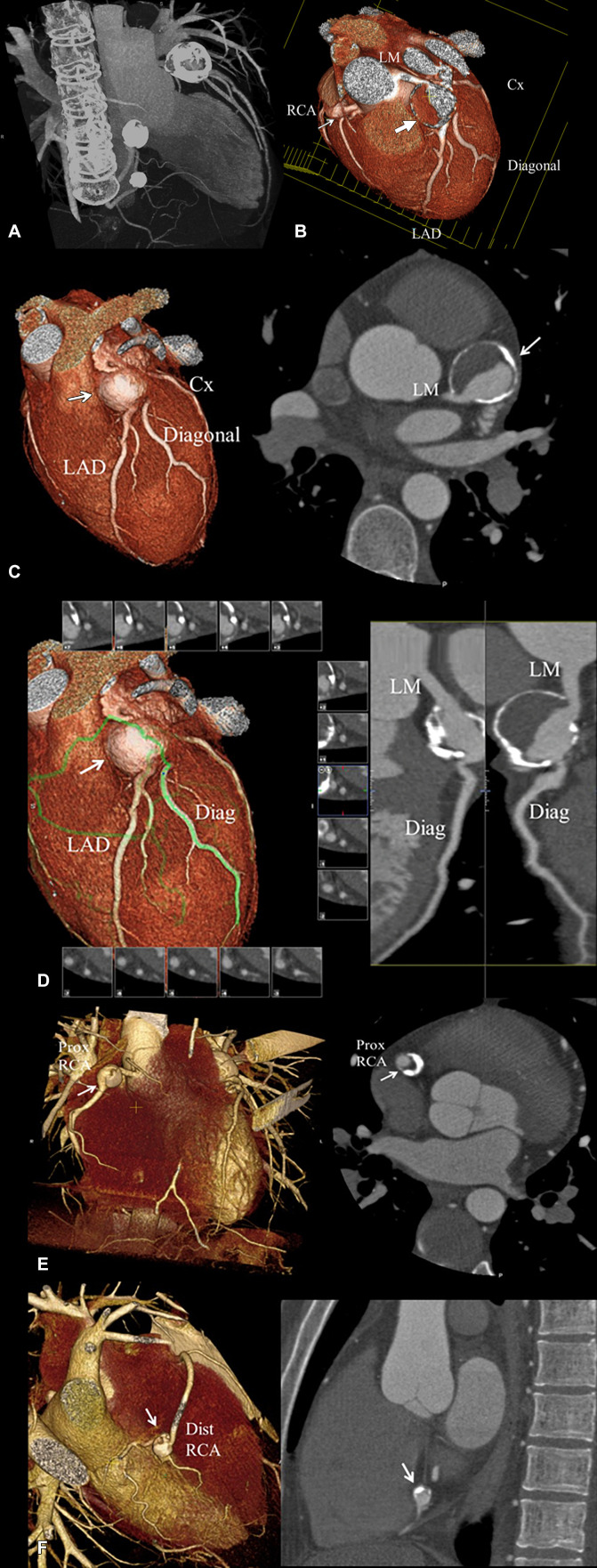
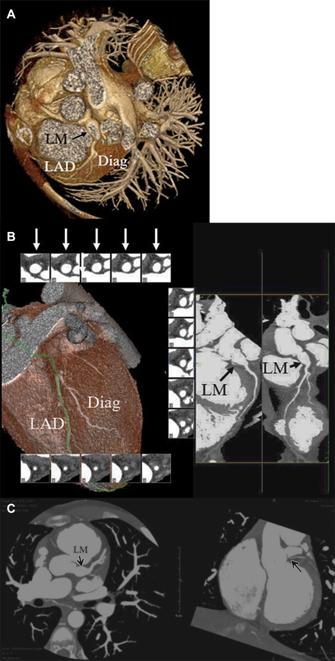
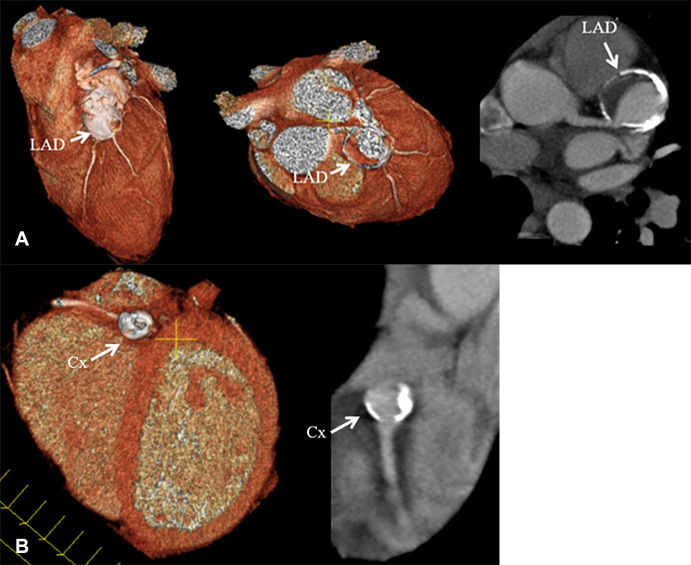
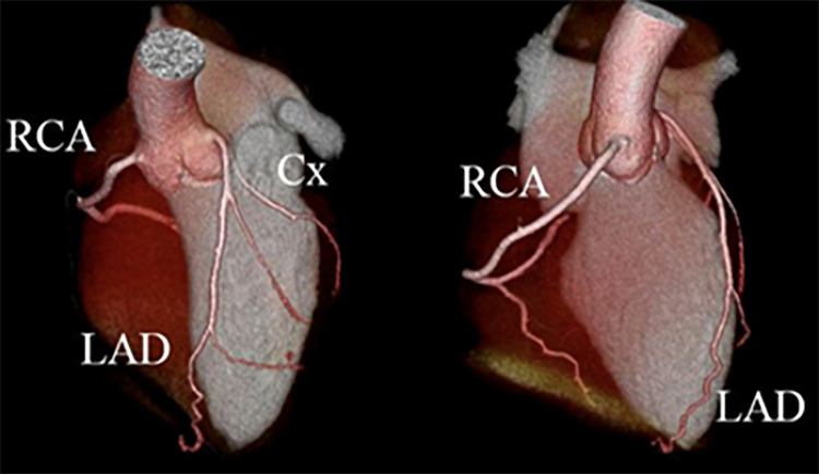
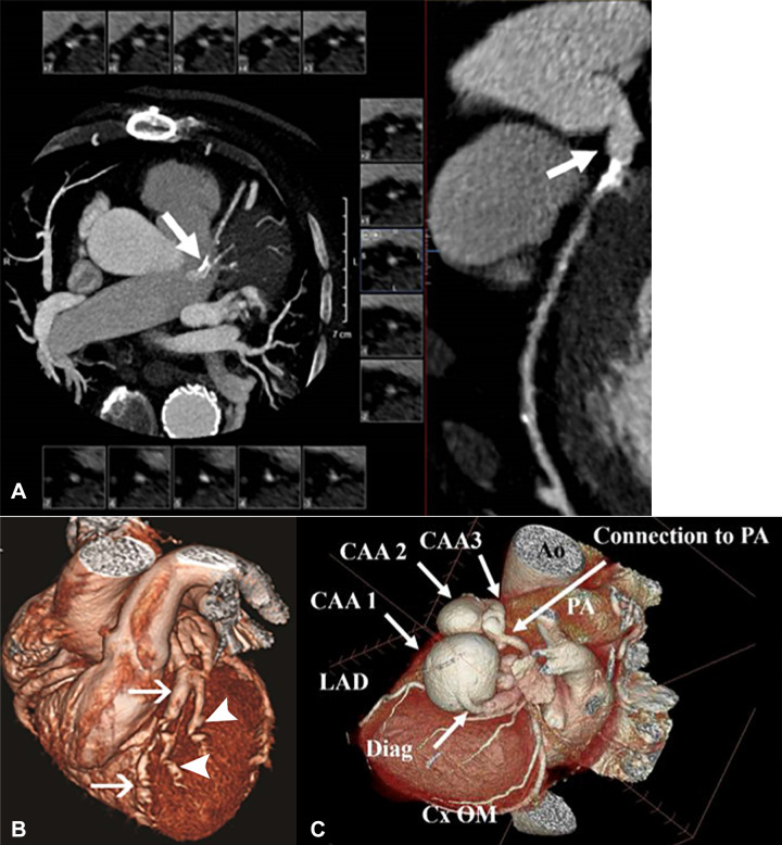
Kawasaki disease (KD) is an inflammatory autoimmune vasculitis affecting the coronary arteries of very young patients, which can result in coronary artery aneurysms (CAAs) with lifelong manifestations. Accurate identification and assessment of CAAs in the acute phase and sequentially during the chronic phase of KD is fundamental to the treatment plan for these patients. The differential diagnosis of CAA includes atherosclerosis, other vasculitic processes, connective tissue disorders, fistulas, mycotic aneurysms, and procedural sequelae. Understanding of the initial pathophysiology and evolutionary arterial changes is important to interpretation of imaging findings. There are multiple applicable imaging modalities, each with its own strengths, limitations, and role at various stages of the disease process. Coronary CT angiography is useful for evaluation of CAAs as it provides assessment of the entire coronary tree, CAA size, structure, wall, and lumen characteristics and visualization of other cardiothoracic vasculature. Knowledge of the natural history of KD, the spectrum of other conditions that can cause CAA, and the strengths and limitations of cardiovascular imaging are all important factors in imaging decisions and interpretation. Keywords: Pediatrics, Coronary Arteries, Angiography, Cardiac © RSNA, 2021.

Keywords: Angiography; Cardiac; Coronary Arteries; Pediatrics.


Conflict of interest statement

Disclosures of Conflicts of Interest: J.T. No relevant relationships. M.K. No relevant relationships. M.T. No relevant relationships. J.S.S. No relevant relationships.

Figures

Coronary CT angiographic images in a 25-year-old man with chronic phase
Kawasaki disease. CAAs are seen in the distal LM coronary artery extending into
the proximal LAD coronary artery, proximal RCA, and distal RCA, for which he was
continued to be given low-dose aspirin and anticoagulation indefinitely.
Features that may enhance long-term risk of myocardial ischemia in this patient
include presence of multiple large aneurysms across multiple coronary artery
branches, distal CAA, and layered mural thrombus. (A) Thick maximum intensity
projection image shows the heavy calcification of the CAA involving the LM
coronary artery extending into the LAD coronary artery, proximal RCA, and distal
RCA. (B) Three-dimensional volume-rendered image of the distal LM coronary
artery extending into the proximal LAD coronary artery (large arrow) viewed in
an editing plane that shows layered mural thrombus with a patent lumen. The
smaller proximal RCA aneurysm is also seen (small arrow). (C) Left:
Three-dimensional volume-rendered image shows the LM coronary artery and LAD CAA
(arrow). Right: Two-dimensional double oblique view shows the tissue
characteristics of the aneurysm with a calcified shell, layered mural thrombus,
and lumen opacified with iodinated contrast material (arrow). The distal LM
connection to the aneurysm is shown to be patent. (D) Left: Three-dimensional
volume-rendered image shows the curved multiplanar reformat path through the
LM-LAD artery aneurysm (arrow) and out the first diagonal (Diag) branch (green
line). Right: Two-dimensional curved multiplanar reformat view shows patency of
the LM into the aneurysms, aneurysm lumen, and diagonal branch ostium extending
from the aneurysm. (E) Left: Three-dimensional volume-rendered image shows the
proximal RCA aneurysm (arrow). Right: Two-dimensional double oblique view shows
the tissue characteristics of the aneurysm with a thick calcified shell, layered
mural thrombus, and lumen opacified with iodinated contrast material (arrow).
(F) Left: Three-dimensional volume-rendered image shows the distal RCA aneurysm
(arrow). Right: Two-dimensional double oblique view shows the tissue
characteristics of the aneurysm with a thick calcified shell, layered mural
thrombus, and lumen opacified with iodinated contrast material (arrow). CAA =
coronary artery aneurysm, Cx = circumflex, Diag = diagonal, Dist = distal, LAD =
left anterior descending, LM = left main, Prox = proximal, RCA = right coronary
artery.
Figure 1:
Coronary CT angiographic images in a 25-year-old man with chronic phase Kawasaki disease. CAAs are seen in the distal LM coronary artery extending into the proximal LAD coronary artery, proximal RCA, and distal RCA, for which he was continued to be given low-dose aspirin and anticoagulation indefinitely. Features that may enhance long-term risk of myocardial ischemia in this patient include presence of multiple large aneurysms across multiple coronary artery branches, distal CAA, and layered mural thrombus. (A) Thick maximum intensity projection image shows the heavy calcification of the CAA involving the LM coronary artery extending into the LAD coronary artery, proximal RCA, and distal RCA. (B) Three-dimensional volume-rendered image of the distal LM coronary artery extending into the proximal LAD coronary artery (large arrow) viewed in an editing plane that shows layered mural thrombus with a patent lumen. The smaller proximal RCA aneurysm is also seen (small arrow). (C) Left: Three-dimensional volume-rendered image shows the LM coronary artery and LAD CAA (arrow). Right: Two-dimensional double oblique view shows the tissue characteristics of the aneurysm with a calcified shell, layered mural thrombus, and lumen opacified with iodinated contrast material (arrow). The distal LM connection to the aneurysm is shown to be patent. (D) Left: Three-dimensional volume-rendered image shows the curved multiplanar reformat path through the LM-LAD artery aneurysm (arrow) and out the first diagonal (Diag) branch (green line). Right: Two-dimensional curved multiplanar reformat view shows patency of the LM into the aneurysms, aneurysm lumen, and diagonal branch ostium extending from the aneurysm. (E) Left: Three-dimensional volume-rendered image shows the proximal RCA aneurysm (arrow). Right: Two-dimensional double oblique view shows the tissue characteristics of the aneurysm with a thick calcified shell, layered mural thrombus, and lumen opacified with iodinated contrast material (arrow). (F) Left: Three-dimensional volume-rendered image shows the distal RCA aneurysm (arrow). Right: Two-dimensional double oblique view shows the tissue characteristics of the aneurysm with a thick calcified shell, layered mural thrombus, and lumen opacified with iodinated contrast material (arrow). CAA = coronary artery aneurysm, Cx = circumflex, Diag = diagonal, Dist = distal, LAD = left anterior descending, LM = left main, Prox = proximal, RCA = right coronary artery.
Coronary CT angiographic images in a 14-year-old boy with chronic phase
Kawasaki disease CAA who has undergone thrombectomy and aneurysm reduction
surgery. A residual fusiform aneurysm of the LM coronary artery, measuring up to
9 mm in diameter over a length of 11 mm was present. (A) Three-dimensional
volume-rendered image shows the LM CAA (arrow). (B) Two-dimensional curved
multiplanar and reformat views show patency of the LM into the aneurysm (long
black arrow), aneurysm lumen (short black arrow), and patency of the LAD and
first diagonal branch. Short-axis views of the LM aneurysm show a patent and
aneurysm lumen (white arrows). (C) Left: Two-dimensional double oblique view
shows patency of the LM into the aneurysm, aneurysm lumen, and LAD artery and
first diagonal branch (arrow). Right: Two-dimensional double oblique view shows
the LM residual aneurysm. The LCX coronary artery is occluded. A small
collateral vessel arises from the proximal LM coronary artery and supplies the
LCX territory (arrow). CAA = coronary artery aneurysm, Diag = diagonal, LAD =
left anterior descending, LCX = left circumflex artery, LM = left
main.
Figure 2:
Coronary CT angiographic images in a 14-year-old boy with chronic phase Kawasaki disease CAA who has undergone thrombectomy and aneurysm reduction surgery. A residual fusiform aneurysm of the LM coronary artery, measuring up to 9 mm in diameter over a length of 11 mm was present. (A) Three-dimensional volume-rendered image shows the LM CAA (arrow). (B) Two-dimensional curved multiplanar and reformat views show patency of the LM into the aneurysm (long black arrow), aneurysm lumen (short black arrow), and patency of the LAD and first diagonal branch. Short-axis views of the LM aneurysm show a patent and aneurysm lumen (white arrows). (C) Left: Two-dimensional double oblique view shows patency of the LM into the aneurysm, aneurysm lumen, and LAD artery and first diagonal branch (arrow). Right: Two-dimensional double oblique view shows the LM residual aneurysm. The LCX coronary artery is occluded. A small collateral vessel arises from the proximal LM coronary artery and supplies the LCX territory (arrow). CAA = coronary artery aneurysm, Diag = diagonal, LAD = left anterior descending, LCX = left circumflex artery, LM = left main.
Coronary CT angiographic images in a 15-year-old boy with chronic phase
Kawasaki disease CAAs show a heavily calcified, fusiform 1.5 × 0.8-cm
aneurysm present within the proximal LAD coronary artery, with prominent mural
thrombus. Contrast is seen in the LAD artery distal to the aneurysm. A more
saccular-shaped, heavily calcified 1.0 × 0.7-cm aneurysm was present
within the proximal LCX artery, without clinically significant mural thrombus.
Patient was continued to be given low-dose aspirin and anticoagulation
indefinitely. Features found in this patient that may enhance long-term risk of
myocardial ischemia include the presence of multiple giant aneurysms across
multiple coronary artery branches that contain layered mural thrombus and heavy
calcification. (A) Left: Three-dimensional volume-rendered image shows the
proximal LAD coronary artery aneurysm (arrow). Middle: Three-dimensional
volume-rendered image shows the LAD coronary artery aneurysm (arrow). The
editing plane shows the lumen of the LAD artery aneurysm with layered
nonocclusive mural thrombus. Right: Two-dimensional double oblique view shows
the tissue characteristics of the aneurysm with a calcified shell, layered mural
thrombus, and lumen opacified with iodinated contrast material (arrow). The LAD
artery connection to the aneurysm is shown to be patent. (B) Left:
Three-dimensional volume-rendered image shows the saccular-shaped, heavily
calcified aneurysm of the proximal LCX coronary artery (arrow). Right:
Two-dimensional double oblique view shows the tissue characteristics of the
aneurysm with a calcified shell, minimal mural thrombus, and patent lumen
opacified with iodinated contrast material (arrow). CAA = coronary artery
aneurysm, Cx = circumflex, LAD = left anterior descending, LCX = left
circumflex.
Figure 3:
Coronary CT angiographic images in a 15-year-old boy with chronic phase Kawasaki disease CAAs show a heavily calcified, fusiform 1.5 × 0.8-cm aneurysm present within the proximal LAD coronary artery, with prominent mural thrombus. Contrast is seen in the LAD artery distal to the aneurysm. A more saccular-shaped, heavily calcified 1.0 × 0.7-cm aneurysm was present within the proximal LCX artery, without clinically significant mural thrombus. Patient was continued to be given low-dose aspirin and anticoagulation indefinitely. Features found in this patient that may enhance long-term risk of myocardial ischemia include the presence of multiple giant aneurysms across multiple coronary artery branches that contain layered mural thrombus and heavy calcification. (A) Left: Three-dimensional volume-rendered image shows the proximal LAD coronary artery aneurysm (arrow). Middle: Three-dimensional volume-rendered image shows the LAD coronary artery aneurysm (arrow). The editing plane shows the lumen of the LAD artery aneurysm with layered nonocclusive mural thrombus. Right: Two-dimensional double oblique view shows the tissue characteristics of the aneurysm with a calcified shell, layered mural thrombus, and lumen opacified with iodinated contrast material (arrow). The LAD artery connection to the aneurysm is shown to be patent. (B) Left: Three-dimensional volume-rendered image shows the saccular-shaped, heavily calcified aneurysm of the proximal LCX coronary artery (arrow). Right: Two-dimensional double oblique view shows the tissue characteristics of the aneurysm with a calcified shell, minimal mural thrombus, and patent lumen opacified with iodinated contrast material (arrow). CAA = coronary artery aneurysm, Cx = circumflex, LAD = left anterior descending, LCX = left circumflex.
Coronary CT angiographic images in a 21-year-old man with a history of
Kawasaki disease in childhood. Three-dimensional reconstructions show no
evidence of coronary artery aneurysm. Cx = circumflex, LAD = left anterior
descending artery, RCA = right coronary artery.
Figure 4:
Coronary CT angiographic images in a 21-year-old man with a history of Kawasaki disease in childhood. Three-dimensional reconstructions show no evidence of coronary artery aneurysm. Cx = circumflex, LAD = left anterior descending artery, RCA = right coronary artery.
Non–Kawasaki disease CAAs. (A) Coronary CT angiographic (CCTA)
images in a 71-year-old man with multivessel coronary artery disease with an
atherosclerotic left main CAA. Left: Curved multiplanar reformat shows an
aneurysm. Right: Two-dimensional double oblique view. (B) CCTA image in a
29-year-old woman with systemic lupus erythematosus vasculitis with multiple
CAAs. Three-dimensional volume-rendered views with inclusion of the background
cardiac anatomy show multiple aneurysms in the LAD coronary artery (arrows) and
diagonal vessels (arrowheads). (Reprinted, with permission, from reference 56.)
(C) CCTA image in a 76-year-old man with multiple potential causes of CAA. A
three-dimensional reconstruction of three serial aneurysms of the diagonal
artery shows three giant aneurysms that were present in the first diagonal
branch of the LAD coronary artery. The largest measured 4 × 4.4 ×
3.6 cm, followed by two additional smaller aneurysms. The artery terminated into
a small (2.5 mm) fistulous tract to the main PA. The RCA ostium was mildly
aneurysmal. A right bronchial artery aneurysm and focal dilated peripheral
branches of the pulmonary arteries in the right upper lobe were also present.
Multiple causes are possible, including previous vasculitis, given the multiple
aneurysms in multiple vascular distributions, atherosclerotic vascular disease,
and the presence of a small arterial to venous fistula in the diagonal artery
aneurysms. Ao = aorta, CAA = coronary artery aneurysm, Cx = circumflex, Diag =
diagonal, LAD = left anterior descending, OM = obtuse marginal, PA = pulmonary
artery, RCA = right coronary artery.
Figure 5:
Non–Kawasaki disease CAAs. (A) Coronary CT angiographic (CCTA) images in a 71-year-old man with multivessel coronary artery disease with an atherosclerotic left main CAA. Left: Curved multiplanar reformat shows an aneurysm. Right: Two-dimensional double oblique view. (B) CCTA image in a 29-year-old woman with systemic lupus erythematosus vasculitis with multiple CAAs. Three-dimensional volume-rendered views with inclusion of the background cardiac anatomy show multiple aneurysms in the LAD coronary artery (arrows) and diagonal vessels (arrowheads). (Reprinted, with permission, from reference 56.) (C) CCTA image in a 76-year-old man with multiple potential causes of CAA. A three-dimensional reconstruction of three serial aneurysms of the diagonal artery shows three giant aneurysms that were present in the first diagonal branch of the LAD coronary artery. The largest measured 4 × 4.4 × 3.6 cm, followed by two additional smaller aneurysms. The artery terminated into a small (2.5 mm) fistulous tract to the main PA. The RCA ostium was mildly aneurysmal. A right bronchial artery aneurysm and focal dilated peripheral branches of the pulmonary arteries in the right upper lobe were also present. Multiple causes are possible, including previous vasculitis, given the multiple aneurysms in multiple vascular distributions, atherosclerotic vascular disease, and the presence of a small arterial to venous fistula in the diagonal artery aneurysms. Ao = aorta, CAA = coronary artery aneurysm, Cx = circumflex, Diag = diagonal, LAD = left anterior descending, OM = obtuse marginal, PA = pulmonary artery, RCA = right coronary artery.

References

    1. Kawasaki T. Acute febrile mucocutaneous syndrome with lymphoid involvement with specific desquamation of the fingers and toes in children [in Japanese]. Arerugi 1967;16(3):178–222. - PubMed
    1. Newburger JW, Takahashi M, Gerber MA, et al. . Diagnosis, treatment, and long-term management of Kawasaki disease: a statement for health professionals from the Committee on Rheumatic Fever, Endocarditis and Kawasaki Disease, Council on Cardiovascular Disease in the Young, American Heart Association. Circulation 2004;110(17):2747–2771. - PubMed
    1. Newburger JW, Takahashi M, Burns JC. Kawasaki Disease. J Am Coll Cardiol 2016;67(14):1738–1749. - PubMed
    1. Nakamura Y. Kawasaki disease: epidemiology and the lessons from it. Int J Rheum Dis 2018;21(1):16–19. - PubMed
    1. Nakamura Y, Yashiro M, Uehara R, Oki I, Watanabe M, Yanagawa H. Monthly observation of the number of patients with Kawasaki disease and its incidence rates in Japan: chronological and geographical observation from nationwide surveys. J Epidemiol 2008;18(6):273–279. - PMC - PubMed

LinkOut - more resources