Review
. 2021 Mar 3:2:219-234.
doi: 10.1016/j.jvssci.2021.01.002. eCollection 2021.

Translating mouse models of abdominal aortic aneurysm to the translational needs of vascular surgery

Affiliations
Review

Translating mouse models of abdominal aortic aneurysm to the translational needs of vascular surgery

Albert Busch et al. JVS Vasc Sci. .

Abstract

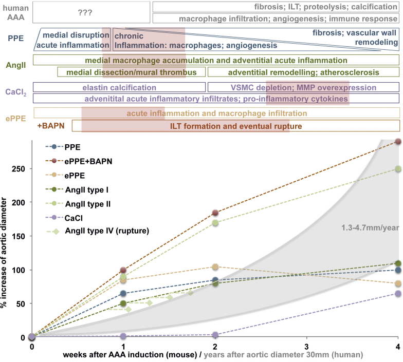
Introduction: Abdominal aortic aneurysm (AAA) is a condition that has considerable socioeconomic impact and an eventual rupture is associated with high mortality and morbidity. Despite decades of research, surgical repair remains the treatment of choice and no medical therapy is currently available. Animal models and, in particular, murine models, of AAA are a vital tool for experimental in vivo research. However, each of the different models has individual limitations and provide only partial mimicry of human disease. This narrative review addresses the translational potential of the available mouse models, highlighting unanswered questions from a clinical perspective. It is based on a thorough presentation of the available literature and more than a decade of personal experience, with most of the available models in experimental and translational AAA research.

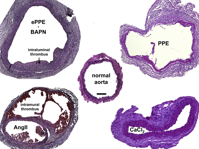
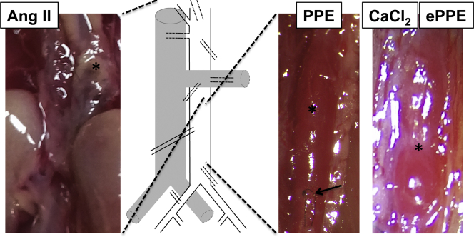
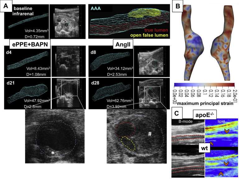
Results: From all the models published, only the four inducible models, namely the angiotensin II model (AngII), the porcine pancreatic elastase perfusion model (PPE), the external periadventitial elastase application (ePPE), and the CaCl2 model have been widely used by different independent research groups. Although the angiotensin II model provides features of dissection and aneurysm formation, the PPE model shows reliable features of human AAA, especially beyond day 7 after induction, but remains technically challenging. The translational value of ePPE as a model and the combination with β-aminopropionitrile to induce rupture and intraluminal thrombus formation is promising, but warrants further mechanistic insights. Finally, the external CaCl2 application is known to produce inflammatory vascular wall thickening. Unmet translational research questions include the origin of AAA development, monitoring aneurysm growth, gender issues, and novel surgical therapies as well as novel nonsurgical therapies.

Conclusion: New imaging techniques, experimental therapeutic alternatives, and endovascular treatment options provide a plethora of research topics to strengthen the individual features of currently available mouse models, creating the possibility of shedding new light on translational research questions.

Keywords: AAA; Abdominal aortic aneurysm; Aneurysm mouse models; Translational research.


Figures

Fig 1
Fig 1
Timeline of abdominal aortic aneurysm (AAA) development in mouse models. Although the data for aortic diameter enlargement are reported for different time points in many studies, the specific molecular characteristics of the aortic wall are mainly reported for the time of sacrifice and based on the specific study. In human disease, the timeline of events is largely unclear, since samples are only available from the time of surgery, normally when diameter exceeds 50 mm and also the initial stimulus for abdominal aortic aneurysm (AAA) development is completely unknown. However, human AAA tends to grow exponentially based on diameter (grey area) in a chronic manner. This graph shows the percentage growth of the aortic diameter for the first 4 weeks after AAA induction for various mouse models. This data are based on the available systematic reviews and leading articles and are semiquantitative only to compare aortic enlargement.,, , , For external periadventitial elastase application (ePPE), the addition of β-aminopropionitrile (BAPN) (red line) results in a marked increase in aneurysm diameter. For the angiotensin II model (AngII), the classification suggested by Daugherty et al in 2011 (see text) is included: type I (dark green; dilation <2 times baseline) and type II (light green: dilation <2 times baseline); type IV (light green rhomb: rupture) can occur at any time, most likely within days 4 to 10 after minipump implantation. The timeline of events in the aortic wall in comparison with the features of human disease can only be assumed for many of the models and specific details warrant further elucidation. The red boxes suggest time frames for interventional studies on AAA mouse models to suggest that not only initial stimulus-based, but human disease mimicking mechanisms are being interfered with. For most models, some aortic diameter data beyond 4 weeks after aneurysm induction is available (for ≤10 weeks) and demonstrates further flattening of the growth curve (not included in this figure). ILT, Intraluminal thrombus; VSMC, vascular smooth muscle cell.
Fig 2
Fig 2
Histologic review of inducible abdominal aortic aneurysm (AAA) murine models. Histologic cross-sections from 28 days after aneurysm induction are presented with hematoxylin and eosin staining. The normal mouse infrarenal aorta is approximately 500 μm in diameter when perfused and contains a medial layer with four to five elastic lamellar units. One layer of endothelial cells lines the inner luminal layer, and a surrounding adventitia is composed of mainly connective tissue. In the external periadventitial elastase application (ePPE) (β-aminopropionitrile [+BAPN]) model, the adventitia and media show cellular enrichment and the medial elastin breakdown. Most notably is the intraluminal thrombus (ILT). In contrast, for the angiotensin II model (AngII) aorta, the media remains mostly intact and cellular enrichment is more prominent in the adventitia. Note the thrombus formation in between the media and the adventitia (washed out in parts owing to fixation). In the PPE model, the media is mostly disrupted, and the adventitia shows increased fibrosis, signs of chronic inflammation and angiogenesis (not shown). Finally, in the CaCl2 model, the elastic fibers remain intact but thicken along with the adventitia and exhibits signs of fibrosis and inflammatory infiltrates in all layers. Scale bar = 100 μm; original magnification ×10; histologic images are courtesy of the authors.
Fig 3
Fig 3
Macroscopic review of inducible abdominal aortic aneurysm (AAA) murine models. The photographs reveal the aneurysm (∗) of individual models in situ. In the angiotensin II model (AngII), the maximum dilation occurs at the thoracoabdominal and visceral sections of the aorta (dashed lines). In the other models, the exact formation of aneurysm is due to the site of exposure of the aorta. Note the suture (arrow) from elastase perfusion in PPE and the vast retroperitoneal adhesion with the surrounding tissue in the CaCl2 and the external periadventitial elastase application (ePPE) (photo shown) model.
Fig 4
Fig 4
Surgical review of inducible murine aortic models. For the angiotensin II model (AngII), a subcutaneous tunnel is prepared through a small dorsal flank incision for the osmotic minipump (approximately 20 × 6 mm; blow-up) to gradually release the AngII over 28 days or more (A). For the other models, the mouse is put in a supine position and via a transabdominal incision the retroperitoneum and the aorta is exposed (blow-up) (B). The aorta is freed from its covering fascia in between the testicular arteries (T) and separated from the inferior vena cava (#) for topical soaking in elastase (external periadventitial elastase application [ePPE]) or CaCl2. For the PPE procedure, the aorta is prepared from the surrounding tissue circumferentially and temporary silk ligatures are placed for the insertion of the perfusion catheter (★) (C). Before restoration of the blood flow, this hole is closed with a 10-0 suture (magnified subfigure) (C). Exposure of the descending aorta (arrow) is achieved in intubated and ventilated mice (+) in a right lateral position after lateral thoracotomy (dotted line) and careful retraction of the left lung (L) (blow-up) (D). Similarly, aneurysm formation of the femoral artery is achieved by exposure of the artery (A) and separation from the femoral nerve (N) and vein (V) (upper blow-up). Topical application of elastase leads to diameter dilation from 200 to 450 μm after 2 weeks (lower blow-up) (E). Corresponding histologic sections (dotted black line) from these two different time points shows an increasing cellular density surrounding the artery (§).
Fig 5
Fig 5
Example data from advanced imaging approaches of the murine abdominal aorta. Volumetric high frequency ultrasound of external periadventitial elastase application (ePPE+) β-aminopropionitrile (BAPN) infrarenal abdominal aortic aneurysm (AAA) with intraluminal thrombus (ILT) () and angiotensin II model (AngII) suprarenal dissections with intramural thrombus (#), true lumen (red), and open false lumen (yellow) (12-mm aortic length measured; outer aortic diameter. D, diameter; d, postoperative day after aneurysm induction; Vol, Volume. (A). Three-dimensional (3D) data can be used to quantify AAA volume and allows for flexibility when measuring the maximum AAA diameter or length. The four-dimensional ultrasound data can be used to create cyclic strain maps using a direct deformation estimation approach that calculates the full 3D Green Lagrange strain tensor. In this AngII dissection, lower maximum first principal component strain values are observed within the dissection where higher amounts of collagen and intramural thrombus were present (blue) (B). Vibrational photoacoustic images of the infrarenal aorta uses endogenous contrast and reveals greater perivascular lipid accumulation in apolipoprotein E-deficient (ApoE-/-) mice compared to wild-type (wt) (C).

Similar articles

Cited by

References

    1. Oliver-Williams C., Sweeting M.J., Turton G., Parkin D., Cooper D., Rodd C. Lessons learned about prevalence and growth rates of abdominal aortic aneurysms from a 25-year ultrasound population screening programme. Br J Surg. 2018;105:68–74. - PubMed
    1. Cosford P.A., Leng G.C. Screening for abdominal aortic aneurysm. Cochrane Database Syst Rev. 2007:CD002945. - PubMed
    1. Lindeman J.H., Matsumura J.S. Pharmacologic management of aneurysms. Circ Res. 2019;124:631–646. - PMC - PubMed
    1. Wanhainen A., Verzini F., Van Herzeele I., Allaire E., Bown M., Cohnert T. Editor's choice - European Society for Vascular Surgery (ESVS) 2019 Clinical Practice Guidelines on the Management of Abdominal Aorto-iliac Artery Aneurysms. Eur J Vasc Endovasc Surg. 2019;57:8–93. - PubMed
    1. Chaikof E.L., Dalman R.L., Eskandari M.K., Jackson B.M., Lee W.A., Mansour M.A. The Society for Vascular Surgery practice guidelines on the care of patients with an abdominal aortic aneurysm. J Vasc Surg. 2018;67:2–77 e2. - PubMed

LinkOut - more resources