Review
. 2021;1(1):5.
doi: 10.1186/s44149-021-00005-9. Epub 2021 Apr 23.

The taxonomy, host range and pathogenicity of coronaviruses and other viruses in the Nidovirales order

Affiliations
Review

The taxonomy, host range and pathogenicity of coronaviruses and other viruses in the Nidovirales order

Zhijian Zhou et al. Anim Dis. 2021.

Abstract

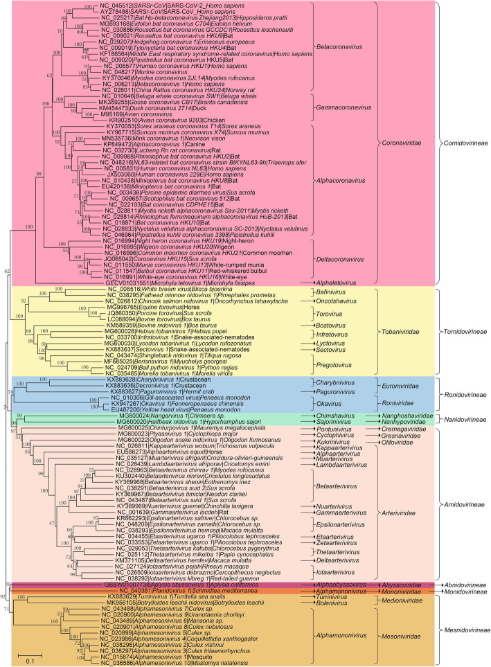
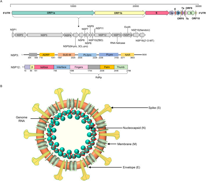
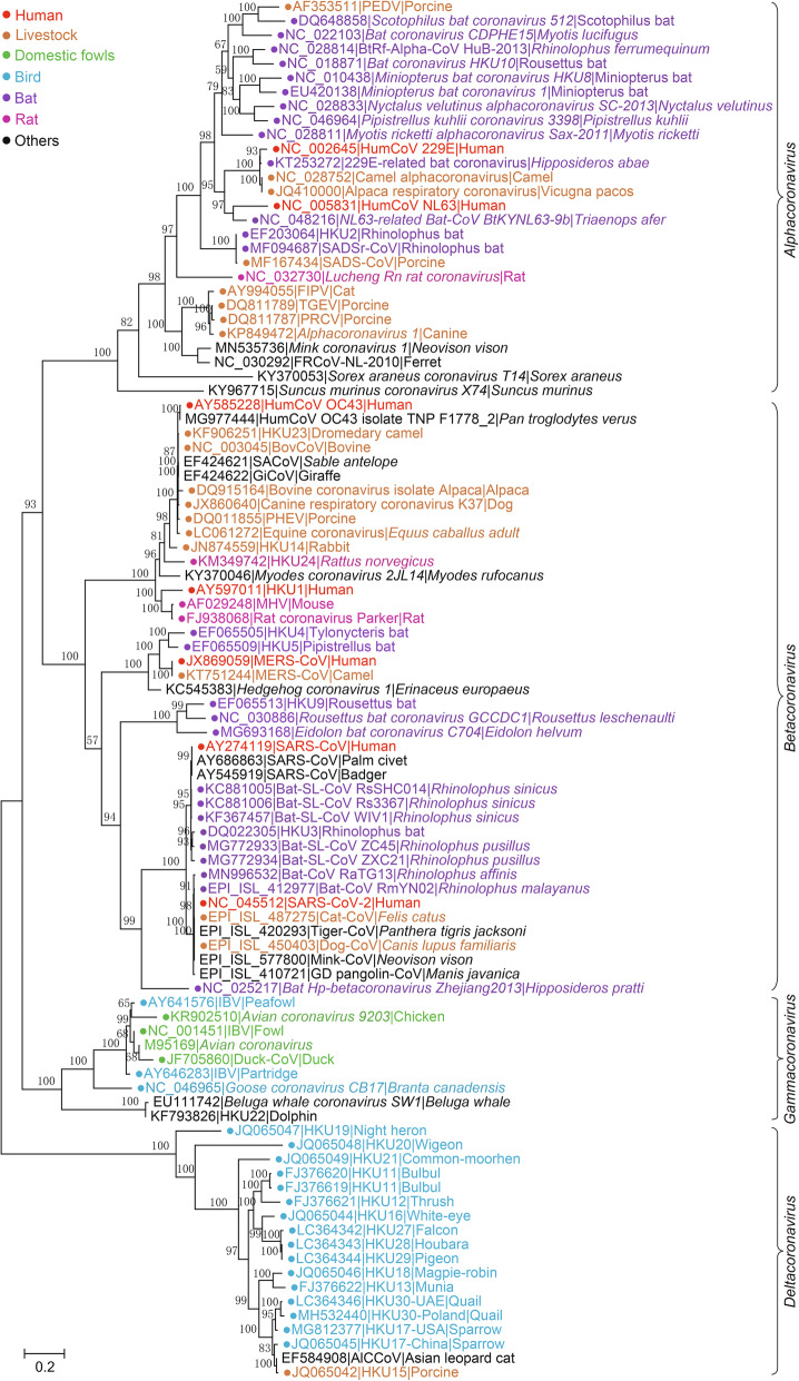
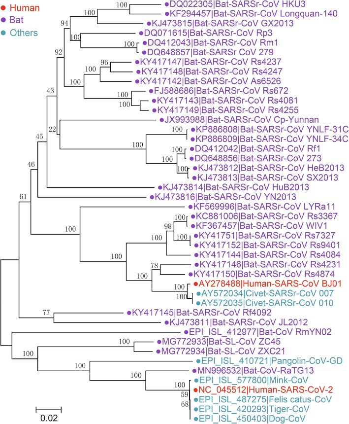
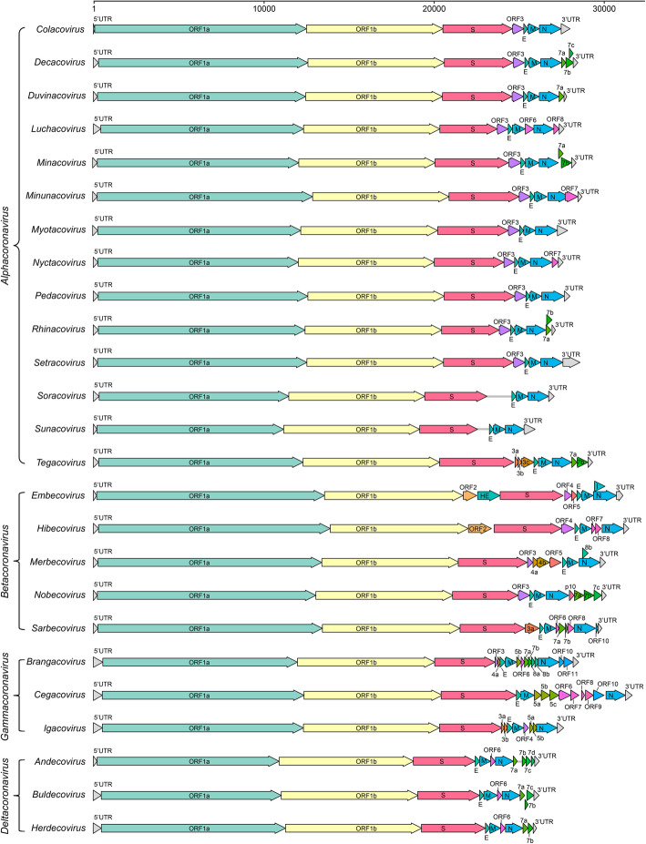
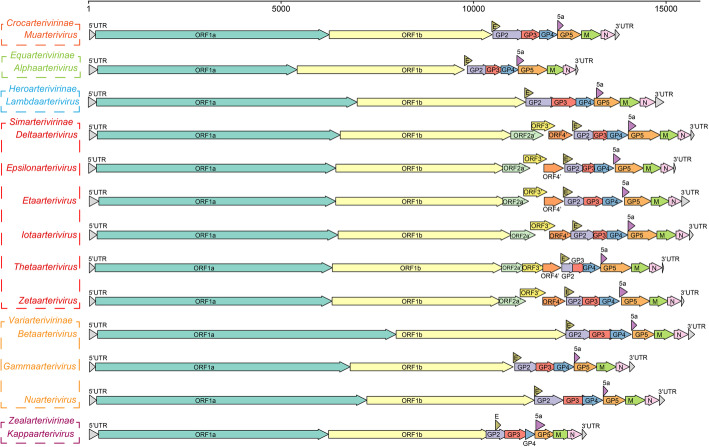
The frequent emergence of coronavirus (CoV) epidemics has seriously threatened public health and stock farming. The major hosts for CoVs are birds and mammals. Although most CoVs inhabit their specific natural hosts, some may occasionally cross the host barrier to infect livestock and even people, causing a variety of diseases. Since the beginning of the new century, increasing attention has been given to research on CoVs due to the emergence of highly pathogenic and genetically diverse CoVs that have caused several epidemics, including the recent COVID-19 pandemic. CoVs belong to the Coronaviridae family of the Nidovirales order. Recently, advanced techniques for viral detection and viral genome analyses have enabled characterization of many new nidoviruses than ever and have greatly expanded the Nidovirales order with new classification and nomenclature. Here, we first provide an overview of the latest research progress in the classification of the Nidovirales order and then introduce the host range, genetic variation, genomic pattern and pathogenic features of epidemic CoVs and other epidemic viruses. This information will promote understanding of the phylogenetic relationship and infectious transmission of various pathogenic nidoviruses, including epidemic CoVs, which will benefit virological research and viral disease control.

Keywords: Coronavirus; Evolution; Genetics; Hosts; Nidovirales; S protein.


Conflict of interest statement

Competing interestsThe authors declare that they have no competing interests.

Figures

Fig. 1
Fig. 1
Phylogenetic tree of RNA-dependent RNA polymerase (RdRp) amino acid sequences of 109 viral species in the Nidovirales order. The tree was constructed using the neighbor-joining method with a p-distance model and 1000 bootstraps in the MEGA V. 7.0.14
Fig. 2
Fig. 2
Genome structure, coding proteins, and viral particle structure diagram of coronavirus represented by SARS-CoV-2. a Genome structure, functional domains and locations of SARS-CoV-2. NSP, nonstructural proteins; 3Clpro, 3C-like protease; NiRAN, nidovirus RdRp-associated nucleotidyltransferase; RdRp, RNA-directed RNA polymerase; ZBD, Zn-binding domain covalently linked to HEL1; HEL1, helicase of superfamily 1; NendoU, endonuclease; 2′-O-MT, 2′-O-methyltransferase; ExoN, 3′-5′ exonuclease; SUD-M, SARS-unique domains; ADRP, ADP-ribose-1″-phosphatase; PLpro, papain-like protease; NAR, nucleic acid-binding domain. b Viral particle diagram of SARS-CoV-2. Structural proteins of S, N, M, and E are labeled, and the red line indicates the RNA genome
Fig. 3
Fig. 3
The maximum likelihood tree of coronaviruses based on amino acid sequences of RdRp gene. The tree was constructed using an IQ-tree with 10,000 ultrafast bootstraps and the most appropriate substitution model of LG+F+I+G4, which was calculated by ModelFinder
Fig. 4
Fig. 4
The neighbor-joining tree of spike gene nucleotide sequences in Sarbecovirus. The tree was constructed using the p-distance model and 1000 bootstraps in the MEGA V. 7.0.14. Viral strains derived from humans, bats, and other animals are indicated by red, purple, and cyan, respectively
Fig. 5
Fig. 5
Gene composition of 25 subgenera in 4 viral genera of Orthocoronavirinae. The reference strains used for mapping in each subgenus are as follows: Colacovirus (Bat-CoV CDPHE15/USA/2006, NC_022103.1), Decacovirus (Bat-CoV HKU10, NC_018871.1), Duvinacovirus (229E-related bat-CoV BtKY229E-1, KY073747.1), Luchacovirus (AcCoV-JC34, NC_034972.1), Minacovirus (FRCoV-NL-2010, NC_030292.1), Minunacovirus (Bat-CoV HKU8, NC_010438.1), Myotacovirus (BtMr-CoV/SAX2011, NC_028811.1), Nyctacovirus (Bat-CoV HKU33, MK720944.1), Pedacovirus (PEDV, NC_003436.1), Rhinacovirus (SADS-CoV, MT199592.1), Setracovirus (NL63-related bat-CoV BtKYNL63-9b, NC_048216.1), Soracovirus (Shrew-CoV/Tibet2014, KY370053.1), Sunacovirus (Wencheng Sm shrew CoV Xingguo-74, KY967715.1), Tegacovirus (CCoV/NTU336/F/2008, GQ477367.1), Embecovirus (Murine CoV, JX169867.1), Hibecovirus (Bat Hp-betacoronavirus/Zhejiang2013, NC_025217.1), Merbecovirus (Human MERS-CoV, NC_019843.3), Nobecovirus (Rousettus bat-CoV GCCDC1 356, NC_030886.1), Sarbecovirus (SARS-CoV-2, NC_045512.2), Brangacovirus (Canada goose CoV Cambridge_Bay_2017, MK359255.1), Cegacovirus (Beluga Whale CoV SW1, NC_010646.1), Igacovirus (Turkey CoV, NC_010800.1), Andecovirus (Wigeon CoV HKU20, NC_016995.1), Buldecovirus (Common-moorhen CoV HKU21, NC_016996.1), Herdecovirus (Night-heron CoV HKU19, NC_016994.1)
Fig. 6
Fig. 6
Gene composition of viruses in 13 viral genera in Arteriviridae. The reference strains used for mapping in each genus are as follows: Muarterivirus (Olivier's shrew virus 1, NC_035127.1), Alphaarterivirus (Equine arteritis virus, NC_002532.2), Lambdaarterivirus (Forest pouched giant rat arterivirus, NC_026439.1), Deltaarterivirus (Simian hemorrhagic fever virus, NC_003092.2), Epsilonarterivirus (Free State vervet virus, NC_029992.1), Etaarterivirus (Kibale red colobus virus 2, NC_034455.1), Iotaarterivirus (DeBrazzas monkey arterivirus, NC_026509.1), Thetaarterivirus (Kafue kinda chacma baboon virus, NC_029053.1), Zetaarterivirus (Kibale red colobus virus 1, NC_033553.1), Betaarterivirus (Rat arterivirus 1, NC_028963.1), Gammaarterivirus (Lactate dehydrogenase-elevating virus, NC_001639.1), Nuarterivirus (Rodent arterivirus, KY369969.1), Kappaarterivirus (Wobbly possum disease virus, NC_026811.2)

Similar articles

Cited by

References

    1. Azhar EI, El-Kafrawy SA, Farraj SA, Hassan AM, Al-Saeed MS, Hashem AM, Madani TA. Evidence for camel-to-human transmission of MERS coronavirus. The New England Journal of Medicine. 2014;370(26):2499–2505. doi: 10.1056/NEJMoa1401505. - DOI - PubMed
    1. Bailey AL, Lauck M, Ghai RR, Nelson CW, Heimbruch K, Hughes AL, Goldberg TL, Kuhn JH, Jasinska AJ, Freimer NB, et al. Arteriviruses, pegiviruses, and lentiviruses are common among wild African monkeys. Journal of Virology. 2016;90(15):6724–6737. doi: 10.1128/jvi.00573-16. - DOI - PMC - PubMed
    1. Bailey AL, Lauck M, Sibley SD, Friedrich TC, Kuhn JH, Freimer NB, Jasinska AJ, Phillips-Conroy JE, Jolly CJ, Marx PA, et al. Zoonotic potential of Simian arteriviruses. Journal of Virology. 2016;90(2):630–635. doi: 10.1128/JVI.01433-15. - DOI - PMC - PubMed
    1. Bailey AL, Lauck M, Weiler A, Sibley SD, Dinis JM, Bergman Z, Nelson CW, Correll M, Gleicher M, Hyeroba D, et al. High genetic diversity and adaptive potential of two simian hemorrhagic fever viruses in a wild primate population. PLoS One. 2014;9(3):e90714. doi: 10.1371/journal.pone.0090714. - DOI - PMC - PubMed
    1. Balasuriya UB, Carossino M. Reproductive effects of arteriviruses: equine arteritis virus and porcine reproductive and respiratory syndrome virus infections. Current Opinion in Virology. 2017;27:57–70. doi: 10.1016/j.coviro.2017.11.005. - DOI - PubMed

LinkOut - more resources