

Fully automated identification of brain abnormality from whole-body FDG-PET imaging using deep learning-based brain extraction and statistical parametric mapping
- PMID: 34778923
- PMCID: PMC8590988
- DOI: 10.1186/s40658-021-00424-0
Fully automated identification of brain abnormality from whole-body FDG-PET imaging using deep learning-based brain extraction and statistical parametric mapping
Abstract
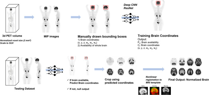
Background: The whole brain is often covered in [18F]Fluorodeoxyglucose positron emission tomography ([18F]FDG-PET) in oncology patients, but the covered brain abnormality is typically screened by visual interpretation without quantitative analysis in clinical practice. In this study, we aimed to develop a fully automated quantitative interpretation pipeline of brain volume from an oncology PET image.
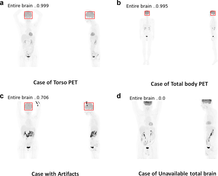
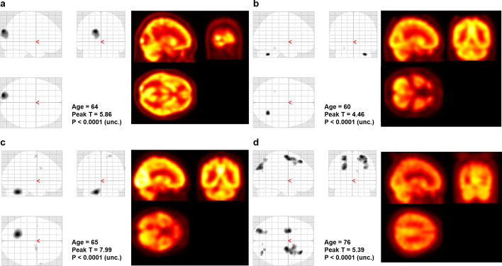
Method: We retrospectively collected 500 oncologic [18F]FDG-PET scans for training and validation of the automated brain extractor. We trained the model for extracting brain volume with two manually drawn bounding boxes on maximal intensity projection images. ResNet-50, a 2-D convolutional neural network (CNN), was used for the model training. The brain volume was automatically extracted using the CNN model and spatially normalized. For validation of the trained model and an application of this automated analytic method, we enrolled 24 subjects with small cell lung cancer (SCLC) and performed voxel-wise two-sample T test for automatic detection of metastatic lesions.
Result: The deep learning-based brain extractor successfully identified the existence of whole-brain volume, with an accuracy of 98% for the validation set. The performance of extracting the brain measured by the intersection-over-union of 3-D bounding boxes was 72.9 ± 12.5% for the validation set. As an example of the application to automatically identify brain abnormality, this approach successfully identified the metastatic lesions in three of the four cases of SCLC patients with brain metastasis.
Conclusion: Based on the deep learning-based model, extraction of the brain volume from whole-body PET was successfully performed. We suggest this fully automated approach could be used for the quantitative analysis of brain metabolic patterns to identify abnormalities during clinical interpretation of oncologic PET studies.
Keywords: Brain FDG-PET; Brain segmentation; Convolutional neural network; Deep learning; FDG-PET; Quantitative PET analysis.
© 2021. The Author(s).
Conflict of interest statement
The authors declare that they have no competing interests.
Figures
Similar articles
-
Deep Learning-Based Feature Extraction from Whole-Body PET/CT Employing Maximum Intensity Projection Images: Preliminary Results of Lung Cancer Data.Nucl Med Mol Imaging. 2023 Oct;57(5):216-222. doi: 10.1007/s13139-023-00802-9. Epub 2023 Apr 19. Nucl Med Mol Imaging. 2023. PMID: 37720886 Free PMC article.
-
18F-FDG PET/CT Uptake Classification in Lymphoma and Lung Cancer by Using Deep Convolutional Neural Networks.Radiology. 2020 Feb;294(2):445-452. doi: 10.1148/radiol.2019191114. Epub 2019 Dec 10. Radiology. 2020. PMID: 31821122
-
Computer-aided detection and segmentation of malignant melanoma lesions on whole-body 18F-FDG PET/CT using an interpretable deep learning approach.Comput Methods Programs Biomed. 2022 Jun;221:106902. doi: 10.1016/j.cmpb.2022.106902. Epub 2022 May 22. Comput Methods Programs Biomed. 2022. PMID: 35636357
-
A deep learning framework for 18F-FDG PET imaging diagnosis in pediatric patients with temporal lobe epilepsy.Eur J Nucl Med Mol Imaging. 2021 Jul;48(8):2476-2485. doi: 10.1007/s00259-020-05108-y. Epub 2021 Jan 9. Eur J Nucl Med Mol Imaging. 2021. PMID: 33420912 Free PMC article.
-
Generation of PET Attenuation Map for Whole-Body Time-of-Flight 18F-FDG PET/MRI Using a Deep Neural Network Trained with Simultaneously Reconstructed Activity and Attenuation Maps.J Nucl Med. 2019 Aug;60(8):1183-1189. doi: 10.2967/jnumed.118.219493. Epub 2019 Jan 25. J Nucl Med. 2019. PMID: 30683763 Free PMC article.
Cited by
-
Artificial intelligence to detect malignant eyelid tumors from photographic images.NPJ Digit Med. 2022 Mar 2;5(1):23. doi: 10.1038/s41746-022-00571-3. NPJ Digit Med. 2022. PMID: 35236921 Free PMC article.
-
Deep Learning-Based Feature Extraction from Whole-Body PET/CT Employing Maximum Intensity Projection Images: Preliminary Results of Lung Cancer Data.Nucl Med Mol Imaging. 2023 Oct;57(5):216-222. doi: 10.1007/s13139-023-00802-9. Epub 2023 Apr 19. Nucl Med Mol Imaging. 2023. PMID: 37720886 Free PMC article.
References
-
- Kamel EM, Zwahlen D, Wyss MT, Stumpe KD, von Schulthess GK, Steinert HC. Whole-body 18F-FDG PET improves the management of patients with small cell lung cancer. J Nucl Med. 2003;44(12):1911–1917. - PubMed
Grants and funding
LinkOut - more resources
Full Text Sources
Research Materials
