. 2022 Jan;96(1):321-334.
doi: 10.1007/s00204-021-03186-x. Epub 2021 Nov 15.

Organophosphorus pesticides exhibit compound specific effects in rat precision-cut lung slices (PCLS): mechanisms involved in airway response, cytotoxicity, inflammatory activation and antioxidative defense

Affiliations

Organophosphorus pesticides exhibit compound specific effects in rat precision-cut lung slices (PCLS): mechanisms involved in airway response, cytotoxicity, inflammatory activation and antioxidative defense

Jonas Tigges et al. Arch Toxicol. 2022 Jan.

Abstract

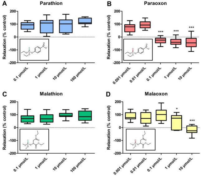
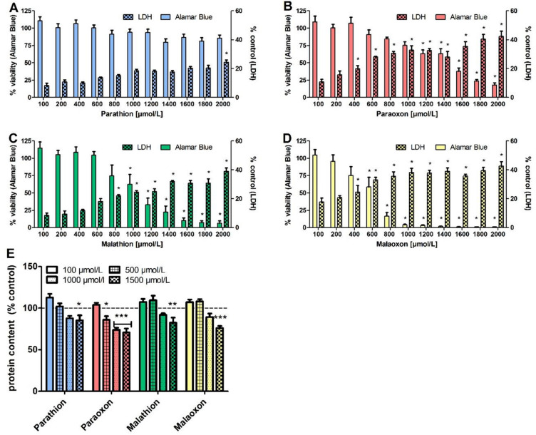
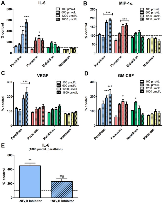
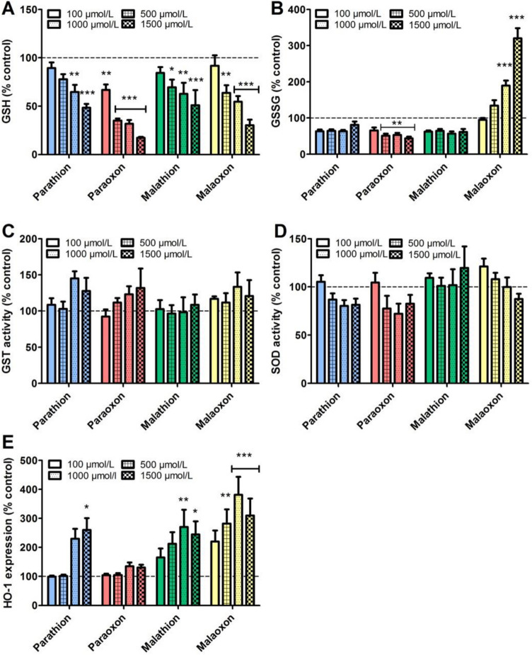
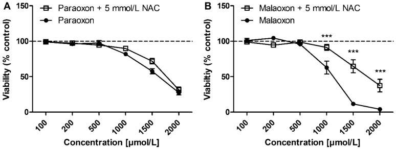
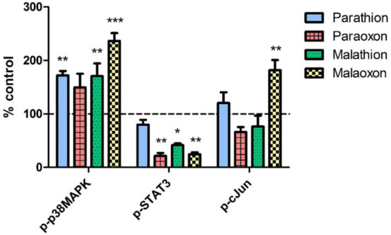
Organophosphorus compound pesticides (OP) are widely used in pest control and might be misused for terrorist attacks. Although acetylcholinesterase (AChE) inhibition is the predominant toxic mechanism, OP may induce pneumonia and formation of lung edema after poisoning and during clinical treatment as life-threatening complication. To investigate the underlying mechanisms, rat precision-cut lung slices (PCLS) were exposed to the OP parathion, malathion and their biotransformation products paraoxon and malaoxon (100-2000 µmol/L). Airway response, metabolic activity, release of LDH, cytokine expression and oxidative stress response were analyzed. A concentration-dependent inhibition of airway relaxation was observed after exposure with the oxon but not with the thion-OP. In contrast, cytotoxic effects were observed for both forms in higher concentrations. Increased cytokine expression was observed after exposure to parathion and paraoxon (IL-6, GM-CSF, MIP-1α) and IL-6 expression was dependent on NFκB activation. Intracellular GSH levels were significantly reduced by all four tested OP but an increase in GSSG and HO-1 expression was predominantly observed after malaoxon exposure. Pretreatment with the antioxidant N-acetylcysteine reduced malaoxon but not paraoxon-induced cytotoxicity. PCLS as a 3D lung model system revealed OP-induced effects depending on the particular OP. The experimental data of this study contribute to a better understanding of OP toxicity on cellular targets and may be a possible explanation for the variety of clinical outcomes induced by different OP.

Keywords: Bronchoconstriction; Inflammation; Organophosphates; Oxidative stress; PCLS.


Conflict of interest statement

The authors declare that they have no conflict of interest.

Figures

Fig. 1
Fig. 1
Inhibitory effect of parathion, paraoxon, malathion and malaoxon on airway relaxation after acetylcholine stimulus. PCLS airways exposed for 3 min to parathion (A), paraoxon (B), malathion (C) or malaoxon (D) in different concentrations (0.001–100 µmol/L). Upon acetylcholine stimulus (0.5 µmol/L; 2 min), airway constriction was recorded. Subsequent airway relaxation was evaluated after 60 min. Relaxation of the control was set as 100%. Boxplots show mean with minimum and maximum values. Asterisks indicate significant differences to the control (*p < 0.05, ***p < 0.001; n = 10 airways from at least three different animals)
Fig. 2
Fig. 2
Effects of parathion, paraoxon, malathion and malaoxon on viability, cell death and protein content. PCLS were exposed for 24 h to different concentrations of parathion (A), paraoxon (B), malathion (C) or malaoxon (D) (10–2000 µmol/L). Viability was analyzed by Alamar Blue assay and effects on cellular death were evaluated by measurement of LDH release into PCLS supernatant. For detection of severe tissue destruction, intracellular protein content was detected by BCA assay (E). Data are presented as % of control (Alamar Blue assay and protein content) or as % of the positive control Triton-X (LDH). Data are shown as mean ± SEM. Asterisk indicate significant differences to control (*p < 0.05; **p < 0.01; ***p < 0.001; n = 6 PCLS from three different animals)
Fig. 3
Fig. 3
Expression of cytokines in PCLS after parathion, paraoxon, malathion and malaoxon exposure. PCLS were exposed to increasing concentrations of paraoxon, parathion, malaoxon, malathion (100–1800 µmol/L) for 8 h. Cytokine expression of IL-6 (A), MIP-1α (B), VEGF (C) and GM-CSF (D) was detected in the supernatant and cytosolic fraction using a Bioplex-system and was afterwards combined to analyze the overall cytokine production. Observed concentrations are corrected for the protein content and are shown as fold increase of control (indicated by dashed line). For evaluation of NF-κB activation, PCLS were exposed to parathion (1800 µmol/L) with or without addition of an NF-κB Activation Inhibitor (10 µmol/L; E). Interleukin-6 was detected in the supernatant and cytosolic fraction by ELISA, and afterwards combined to analyze the overall cytokine production. Data are shown as mean ± SEM. Asterisk indicate significant differences to the control (*p < 0.05; **p < 0.01; ***p < 0.001; n = 8 samples from four different animals) Hash characters indicate significant differences to parathion exposure without inhibitor (##p < 0.01; n = 8 samples from four different animals)
Fig. 4
Fig. 4
Effect of parathion, paraoxon, malathion and malaoxon exposure on reduced and oxidized glutathione content, GST- and SOD activity and expression of HO-1. PCLS were exposed for 8 h to different concentrations of paraoxon, parathion, malaoxon, malathion (100–1500 µmol/L). Afterwards the intracellular level of reduced (A) and oxidized (B) glutathione was detected. Furthermore, intracellular activity of GST (C) and SOD (D) was evaluated. Expression of intracellular HO-1 was evaluated by ELISA and corrected for the protein concentration. All results are shown as % of control (indicated by dashed line). Data are shown as mean ± SEM. Asterisk indicate significant differences to the control (*< 0.05; **p < 0.01; ***p < 0.001; n = 6 PCLS from three different animals (GST and SOD Activity Assay) n = 8 PCLS from four different animals (GSH, GSSG, HO-1))
Fig. 5
Fig. 5
Effects of N-acetylcysteine (NAC) pre-treatment on PCLS viability. PCLS were pre-treated with 5 mmol/L of NAC for 4 h. Afterwards medium was replaced by paraoxon (A) or malaoxon (B) containing medium for 24 h and viability was analyzed by Alamar Blue assay. Viability is shown as % of control (indicated by dashed line). Data are shown as mean ± SEM. Asterisk indicate significant differences to PCLS without NAC pre-treatment (*p < 0.05; n = 6 PCLS from three different animals)
Fig. 6
Fig. 6
Phosphorylation of signaling pathways after OP exposure. PCLS were exposed to 1000 µmol/L of parathion, paraoxon, malathion or malaoxon for 8 h. Intracellular levels of the phosphorylated proteins p-p38MAPK, p-STAT3 and p-c-Jun were evaluated using a Bioplex system. Data are calculated as % of control (indicated by dashed line) and are shown as mean ± SEM. Asterisks indicate significant differences to the control (*p < 0.05; n = 4 samples from four different animals)

Similar articles

Cited by

References

    1. Abdo W, Elmadawy MA, Abdelhiee EY, Abdel-Kareem MA, Farag A, Aboubakr M, Ghazy E, Fadl SE. Protective effect of thymoquinone against lung intoxication induced by malathion inhalation. Sci Rep. 2021;11(1):2498. doi: 10.1038/s41598-021-82083-w. - DOI - PMC - PubMed
    1. Afonso V, Champy R, Mitrovic D, Collin P, Lomri A. Reactive oxygen species and superoxide dismutases: role in joint diseases. Jt Bone Spine. 2007;74(4):324–329. doi: 10.1016/j.jbspin.2007.02.002. - DOI - PubMed
    1. Angelini DJ, Moyer RA, Cole S, Willis KL, Oyler J, Dorsey RM, Salem H. The pesticide metabolites paraoxon and malaoxon induce cellular death by different mechanisms in cultured human pulmonary cells. Int J Toxicol. 2015;34(5):433–441. doi: 10.1177/1091581815593933. - DOI - PubMed
    1. Banerjee BD, Seth V, Bhattacharya A, Pasha ST, Chakraborty AK. Biochemical effects of some pesticides on lipid peroxidation and free-radical scavengers. Toxicol Lett. 1999;107(1–3):33–47. doi: 10.1016/S0378-4274(99)00029-6. - DOI - PubMed
    1. Behrsing HP, Furniss MJ, Davis M, Tomaszewski JE, Parchment RE. In vitro exposure of precision-cut lung slices to 2-(4-amino-3-methylphenyl)-5-fluorobenzothiazole lysylamide dihydrochloride (NSC 710305, Phortress) increases inflammatory cytokine content and tissue damage. Toxicol Sci. 2013;131(2):470–479. doi: 10.1093/toxsci/kfs319. - DOI - PMC - PubMed

Publication types

Grants and funding