A Case of Erythema Elevatum Diutinum (EED) Exhibiting A Keloid-Like Appearance
- PMID: 34778936
- PMCID: PMC8611145
- DOI: 10.1007/s13555-021-00639-0
A Case of Erythema Elevatum Diutinum (EED) Exhibiting A Keloid-Like Appearance
Abstract
Introduction: Most severe-appearing keloids tend to occur around joints because of the increased extensional stimulation of the scar in those areas. However, erythema elevatum diutinum (EED) appears more commonly on friction sites including extensor surfaces of the extremities and dorsal surfaces of joints. EEDs also presents as red-brown and elevated lesions.
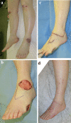
Case presentation: In this report, we describe a 42-year-old female who presented with firm, sporadic, brown-colored raised nodules on her bilateral lower extremities. As the appearance of these nodules resembled keloids, resection of the affected area with subsequent radiation therapy was initiated. However, histopathologic examination performed after treatment revealed tuberous lesions in the dermis, increased wired collagen fibers, neutrophilic infiltrate with nuclear dust, and edematous endothelial cells in the small vessels. Consequently, the patient was later diagnosed with EED. Post-surgery, no recurrence or abnormal scars appeared.
Discussion: Whereas clinical findings of EED are similar to that of keloids, the mechanisms of the two conditions differ considerably, leading to varying management strategies. EEDs can be misdiagnosed as keloids on several grounds; they can both appear morphologically similar, exhibit as stiff lesions, demonstrate chronic inflammation of the reticular dermis, and appear anywhere on the body. The only definitive method of differentiating between the two is through histopathologic examination.
Conclusion: EED should be considered as one of the differential diagnoses for any patients presenting with keloid-like lesions on friction sites and biopsy should be performed prior to resection and radiotherapy.
Keywords: Abnormal scars; Erythema elevatum diutinum; Keloids; Nodules.
© 2021. The Author(s).
Figures


Similar articles
-
[Erythema elevatum diutinum associated with pyoderma gangrenosum in an HIV-positive patient].Ann Dermatol Venereol. 2010 May;137(5):386-90. doi: 10.1016/j.annder.2010.03.025. Ann Dermatol Venereol. 2010. PMID: 20470922 French.
-
Atypical Presentation of Erythema Elevatum Diutinum in a Patient With Hashimoto's Disease.Cureus. 2021 Sep 23;13(9):e18214. doi: 10.7759/cureus.18214. eCollection 2021 Sep. Cureus. 2021. PMID: 34722026 Free PMC article.
-
Cutaneous Leiomyoma Mimicking a Keloid.Acta Dermatovenerol Croat. 2020 Aug;28(2):116. Acta Dermatovenerol Croat. 2020. PMID: 32876039
-
[Erythema elevatum diutinum. A rare dermatosis with a broad spectrum of associated illnesses].Hautarzt. 1997 Feb;48(2):113-7. doi: 10.1007/s001050050556. Hautarzt. 1997. PMID: 9173057 Review. German.
-
HIV-associated erythema elevatum diutinum: a case report and review of a clinically distinct variant.Dermatol Online J. 2018 May 15;24(5):13030/qt38c4c9g3. Dermatol Online J. 2018. PMID: 30142732 Review.
References
-
- Berman B, Maderal A, Raphael B. Keloids and hypertrophic scars: pathophysiology, classification, and treatment. 2017;S3–18. 10.1097/DSS.0000000000000819 - PubMed
LinkOut - more resources
Full Text Sources
Research Materials

