

Identification of multinucleated cells in human kidney cortex: A way for tissue repairing?
- PMID: 34778949
- PMCID: PMC9005679
- DOI: 10.1111/joa.13595
Identification of multinucleated cells in human kidney cortex: A way for tissue repairing?
Abstract
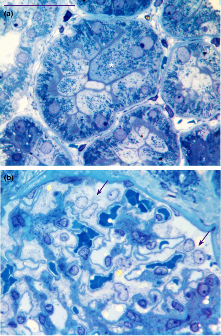
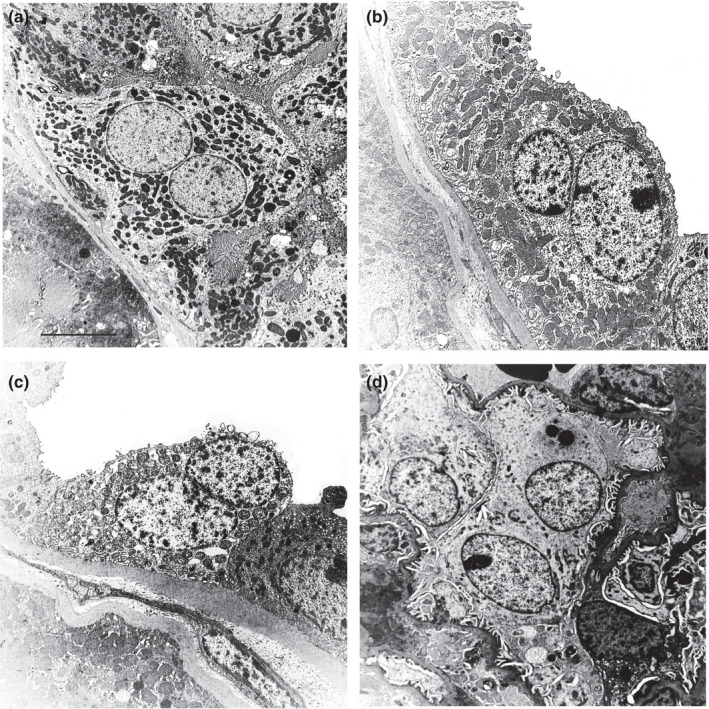
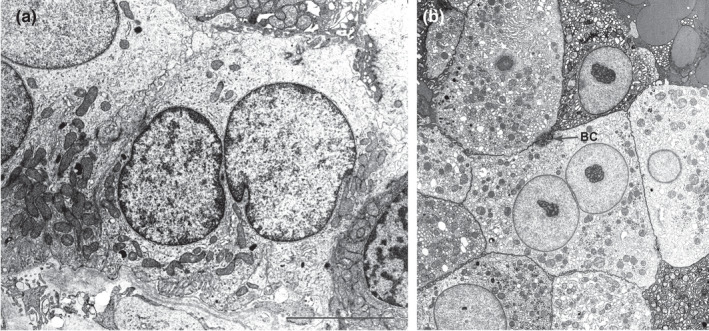
The presence of multinucleated cells has never been demonstrated in renal tissue, although, polyploid cells were recently observed in the tubules of normal and pathological human kidney. Therefore, the aim of the present study is to identify and quantify, by electron microscopy, multinucleated cells in the cortical tissue of normal human kidney i.e., in the three compartments of renal tubule: the proximal tubule (PT), the distal tubule (DT), and the collecting duct (CD), as well as, in the glomerulus (podocytes). The percentage of the multinucleated cells observed was 5% (95%CI: 3.6%-6.7%) in renal cortical tubules with distribution in each tubular compartment of 6% in PT, 4% in DT and 3% in CD with no statistically significant difference in the distribution of multinucleated cells according to tubular compartments. Four percent of analysed podocytes (in total 149 podocytes) were multinucleated (95%CI: 1.5%-8.6%). In conclusion, multinucleated cells were identified and quantified in functionally normal kidneys, as previously demonstrated in other organs such as the liver.
Keywords: electron microscopy; kidney; multinucleate cells; podocytes; quantitative analysis.
© 2021 The Authors. Journal of Anatomy published by John Wiley & Sons Ltd on behalf of Anatomical Society.
Conflict of interest statement
The authors report no relationships with companies that may have a financial interest in the information contained in the manuscript.
Figures
References
-
- Angelotti, M.L. , Ronconi, E. , Ballerini, L. , Peired, A. , Mazzinghi, B. , Sagrinati, C. et al. (2012) Characterization of renal progenitors committed toward tubular lineage and their regenerative potential in renal tubular injury. Stem Cells, 30, 1714–1725. - PubMed
-
- Bonsib, S.M. & Horvath, F. Jr (1999) Multinucleated podocytes in a child with nephrotic syndrome and Fanconi's syndrome: a unique clue to the diagnosis. American Journal of Kidney Diseases, 34, 966–971. - PubMed
-
- Cangiotti, A.M. , Lorenzi, T. , Zingaretti, M.C. , Fabri, M. & Morroni, M. (2018) Polarized ends of human macula densa cells: ultrastructural investigation and morphofunctional correlations. Anatomical Record (Hoboken), 301, 922–931. - PubMed
-
- Chandra, M. , Stokes, M.B. & Kaskel, F. (2010) Multinucleated podocytes: a diagnostic clue to cystinosis. Kidney International, 78, 1052. - PubMed
-
- Diril, M.K. , Ratnacaram, C.K. , Padmakumar, V.C. , Du, T. , Wasser, M. , Coppola, V. et al. (2012) Cyclin‐dependent kinase 1 (Cdk1) is essential for cell division and suppression of DNA re‐replication but not for liver regeneration. Proceedings of the National Academy of Sciences, 109, 3826–3831. - PMC - PubMed
Publication types
MeSH terms
Grants and funding
LinkOut - more resources
Full Text Sources
