

Chest radiography findings of COVID-19 pneumonia: a specific pattern for a confident differential diagnosis
- PMID: 34779269
- PMCID: PMC9659507
- DOI: 10.1177/02841851211055163
Chest radiography findings of COVID-19 pneumonia: a specific pattern for a confident differential diagnosis
Abstract
Background: Chest radiography (CR) patterns for the diagnosis of COVID-19 have been established. However, they were not ideated comparing CR features with those of other pulmonary diseases.
Purpose: To create the most accurate COVID-19 pneumonia pattern comparing CR findings of COVID-19 and non-COVID-19 pulmonary diseases and to test the model against the British Society of Thoracic Imaging (BSTI) criteria.
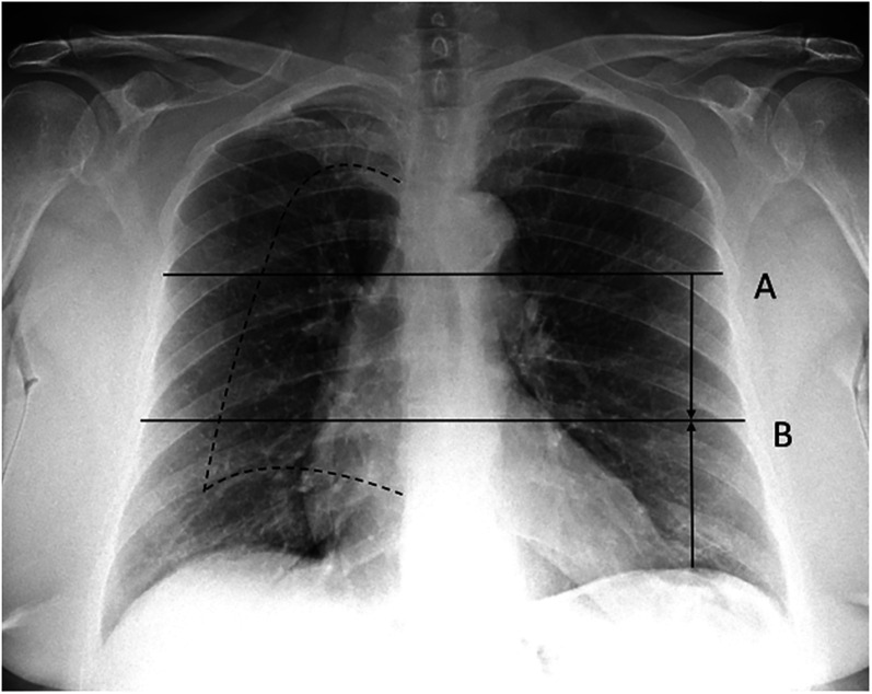
Material and methods: CR of COVID-19 and non-COVID-19 pulmonary diseases, admitted to the emergency department, were evaluated. Assessed features were interstitial opacities, ground glass opacities, and/or consolidations and the predominant lung alteration. We also assessed uni-/bilaterality, location (upper/middle/lower), and distribution (peripheral/perihilar), as well as pleural effusion and perihilar vessels blurring. A binary logistic regression was adopted to obtain the most accurate CR COVID-19 pattern, and sensitivity and specificity were computed. The newly defined pattern was compared to BSTI criteria.
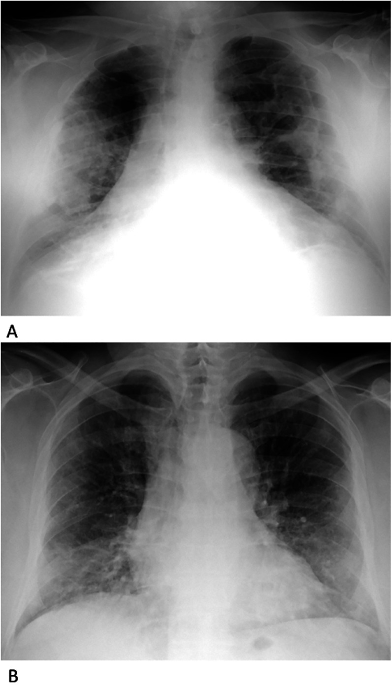
Results: CR of 274 patients were evaluated (146 COVID-19, 128 non-COVID-19). The most accurate COVID-19 pneumonia pattern consisted of four features: bilateral alterations (Expß=2.8, P=0.002), peripheral distribution of the predominant (Expß=2.3, P=0.013), no pleural effusion (Expß=0.4, P=0.009), and perihilar vessels' contour not blurred (Expß=0.3, P=0.002). The pattern showed 49% sensitivity, 81% specificity, and 64% accuracy, while BSTI criteria showed 51%, 77%, and 63%, respectively.
Conclusion: Bilaterality, peripheral distribution of the predominant lung alteration, no pleural effusion, and perihilar vessels contour not blurred determine the most accurate COVID-19 pneumonia pattern. Lower field involvement, proposed by BSTI criteria, was not a distinctive finding. The BSTI criteria has lower specificity.
Keywords: COVID 19; SARS-CoV-2 virus; diagnostic X-ray; differential diagnosis; pneumonitis.
Conflict of interest statement
The author(s) declared no potential conflicts of interest with respect to the research, authorship, and/or publication of this article.
Figures
Similar articles
-
Possible Alterations of Imaging Patterns in Computed Tomography for Delta-VOC of SARS-CoV-2.Rofo. 2022 Nov;194(11):1229-1241. doi: 10.1055/a-1826-0436. Epub 2022 Jul 18. Rofo. 2022. PMID: 35850138 English.
-
Typical Imaging Patterns in COVID-19 Infections of the Lung on Plain Chest Radiographs to Aid Early Triage.Rofo. 2021 Oct;193(10):1189-1196. doi: 10.1055/a-1388-8147. Epub 2021 Mar 10. Rofo. 2021. PMID: 33694145 English.
-
COVIDiag: a clinical CAD system to diagnose COVID-19 pneumonia based on CT findings.Eur Radiol. 2021 Jan;31(1):121-130. doi: 10.1007/s00330-020-07087-y. Epub 2020 Aug 1. Eur Radiol. 2021. PMID: 32740817 Free PMC article.
-
Thoracic imaging tests for the diagnosis of COVID-19.Cochrane Database Syst Rev. 2020 Nov 26;11:CD013639. doi: 10.1002/14651858.CD013639.pub3. Cochrane Database Syst Rev. 2020. Update in: Cochrane Database Syst Rev. 2021 Mar 16;3:CD013639. doi: 10.1002/14651858.CD013639.pub4. PMID: 33242342 Updated.
-
Comparison of the computed tomography findings in COVID-19 and other viral pneumonia in immunocompetent adults: a systematic review and meta-analysis.Eur Radiol. 2020 Dec;30(12):6485-6496. doi: 10.1007/s00330-020-07018-x. Epub 2020 Jun 27. Eur Radiol. 2020. PMID: 32594211 Free PMC article.
References
-
- Litmanovich DE, Chung M, Kirkbride RR, et al.Review of chest radiograph findings of COVID-19 pneumonia and suggested reporting language. J Thorac Imaging 2020;35:354–360. - PubMed
MeSH terms
LinkOut - more resources
Full Text Sources
Medical
Miscellaneous
