Review
. 2022 Jan;25(1):13.
doi: 10.3892/mmr.2021.12529. Epub 2021 Nov 15.

Efferocytosis in multisystem diseases (Review)

Affiliations
Review

Efferocytosis in multisystem diseases (Review)

Yifan Zhang et al. Mol Med Rep. 2022 Jan.

Abstract

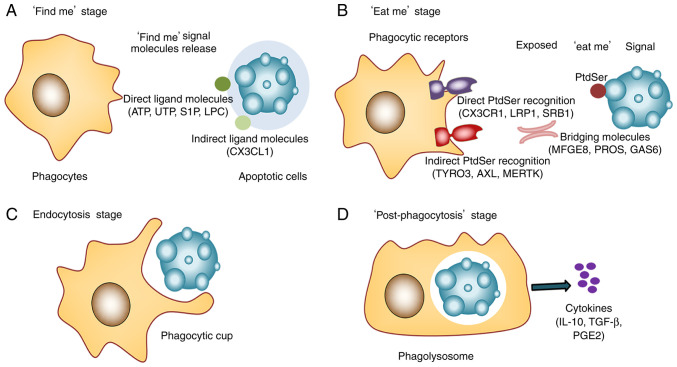
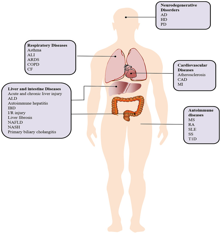
Efferocytosis, the phagocytosis of apoptotic cells performed by both specialized phagocytes (such as macrophages) and non‑specialized phagocytes (such as epithelial cells), is involved in tissue repair and homeostasis. Effective efferocytosis prevents secondary necrosis, terminates inflammatory responses, promotes self‑tolerance and activates pro‑resolving pathways to maintain homeostasis. When efferocytosis is impaired, apoptotic cells that could not be cleared in time aggregate, resulting in the necrosis of apoptotic cells and release of pro‑inflammatory factors. In addition, defective efferocytosis inhibits the intracellular cholesterol reverse transportation pathways, which may lead to atherosclerosis, lung damage, non‑alcoholic fatty liver disease and neurodegenerative diseases. The uncleared apoptotic cells can also release autoantigens, which can cause autoimmune diseases. Cancer cells escape from phagocytosis via efferocytosis. Therefore, new treatment strategies for diseases related to defective efferocytosis are proposed. This review illustrated the mechanisms of efferocytosis in multisystem diseases and organismal homeostasis and the pathophysiological consequences of defective efferocytosis. Several drugs and treatments available to enhance efferocytosis are also mentioned in the review, serving as new evidence for clinical application.

Keywords: autoimmune diseases; cardiovascular diseases; efferocytosis; liver and intestine diseases; neurodegenerative diseases; respiratory diseases.


Conflict of interest statement

The authors declare that they have no competing interests.

Figures

Figure 1.
Figure 1.
Four stages of efferocytosis. (A) ‘Find me’ stage. Chemotactic factors induce macrophages to recognize and surround ACs. The ‘find me’ signal molecules released by ACs are recognized by phagocytes, inducing the migration and recruitment of phagocytes to ACs. (B) ‘Eat me’ stage. Phagocytic receptors of macrophages recognize and bind to the ‘eat me’ signal molecules of ACs. (C) Endocytosis stage. Forming ‘a phagocytic cup’ completes the endocytosis of ACs. (D) ‘Post-phagocytosis’ stage. Macrophages further digest and degrade apoptotic cell debris, activating multiple metabolic signaling pathways. ATP, triphosphate nucleotides adenosine triphosphate; UTP, uridine-5′-triphosphate; S1P, sphingosine-1-phosphate; LPC, lyso-phosphatidylcholine; CX3CL1, CX3C chemokine ligand 1; CX3CR1, CX3C chemokine receptor 1; LRP1, low-density lipoprotein receptor related proteins 1; SRB1, scavenger receptor 1; MFGE8, milk fat globule-epidermal growth factor; ProS, protein S; PtdSer, phosphatidylserine; GAS6, growth arrest specific protein 6; TYRO3/AXL/MERTK, TAM receptors.
Figure 2.
Figure 2.
Efferocytosis in multisystem diseases. AD, Alzheimer's disease; ALD, alcoholic liver disease; ALI, acute lung injury; ARDS, acute respiratory distress syndrome; CAD, Coronary artery disease; CF, cystic fibrosis; COPD, chronic obstructive pulmonary disease; HD, Huntington's disease; IBD, inflammatory bowel disease; I/R injury, ischemia-reperfusion injury; MI, Myocardial infarction; MS, multiple sclerosis; NAFLD, non-alcoholic fatty liver disease; NASH, non-alcoholic steatohepatitis; PD, Parkinson's disease; RA, rheumatoid arthritis; SLE, systemic lupus erythematosus; SS, Sjogren's syndrome; T1D, type 1 diabetes.

References

    1. Thorp E, Tabas I. Mechanisms and consequences of efferocytosis in advanced atherosclerosis. J Leukoc Biol. 2009;86:1089–1095. doi: 10.1189/jlb.0209115. - DOI - PMC - PubMed
    1. Boada-Romero E, Martinez J, Heckmann BL, Green DR. The clearance of dead cells by efferocytosis. Nat Rev Mol Cell Biol. 2020;21:398–414. doi: 10.1038/s41580-020-0232-1. - DOI - PMC - PubMed
    1. Trahtemberg U, Mevorach D. Apoptotic cells induced signaling for immune homeostasis in macrophages and dendritic cells. Front Immunol. 2017;8:1356. doi: 10.3389/fimmu.2017.01356. - DOI - PMC - PubMed
    1. Segawa K, Nagata S. An apoptotic ‘Eat Me’ Signal: Phosphatidylserine exposure. Trends Cell Biol. 2015;25:639–650. doi: 10.1016/j.tcb.2015.08.003. - DOI - PubMed
    1. Kourtzelis I, Hajishengallis G, Chavakis T. Phagocytosis of apoptotic cells in resolution of inflammation. Front Immunol. 2020;11:553. doi: 10.3389/fimmu.2020.00553. - DOI - PMC - PubMed