Review
. 2022 Feb 10:84:59-85.
doi: 10.1146/annurev-physiol-061121-035914. Epub 2021 Nov 15.

Running the Female Power Grid Across Lifespan Through Brain Estrogen Signaling

Affiliations
Review

Running the Female Power Grid Across Lifespan Through Brain Estrogen Signaling

Holly A Ingraham et al. Annu Rev Physiol. .

Abstract

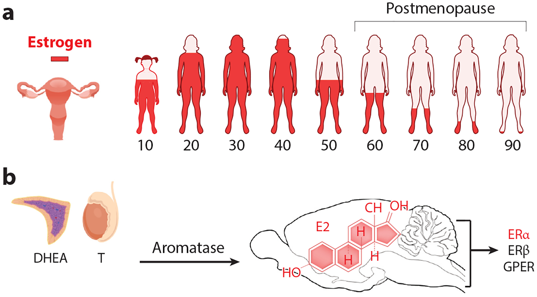
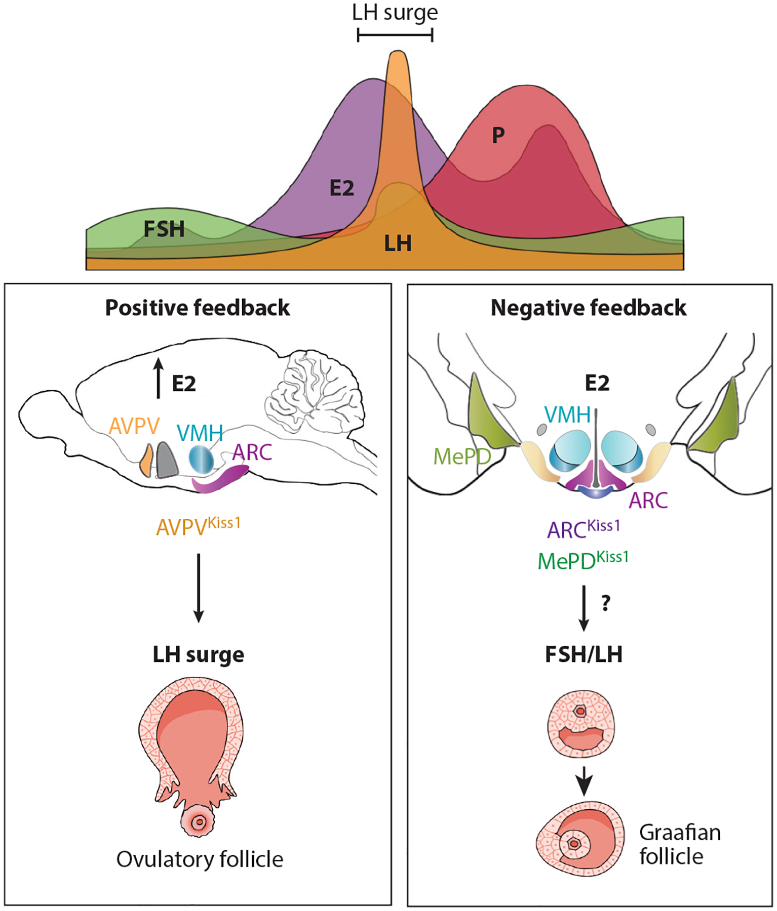
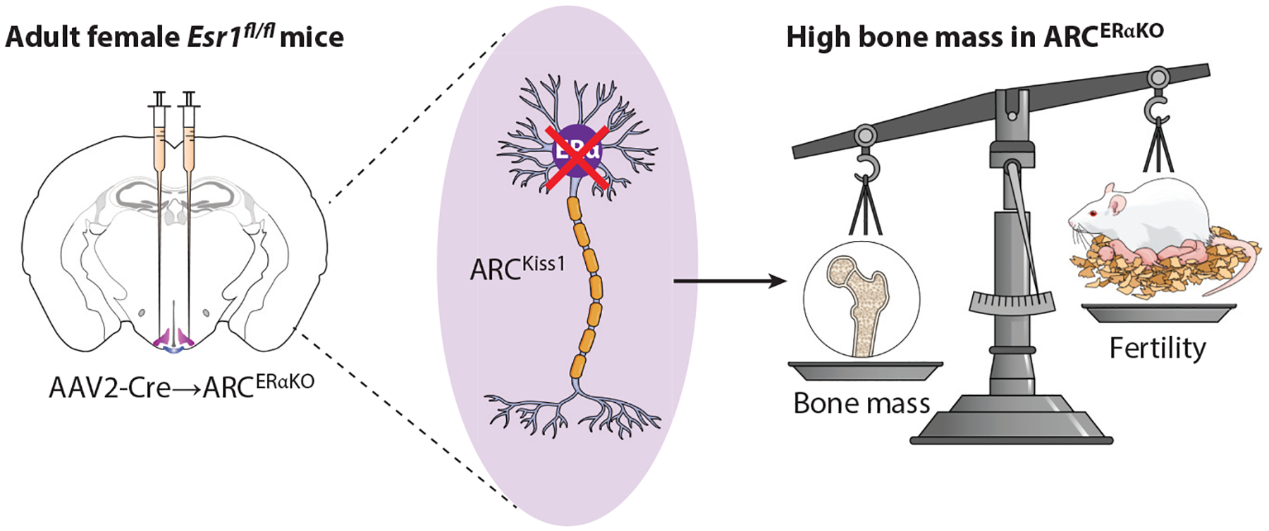
The role of central estrogen in cognitive, metabolic, and reproductive health has long fascinated the lay public and scientists alike. In the last two decades, insight into estrogen signaling in the brain and its impact on female physiology is beginning to catch up with the vast information already established for its actions on peripheral tissues. Using newer methods to manipulate estrogen signaling in hormone-sensitive brain regions, neuroscientists are now identifying the molecular pathways and neuronal subtypes required for controlling sex-dependent energy allocation. However, the immense cellular complexity of these hormone-sensitive brain regions makes it clear that more research is needed to fully appreciate how estrogen modulates neural circuits to regulate physiological and behavioral end points. Such insight is essential for understanding how natural or drug-induced hormone fluctuations across lifespan affect women's health.

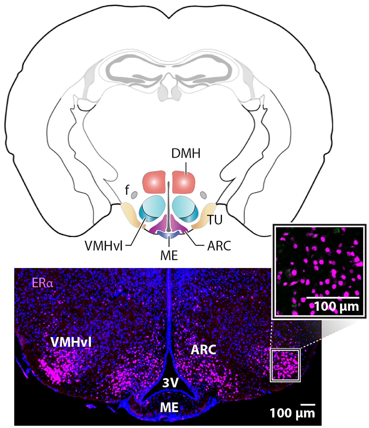
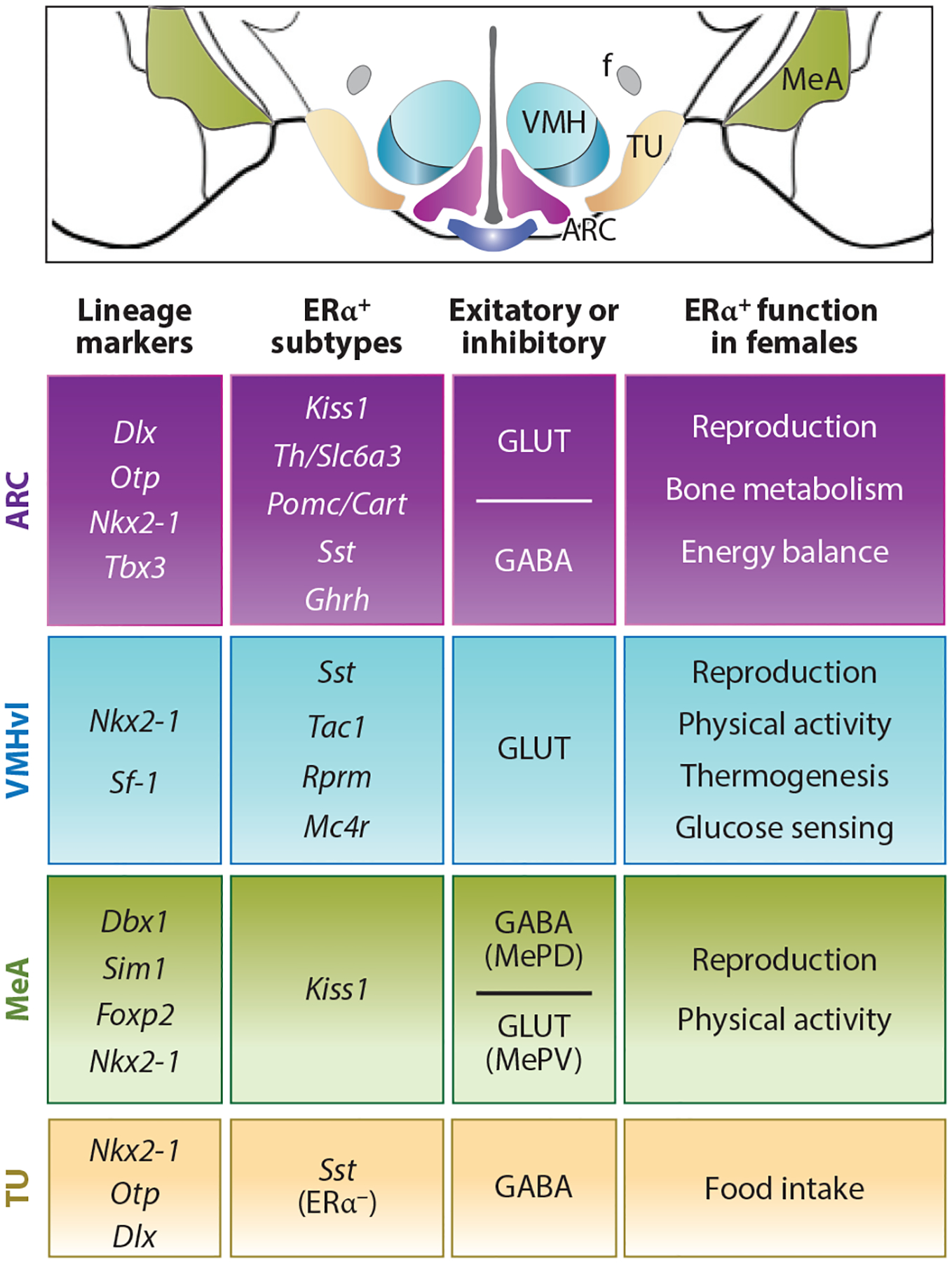
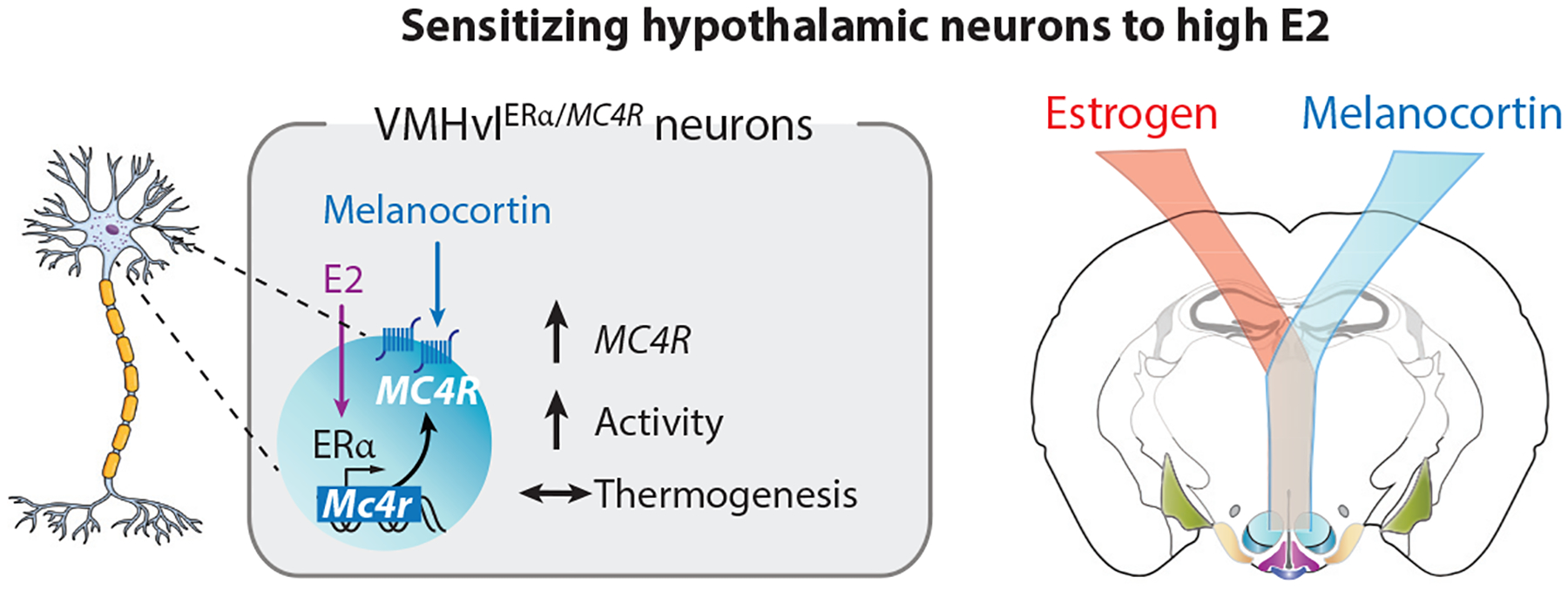
Keywords: arcuate nucleus; brain-bone connection; central estrogen signaling; estrogen receptor alpha; female physiology; hypothalamus; menopause; reproduction; sex-dependent neurocircuits; ventromedial hypothalamus; women's health.


Figures

Figure 1
Figure 1
Estrogen signaling in females and the medial basal hypothalamus. (a) Levels of ovarian estrogen during lifespan in women with significant depletion beginning before menopause (~47 years old). (b) Sources of androgens from the testes or adrenals can be converted to estradiol in the gonads, tissues, or in specific brain regions expressing Cyp19A1. E2 binds three estrogen receptors, all of which are found in the brain. Abbreviations: DHEA, dehydroepiandrosterone; E2, estradiol; ERα, estrogen receptor alpha; ERβ, estrogen receptor beta; GPER, G protein–coupled transmembrane estrogen receptor.
Figure 2
Figure 2
Enriched expression of estrogen receptor alpha (ERα) in the female medial basal hypothalamus is independent of gonadal hormones. Coronal mouse brain schematic with colored regions of interest, including the ventral lateral region of the ventromedial hypothalamus (VMHvl), the arcuate nucleus (ARC), and the tuberal nucleus (TU). Other landmarks include the fornix (f), dorsal medial hypothalamus (DMH), median eminence (ME), and third ventricle (3V). Lower panels show the expression of ERα in the VMHvl and ARC. The higher magnification panel shows nuclear staining at basal hormone levels or in estrogen-depleted ovariectomized females.
Figure 3
Figure 3
Neurons in the female reproductive cycle. The top panel shows a typical cycle of hormone fluctuations with E2 levels peaking as LH surges to then trigger ovulation. Collective work, including specific targeting of AVPVKiss1 neurons, shows that this population is critical for generating an LH surge during high E2 levels for positive feedback (left lower box). Estrogen signaling in ARCKiss1 neurons is important for maximizing the number of continuous estrous cycles: estrus, diestrus, and proestrus. The number of LH pulses or levels of LH are unchanged after deleting ERα in the ARC or ARCKiss1 neurons. Thus, other estrogen-sensitive brain regions, such as the MeA, must contribute to negative feedback when E2 and LH are low (right lower box). Abbreviations: ARC, arcuate nucleus; AVPV, anteroventral periventricular (nucleus); E2, estradiol; ERα, estrogen receptor alpha; FSH, follicle-stimulating hormone; Kiss, kisspeptin; LH, luteinizing hormone; MeA, medial amygdala; MePD, posterodorsal medial amygdala; P, progesterone; VMH, ventromedial hypothalamus.
Figure 4
Figure 4
Estrogen signaling in female ARCKiss1 neurons restrains bone. Scheme showing stereotaxic elimination of Esr1fl/fl in the ARC. Other genetic methods have also been used and show a significant increase in bone mass or high bone mass phenotype with drop infertility. The underlying molecular mechanism for this brain-to-bone connection remains to be determined. Abbreviations: AAV, adeno-associated virus; ARC, arcuate nucleus; Kiss, kisspeptin; KO, knockout; VMHvl, ventral lateral region of the ventromedial hypothalamus.
Figure 5
Figure 5
Diversity in estrogen-responsive ARC and the VMHvl neurons. Schematized coronal section through the MBH brain region. Table summarizing lineage markers, defined ERα neuron subtypes, excitatory (GLUT) or inhibitory (GABA) neurotransmitter type, and functional changes in female mice following regional knockout of ERα for the ARC, VMHvl, MeA, and TU. All four areas share Nkx2-1 as a common developmental marker, and as such, Nkx2-1-Cre will eliminate the expression of genes in these and other brain nuclei. Note that Pomc/Cart neurons include both excitatory and inhibitory subtypes. Abbreviations: ARC, arcuate nucleus; Cart, cocaine and amphetamine-regulated transcript; ERα, estrogen receptor alpha; f, fornix; GABA, gamma-aminobutyric acid; Ghrh, growth hormone–releasing hormone; GLUT, glutamate; Kiss, kisspeptin; MBH, medial basal hypothalamus; Mc4r, melanocortin-4 receptor; MeA, medial amygdala; MePD, posterodorsal medial amygdala; MePV, posteroventral medial amygdala; Pomc, proopiomelanocortin; Rprm, Reprimo; Sst, somatostatin; Th, tyrosine hydroxylase; TU, tuberal nucleus; VMHvl, ventral lateral region of the ventromedial hypothalamus.
Figure 6
Figure 6
Estrogen signaling in female VMHvlERα/MC4R neurons promotes locomotor activity. High estrogen levels during proestrus or after E2 treatment increase MC4R expression in about 200 VMHvl neurons, which, when stimulated, promote physical activity. This molecular pathway demonstrates how the estrogen receptor can sensitize adaptive behaviors to prioritize energy utilization in service of reproductive fitness. Abbreviations: E2, estradiol; ERα, estrogen receptor α; MC4R, melanocortin-4 receptor; VMHvl, ventral lateral region of the ventromedial hypothalamus.

References

    1. Colditz GA, Hankinson SE, Hunter DJ, Willett WC, Manson JE, et al. 1995. The use of estrogens and progestins and the risk of breast cancer in postmenopausal women. N. Engl. J. Med 332:1589–93 - PubMed
    1. Arnold AP. 2004. Sex chromosomes and brain gender. Nat. Rev. Neurosci 5:701–8 - PubMed
    1. Cooke PS, Nanjappa MK, Ko C, Prins GS, Hess RA. 2017. Estrogens in male physiology. Physiol. Rev 97:995–1043 - PMC - PubMed
    1. Wu MV, Manoli DS, Fraser EJ, Coats JK, Tollkuhn J, et al. 2009. Estrogen masculinizes neural pathways and sex-specific behaviors. Cell 139:61–72 - PMC - PubMed
    1. Brooks DC, Coon VJ, Ercan CM, Xu X, Dong H, et al. 2020. Brain aromatase and the regulation of sexual activity in male mice. Endocrinology 161:bqaa137. - PMC - PubMed

Publication types