. 2022 Feb:348:113923.
doi: 10.1016/j.expneurol.2021.113923. Epub 2021 Nov 12.

Preclinical investigation of Pegylated arginase 1 as a treatment for retina and brain injury

Affiliations

Preclinical investigation of Pegylated arginase 1 as a treatment for retina and brain injury

Abdelrahman Y Fouda et al. Exp Neurol. 2022 Feb.

Abstract

Arginase 1 (A1) is the enzyme that hydrolyzes the amino acid, L-arginine, to ornithine and urea. We have previously shown that A1 deletion worsens retinal ischemic injury, suggesting a protective role of A1. In this translational study, we aimed to study the utility of systemic pegylated A1 (PEG-A1, recombinant human arginase linked to polyethylene glycol) treatment in mouse models of acute retinal and brain injury. Cohorts of WT mice were subjected to retinal ischemia-reperfusion (IR) injury, traumatic optic neuropathy (TON) or brain cerebral ischemia via middle cerebral artery occlusion (MCAO) and treated with intraperitoneal injections of PEG-A1 or vehicle (PEG only). Drug penetration into retina and brain tissues was measured by western blotting and immunolabeling for PEG. Neuroprotection was measured in a blinded fashion by quantitation of NeuN (neuronal marker) immunolabeling of retina flat-mounts and brain infarct area using triphenyl tetrazolium chloride (TTC) staining. Furthermore, ex vivo retina explants and in vitro retina neuron cultures were subjected to oxygen-glucose deprivation (OGD) followed by reoxygenation (R) and treated with PEG-A1. PEG-A1 given systemically did not cross the intact blood-retina/brain barriers in sham controls but reached the retina and brain after injury. PEG-A1 provided neuroprotection after retinal IR injury, TON and cerebral ischemia. PEG-A1 treatment was also neuroprotective in retina explants subjected to OGD/R but did not improve survival in retinal neuronal cultures exposed to OGD/R. In summary, systemic PEG-A1 administration is neuroprotective and provides an excellent route to deliver the drug to the retina and the brain after acute injury.

Keywords: Arginase; Ischemia-reperfusion injury; Neurodegeneration; Neuroprotection; Retinal ischemia; Stroke.


Conflict of interest statement

CONFLICT OF INTEREST

AF, RBC, and RWC have a pending patent on the use of arginase 1 as a treatment for ischemic retinopathies. PNMC is the chief executive officer of Bio-Cancer Treatment International Limited and holds stocks or shares in Bio-Cancer Treatment International Limited. The other authors declare no conflict of interest.

Figures

Figure 1.
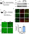
Figure 1.. Systemic PEG-A1 treatment is neuroprotective and crosses the blood-retina barrier after IR injury.
A) Schematic representation of the experimental protocol for figures B and C. B) PEGA1 treated mice show a strong PEG-positive band in western blots of perfused liver homogenates collected at 24 hours. C) Western blotting on perfused retinas collected at 24 hours after treatment show a strong PEG positive band in the IR injured retinas but not the sham retinas from the same mice. IR injured retinas also show a stronger albumin band, an indicator of increased vascular permeability. D) Schematic representation of the experimental protocol for figures E, F and G. E). Flat-mounts of retinas from PEG-A1 treated mice collected without perfusion at 7 days after treatment show an immunofluorescent signal that is confined to the retina vessel network in the sham eye with a more diffuse signal in the IR injured retinas. F, G) Neun staining and quantification of retina flat-mounts collected at 7 days after retinal IR show significant neuronal preservation after injury with PEG-A1 treatment as compared to vehicle treatment, n=6–8, *p<0.05 vs vehicle group.
Figure 2.
Figure 2.. Systemic PEG-A1 treatment is neuroprotective and crosses the blood-retina barrier after cerebral ischemia.
A) Schematic representation of the experimental protocol for figures B, C, D and E. B) Western blotting on perfused brain homogenates shows a strong band for anti-PEG after middle cerebral artery occlusion (MCAO) but not in the sham brains. C) Representative triphenyltetrazolium chloride (TTC) red staining of the viable tissue in brain tissue sections highlights the unstained infarct area in white color in mice with no treatment (ttt) and vehicle or PEG-A1 treated mice. D) Quantification of the infarct size as a percent of contralesional hemisphere shows significant reduction with PEG-A1 treatment (*p<0.05 vs control). E) Bederson score of behavioral function after cerebral ischemia shows a trend towards improvement with PEG-A1 but the difference did not reach statistical significance. There was no difference in the infarct size or Bederson score between the untreated and the vehicle treated mice and therefore these groups were combined and identified by open and closed circles, respectively, n=6–11, *p<0.05 vs control group.
Figure 3.
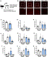
Figure 3.. Systemic PEG-A1 treatment is neuroprotective and crosses the blood-retina barrier after ONC.
A) Schematic representation of the experimental protocol for figure B. B) Western blotting on perfused retina and brain homogenates shows a strong band for anti-PEG in the retina/optic nerve (ON) subjected to ONC. Much less is present in brain or sham retina/ON which could be due to residual blood remaining after perfusion. C) Schematic representation of the experimental protocol for figures D, E and F. D) Flat-mount immunolabeling using anti-PEG antibody and co-stained with lectin (stains vessels and some microglia/macrophages) shows PEG-A1 extravasation after ONC (white arrows). E, F) Flat-mount immunolabeling for the neuronal marker (NeuN) and quantification show significant neuronal preservation with PEG-A1 treatment after ONC, n=4, *p<0.05 vs vehicle group.
Figure 4:
Figure 4:. PEG-A1 treatment mitigates retinal inflammatory response after ONC.
A) Schematic representation of the experimental protocol for figures B through K. B, C) Immunolabeling of retina flatmounts with the microglia/macrophage marker, Iba-1, and quantification showed a reduction in number with PEG-A1 treatment. n=4–5, *p<0.05 vs vehicle group. D-K) RT-PCR analysis of different cytokines at day 4 after ONC showed reduction in IL-1β, and increase in IL-6, IL-10 and BDNF with PEG-A1 treatment. n=4–5, *p<0.05 vs sham vehicle,#p<0.05 vs ONC vehicle.
Figure 5:
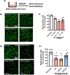
Figure 5:. PEG-A1 treatment protects retinal explants from oxygen-glucose deprivation/reoxygenation (OGD/R) injury ex vivo.
A) Schematic representation of the experimental protocol for figures B, C, D and E. B, C) Time course of exposing retina explants to OGD/R ex vivo followed by fixation and flat-mount immunolabeling with Neun showed neuronal degeneration after 1, 3 or 5 hours of OGD then reoxygenation (R), with maximum neuronal loss (about 40%) after 3 hours of OGD and 21 hours of reoxygenation, n=3 per group, *p<0.05 vs normoxia, **p<0.01 vs normoxia. D, E) PEG-A1 treatment at reoxygenation significantly protected the retina explant neurons against OGD/R (3 hours/21 hours) as compared to control (OGD/R without treatment) or vehicle treated (PEG only) explants, n=8–10, *p<0.05 vs normoxia,#p<0.05 vs OGD/R control and vehicle groups.
Figure 6:
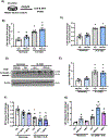
Figure 6:. PEG-A1 treatment does not provide neuroprotection to retinal neurons in vitro after OGD.
A) Schematic representation of the experimental protocol for figures B, C, D and E. B, C) R28 retinal neurons were subjected to OGD for 6 or 24 hours with or without PEG-A1 treatment (1 μg/mL) followed by LDH release assay on the supernatant. OGD increased cell death as evident by a two-fold increase in LDH release while PEG-A1 did not alter this effect, *p<0.05 vs control normoxia group. D, E) Western blotting on R28 cell lysates showed upregulation of cleaved PARP after 6 hours of OGD which was not rescued by PEG-A1 treatment. F) MTT assay on R28 cells showed a 20 % reduction in cell viability after 3 hours of OGD and 3 hours of reoxygenation while PEG-A1 treatment (0.1 or 1 μg/mL) did not show improvement in viability, *p<0.05 vs control normoxia group. G) Primary rat retinal mixed neurons were subjected to OGD/R (6 hours/18 hours) and treated at reperfusion with PEG-A1 (1 μg/mL) or ABH (100 μM). Neither treatment with PEG-A1 or ABH promoted protection as measured by LDH release. *p<0.05 vs control and PEG-A1 normoxia groups and ctrl OGD group,#p<0.05 vs control and ABH normoxia groups.
Figure 7:
Figure 7:. Explants from control and myeloid A1 KO mice show no difference in neuronal cell death after OGD/R.
A) Schematic representation of the experimental protocol for figures B and C. B, C) Exposing retina explants from Ctrl and M-A1KO mice to OGD/R ex vivo showed similar neuronal degeneration, n=3–4, *p<0.05 vs normoxia. D) Western blotting on primary mouse macrophages shows a strong band for anti-PEG in cells treated with PEG-A1 for 18 hours suggesting possible uptake of the drug into the cells.

References

    1. Fouda AY, Eldahshan W, Narayanan SP, Caldwell RW, Caldwell RB. Arginase pathway in acute retina and brain injury: Therapeutic opportunities and unexplored avenues. Front Pharmacol. 2020;11:277. - PMC - PubMed
    1. London A, Benhar I, Schwartz M. The retina as a window to the brain—from eye research to cns disorders. Nature Reviews Neurology. 2013;9:44–53 - PubMed
    1. Jindal V Interconnection between brain and retinal neurodegenerations. Mol Neurobiol. 2015;51:885–892 - PubMed
    1. Fouda AY, Xu Z, Shosha E, Lemtalsi T, Chen J, Toque HA, et al. Arginase 1 promotes retinal neurovascular protection from ischemia through suppression of macrophage inflammatory responses. Cell death & disease. 2018;9:1001. - PMC - PubMed
    1. Shosha E, Xu Z, Yokota H, Saul A, Rojas M, Caldwell RW, et al. Arginase 2 promotes neurovascular degeneration during ischemia/reperfusion injury. Cell death & disease. 2016;7:e2483–e2483 - PMC - PubMed

Publication types

MeSH terms