Review
. 2022 Jan:23:27-39.
doi: 10.1016/j.jtos.2021.11.003. Epub 2021 Nov 13.

Diagnostic armamentarium of infectious keratitis: A comprehensive review

Affiliations
Review

Diagnostic armamentarium of infectious keratitis: A comprehensive review

Darren S J Ting et al. Ocul Surf. 2022 Jan.

Abstract

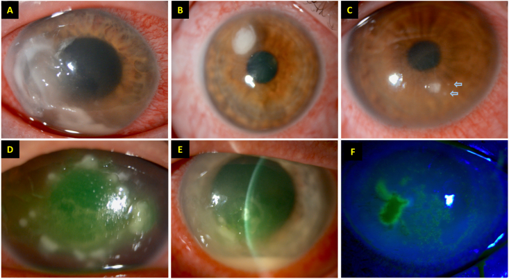
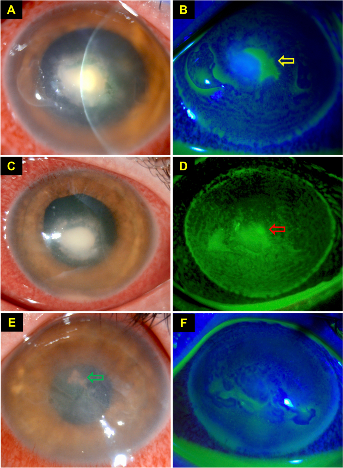
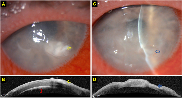
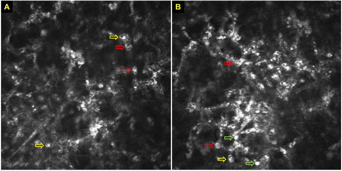
Infectious keratitis (IK) represents the leading cause of corneal blindness worldwide, particularly in developing countries. A good outcome of IK is contingent upon timely and accurate diagnosis followed by appropriate interventions. Currently, IK is primarily diagnosed on clinical grounds supplemented by microbiological investigations such as microscopic examination with stains, and culture and sensitivity testing. Although this is the most widely accepted practice adopted in most regions, such an approach is challenged by several factors, including indistinguishable clinical features shared among different causative organisms, polymicrobial infection, long diagnostic turnaround time, and variably low culture positivity rate. In this review, we aim to provide a comprehensive overview of the current diagnostic armamentarium of IK, encompassing conventional microbiological investigations, molecular diagnostics (including polymerase chain reaction and mass spectrometry), and imaging modalities (including anterior segment optical coherence tomography and in vivo confocal microscopy). We also highlight the potential roles of emerging technologies such as next-generation sequencing, artificial intelligence-assisted platforms. and tele-medicine in shaping the future diagnostic landscape of IK.

Keywords: Artificial intelligence; Confocal microscopy; Corneal infection; Corneal ulcer; Diagnosis; Mass spectrometry; Next-generation sequencing; Polymerase chain reaction.


Figures

Fig. 1
Fig. 1
Slit-lamp photographs demonstrating different types of infectious keratitis (IK), highlighting the clinical challenges of diagnosing IK without any microbiological investigation. (AC) Three separate cases of IK caused by Pseudomonas aeruginosa. Note the difference in the severity and clinical appearance among the three cases. The two “satellite lesions” (blue arrows) depicted in (C) image may give a false impression of fungal keratitis. (D) A case of IK caused by Staphylococcus aureus two days after corneal cross-linking treatment. (EF) A case of polymicrobial IK, caused by S. aureus and herpes simplex keratitis, in a patient with atopic keratoconjunctivitis. (For interpretation of the references to colour in this figure legend, the reader is referred to the Web version of this article.)
Fig. 2
Fig. 2
This figure highlights the diagnostic dilemma of infectious keratitis, even in the presence of positive microbiological culture result. This is a case of polymicrobial keratitis which was initially treated for a culture-proven Staphylococcus aureus keratitis with hourly topical antibiotics at day 0. (AB) At day 4, slit-lamp photograph demonstrating a dense central infiltrate with healing epithelial defect (yellow arrow). (CD) At day 5, slit-lamp photograph demonstrating continued improvement of the ulcer, with gradual contraction of the infiltrate and reduction of epithelial defect (red arrow). However, the deep-seated infiltrate failed to resolve after 10 days of intensive antibiotic treatment, raising the suspicion of co-existing fungal infection (confirmed on in vivo confocal microscopy). Topical antibiotics was then switched to topical voriconazole 1% hourly. (EF) At day 24, complete healing of ulcer was achieved with intensive topical voriconazole drops. Deep central pigmented keratic precipitates (green arrow) and bullous keratopathy secondary to endothelial damage from IK were noted. (For interpretation of the references to colour in this figure legend, the reader is referred to the Web version of this article.)
Fig. 3
Fig. 3
This figure highlights the clinical value of anterior segment optical coherence tomography (AS-OCT) in assessing and monitoring infectious keratitis. (A) Slit-lamp photography demonstrating a case of culture-negative, presumed right fungal keratitis with a moderate-size infiltrate (yellow arrow) at the inferonasal aspect of the cornea. (B) AS-OCT clearly delineates the margin and depth of the infiltrate (yellow arrow), located at the anterior ½ of the stroma, and highlights the presence of a retrocorneal membrane (red arrow), highly suggestive of fungal keratitis. (CD) AS-OCT demonstrating a significant reduction in the corneal infiltrate with moderate corneal thinning (blue arrows) after one month of intensive topical antifungal treatment. (For interpretation of the references to colour in this figure legend, the reader is referred to the Web version of this article.)
Fig. 4
Fig. 4
(AB). Various characteristic features of Acanthamoeba cysts on in vivo confocal microscopy (IVCM), including double-wall cysts (red arrows), signet rings (yellow arrows), and bright spots (green arrows). (For interpretation of the references to colour in this figure legend, the reader is referred to the Web version of this article.)

References

    1. Ting D.S.J., Ho C.S., Deshmukh R., Said D.G., Dua H.S. Infectious keratitis: an update on epidemiology, causative microorganisms, risk factors, and antimicrobial resistance. Eye. 2021;35(4):1084–1101. - PMC - PubMed
    1. Collier S.A., Gronostaj M.P., MacGurn A.K., Cope J.R., Awsumb K.L., Yoder J.S., et al. Estimated burden of keratitis--United States, 2010. MMWR Morb Mortal Wkly Rep. 2014;63(45):1027–1030. - PMC - PubMed
    1. Whitcher J.P., Srinivasan M. Corneal ulceration in the developing world--a silent epidemic. Br J Ophthalmol. 1997;81(8):622–623. - PMC - PubMed
    1. Walker D.H. Principles of diagnosis of infectious diseases. Pathobiology of Human Disease. 2014:222–225.
    1. Ting D.S.J., Settle C., Morgan S.J., Baylis O., Ghosh S. A 10-year analysis of microbiological profiles of microbial keratitis: the North East England Study. Eye. 2018;32(8):1416–1417. - PMC - PubMed

Publication types