Magnetic resonance imaging analysis of screw in-type lateral anchor pull-out in large to massive rotator cuff repair in patients older than 60 years
- PMID: 34781601
- PMCID: PMC8907505
- DOI: 10.5397/cise.2021.00374
Magnetic resonance imaging analysis of screw in-type lateral anchor pull-out in large to massive rotator cuff repair in patients older than 60 years
Abstract
Background: This study was performed to identify the incidence of screw in-type lateral anchor pull-out in patients older than 60 years who underwent rotator cuff repair for large to massive rotator cuff tear (RCT).
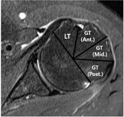
Methods: We reviewed 25 patients over 60 who were diagnosed with large to massive RCT and underwent arthroscopic rotator cuff repair in our hospital from March 2017 to February 2021. Preoperative tear size (anterior to posterior, medial to lateral) was measured via preoperative magnetic resonance imaging (MRI). All 25 patients underwent MRI scanning on postoperative day 1 and at 3 months after surgery. The change of anchor position was measured in axial views on MRI images postoperative day 1 and 3 months after surgery. And it was statistically compared according to bone mineral density (BMD), sex, and number of lateral anchors.
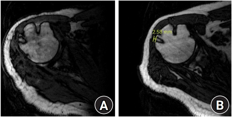
Results: Two MRIs (postoperative day 1 and 3 months) in 25 patients were compared. Anchor pull-out occurred in six patients during 3 months (6.7%), and the mean pull-out length difference was 1.56 mm (range, 0.16-2.58 mm). There was no significant difference in the number of pull-out anchors, degree of pull-out difference by comparing BMD (A, BMD≤-2.5; B, BMD>-2.5), sex, or number of anchors used in each surgery (C, two anchors; D, three anchors) (p>0.05).
Conclusions: Pull-out of screw in-type anchors was rarely observed and the mean pull-out length difference was negligibly small in our study. The screw in-type lateral anchor seems to be a decent option without concern of anchor pull-out even in elderly patients.
Keywords: Magnetic resonance imaging; Pull-out; Rotator cuff; Lateral anchor.
Conflict of interest statement
None.
Figures
Similar articles
-
All-Suture Anchor Settling After Arthroscopic Repair of Small and Medium Rotator Cuff Tears.Am J Sports Med. 2019 Dec;47(14):3483-3490. doi: 10.1177/0363546519886547. Epub 2019 Nov 13. Am J Sports Med. 2019. PMID: 31718248
-
Osteolysis is observed around both bioabsorbable and nonabsorbable anchors on serial magnetic resonance images of patients undergoing arthroscopic rotator cuff repair.Acta Orthop Traumatol Turc. 2019 Nov;53(6):414-419. doi: 10.1016/j.aott.2019.08.015. Epub 2019 Sep 25. Acta Orthop Traumatol Turc. 2019. PMID: 31563430 Free PMC article.
-
Delayed Lateral Row Anchor Failure in Suture Bridge Rotator Cuff Repair: A Report of 3 Cases.Clin Shoulder Elb. 2018 Dec 1;21(4):246-251. doi: 10.5397/cise.2018.21.4.246. eCollection 2018 Dec. Clin Shoulder Elb. 2018. PMID: 33330184 Free PMC article.
-
Perianchor Cyst Formation After Arthroscopic Rotator Cuff Repair Using All-Suture-Type, Bioabsorbable-Type, and PEEK-Type Anchors.Arthroscopy. 2019 Aug;35(8):2284-2292. doi: 10.1016/j.arthro.2019.03.032. Epub 2019 Jul 23. Arthroscopy. 2019. PMID: 31350085
-
Arthroscopic technique for patch augmentation of rotator cuff repairs.Arthroscopy. 2006 Oct;22(10):1136.e1-6. doi: 10.1016/j.arthro.2006.03.022. Arthroscopy. 2006. PMID: 17027416 Review.
References
-
- Lehman C, Cuomo F, Kummer FJ, Zuckerman JD. The incidence of full thickness rotator cuff tears in a large cadaveric population. Bull Hosp Jt Dis. 1995;54:30–1. - PubMed
-
- Milgrom C, Schaffler M, Gilbert S, van Holsbeeck M. Rotator-cuff changes in asymptomatic adults: the effect of age, hand dominance and gender. J Bone Joint Surg Br. 1995;77:296–8. - PubMed
-
- Sher JS, Uribe JW, Posada A, Murphy BJ, Zlatkin MB. Abnormal findings on magnetic resonance images of asymptomatic shoulders. J Bone Joint Surg Am. 1995;77:10–5. - PubMed
-
- Mihata T, Watanabe C, Fukunishi K, et al. Functional and structural outcomes of single-row versus double-row versus combined double-row and suture-bridge repair for rotator cuff tears. Am J Sports Med. 2011;39:2091–8. - PubMed
LinkOut - more resources
Full Text Sources