. 2021 Jan-Dec;13(1):1987780.
doi: 10.1080/19490976.2021.1987780.

Fusobacterium nucleatum drives a pro-inflammatory intestinal microenvironment through metabolite receptor-dependent modulation of IL-17 expression

Affiliations

Fusobacterium nucleatum drives a pro-inflammatory intestinal microenvironment through metabolite receptor-dependent modulation of IL-17 expression

Caitlin A Brennan et al. Gut Microbes. 2021 Jan-Dec.

Abstract

The colorectal cancer (CRC)-associated microbiota creates a pro-tumorigenic intestinal milieu and shapes immune responses within the tumor microenvironment. However, how oncomicrobes - like Fusobacterium nucleatum, found in the oral cavity and associated with CRC tissues- affect these distinct aspects of tumorigenesis is difficult to parse. Herein, we found that neonatal inoculation of ApcMin/+ mice with F. nucleatum strain Fn7-1 circumvents technical barriers preventing its intestinal colonization, drives colonic Il17a expression prior to tumor formation, and potentiates intestinal tumorigenesis. Using gnotobiotic mice colonized with a minimal complexity microbiota (the altered Schaedler's flora), we observed that intestinal Fn7-1 colonization increases colonic Th17 cell frequency and their IL-17A and IL-17F expression, along with a concurrent increase in colonic lamina propria Il23p19 expression. As Fn7-1 stably colonizes the intestinal tract in our models, we posited that microbial metabolites, specifically short-chain fatty acids (SCFA) that F. nucleatum abundantly produces in culture and, as we demonstrate, in the intestinal tract, might mediate part of its immunomodulatory effects in vivo. Supporting this hypothesis, we found that Fn7-1 did not alter RORγt+ CD4+T cell frequency in the absence of the SCFA receptor FFAR2. Taken together, our work suggests that F. nucleatum influences intestinal immunity by shaping Th17 responses in an FFAR2-dependent manner, although further studies are necessary to clarify the precise and multifaceted roles of FFAR2. The potential to increase intestinal Th17 responses is shared by another oncomicrobe, enterotoxigenic Bacteroides fragilis, highlighting a conserved pathway that could potentially be targeted to slow oncomicrobe-mediated CRC.

Keywords: Fusobacterium nucleatum; Microbiome; Th17 cells; altered Schaedler’s flora; colorectal cancer; gnotobiotics; interleukin 17.


Conflict of interest statement

W.S.G. is on the Scientific Advisory Boards of Senda Biosciences, Evelo Biosciences, SanaRx and Tenza Inc. and has consulted for BioMx, Empress Biosciences, GSK, X-Biotix, Janssen, and Merck.

Figures
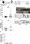
Figure 1.
Figure 1.
Neonatal exposure of SPF ApcMin/+ mice with Fn7-1 promotes intestinal tumor formation. (a) Neonatal exposure model schematic. (b) Colonic tumor burdens from sham or Fn7-1 neonatally exposed ApcMin/+ mice at 14 weeks. (c) Representative photographs of the colons of sham or Fn7-1 neonatally exposed ApcMin/+ mice. (d) Blinded assessment of H&E-stained colonic tissues from sham or Fn7-1 neonatally exposed ApcMin/+ mice to identify aberrant crypt foci (ACF), adenomas (ADE), and adenocarcinomas (ACA). Each column represents an individual mouse. (e) Small intestinal tumor burdens from sham or Fn7-1 neonatally exposed ApcMin/+ mice at 14 weeks. (f) Blinded assessment of H&E-stained small intestinal tissues from sham or Fn7-1 neonatally exposed ApcMin/+ mice to identify ACF, ADE, and ACA. Each column represents an individual mouse. (g) Fusobacterial abundance, determined by probe-based qPCR on DNA from colonic normal and tumor tissue of Fn7-1 neonatally exposed ApcMin/+ mice and normalized to mouse ActB copies, using primers listed in Supplemental Table S1. Data presented are calculated as 2^-(CTF. nucleatum nusG CTmActB). Samples with no detectable Fusobacterium, as determined by the F. nucleatum nusG cycle threshold (CT) compared to a control without F. nucleatum, are indicated by a triangle. All data points reflect data from an individual mouse or sample, with error bars indicating mean ± standard error of the mean (SEM), and statistics were calculated using a Mann-Whitney test. ** indicates P < .01; *** indicates P < .001
Figure 2.
Figure 2.
Neonatal inoculation with Fn7-1 leads to increased Il17a expression in the colonic lamina propria prior to tumor formation but does not alter gene expression of the F. nucleatum-associated pro-inflammatory signature. (a) Relative expression of Il6, Scyb1 (Il8), Tnf, Ccl2 and Ptgs2 (COX-2), as determined by RT-qPCR, from colonic epithelial and LP fractions of Fn7-1 or sham neonatally inoculated ApcMin/+ mice at 7–9 weeks of age. (b) Relative expression of Il23p19 and Il17a, as determined by RT-qPCR, in colonic epithelial and LP fractions of Fn7-1 or sham neonatally inoculated ApcMin/+ mice. (c) Relative fusobacterial abundance, normalized to total 16S, as determined by SYBR qPCR on DNA isolated from the feces of Fn7-1-neonatally inoculated mice at the indicated time points. Data presented are calculated as 2^-(CTFusobacterium 16S – CTeubacterial 16S). Fecal samples with no detectable Fusobacterium, as determined by the Fusobacterium 16S CT compared to a control without F. nucleatum, are indicated by a triangle. All data points reflect the data from an individual mouse or sample, with error bars indicating mean ± SEM, and statistics were calculated using a Wilcoxon signed-rank test compared to a null hypothesis of 1. * indicates P < .05; ns indicates not significant
Figure 3.
Figure 3.
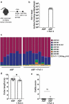
Fn7-1 colonizes ASF mice without altering community structure or inducing colitis. (a) ASF+Fn7-1 experimental schematic. (b) Colony-forming units of Fn7-1 per g feces after 2 weeks of colonization of ASF mice. (c) Fecal community structure from ASF and ASF+Fn7-1 mice as determined by 16S rRNA amplicon sequencing and analyzed by QIIME2 with DADA2. Each column reflects the proportion of different community members from an individual mouse (additional data in Supplemental Table S2). Only F. nucleatum abundance significantly differs across the two groups (LEfSe analysis, LDA(log10) > 4.0). (ASF519: Parabacteroides sp.; ASF457: Mucispirillum sp.; ASF360 and ASF361: Lactobacillus spp.; ASF356 and ASF502: Lachnospiraceae family members; ASF492: Eubacterium sp.; and ASF500: related to Colidextribacter sp.) (d) Colon length from ASF and ASF+Fn7-1 mice. (e) Histological colitis analysis of colons from ASF and ASF+Fn7-1 mice. All data points reflect the data from an individual mouse or sample, with error bars indicating mean ± SEM, and statistics were calculated using a Mann-Whitney test. * indicates P < .05; ns indicates not significant
Figure 4.
Figure 4.
Fn7-1 leads to a specific increase in Th17 cell frequency and IL-17A expression in the colonic lamina propria of ASF mice. (a) Il17a gene expression in the colonic epithelium and LP of ASF+Fn7-1 relative to ASF mice, by RT-qPCR. (b) Frequency of T helper cell subsets in the colonic LP of ASF and ASF+Fn7-1 mice, by intracellular transcription factor staining and flow cytometry. (c) FOXP3 expression within RORγt+ CD4+ T cells distinguishes between Fn7-1 induction of Th17 and RORγt+ T regulatory cells in the colonic LP of ASF mice, demonstrated by the frequency of FOXP3+RORγt+ and FOXP3RORγt+ cells within CD4+ T cells. (d & e) IL-17A expression, by frequency within stimulated CD4+ T cells (d) and mean fluorescence intensity of IL-17A within IL-17A-expressing CD4 + T cells (e) in the colonic LP of ASF and ASF+Fn7-1 mice. (f) Relative colonic epithelial expression of Saa1, Saa2, and Saa3 in ASF or ASF+Fn7-1 mice, by RT-qPCR. (g) Relative colonic epithelial and LP expression of Il23p19 in ASF or ASF+Fn7-1 mice, by RT-qPCR. For b, c, and d, CD4+ T cells are defined by gating on live CD45+CD3+CD4+ lymphocytes. All data points reflect the data from an individual mouse or sample, with error bars indicating mean ± SEM, and statistics were calculated using a Mann-Whitney test (for b, c, d, and e) and a Wilcoxon signed-rank test compared to a null hypothesis of 1 (for a, b, and g). * indicates P < .05, ** indicates P < .01; ns indicates not significant
Figure 5.
Figure 5.
FFAR2, a receptor for the microbial metabolites SCFA, is required for Fn7-1 induction of RORγt+ CD4+ T cells in the colonic lamina propria of ASF mice and is expressed in F. nucleatum-associated CRC tissues. (a) SCFA profiles from extracted cecal contents of ASF or ASF+Fn7-1 mice and analyzed by HPLC. (b) Frequency of RORγt+ cells, within the CD4+ T cell population, in the colonic lamina propria of C57Bl/6J or Ffar2−/− mice colonized with ASF or ASF+Fn7-1, by flow cytometry. (c) Spearman’s rank correlation of SCFA receptors in human CRC tissues with intratumoral F. nucleatum abundance, analyzed from The Cancer Genome Atlas colon adenocarcinoma (TCGA COAD) RNA-seq data and presented as -log10(False Discovery Rate). For b, CD4+ T cells are defined by gating on live CD45+CD3+CD4+ lymphocytes. For a and b, all data points reflect the data from an individual mouse or sample, with error bars indicating mean ± SEM, and statistics were calculated using a Mann-Whitney test. ** indicates P < .01; ns indicates not significant

References

    1. Kostic AD, Chun E, Robertson L, Glickman JN, Gallini CA, Michaud M, Clancy TE, Chung DC, Lochhead P, Hold GL, et al. Fusobacterium nucleatum potentiates intestinal tumorigenesis and modulates the tumor-immune microenvironment. Cell Host Microbe. 2013;14(2):207–19. doi:10.1016/j.chom.2013.07.007. - DOI - PMC - PubMed
    1. Wu S, Rhee K-J, Albesiano E, Rabizadeh S, Wu X, Yen H-R, Huso DL, Brancati FL, Wick E, McAllister F, et al. A human colonic commensal promotes colon tumorigenesis via activation of T helper type 17 T cell responses. Nat Med. 2009;15(9):1016–1022. doi:10.1038/nm.2015. - DOI - PMC - PubMed
    1. Arthur JC, Perez-Chanona E, Mühlbauer M, Tomkovich S, Uronis JM, Fan T-J, Campbell BJ, Abujamel T, Dogan B, Rogers AB, et al. Intestinal inflammation targets cancer-inducing activity of the microbiota. Science. 2012;338(6103):120–123. doi:10.1126/science.1224820. - DOI - PMC - PubMed
    1. Pleguezuelos-Manzano C, Puschhof J, Rosendahl Huber A, Van Hoeck A, Wood HM, Nomburg J, Gurjao C, Manders F, Dalmasso G, Stege PB, et al. Mutational signature in colorectal cancer caused by genotoxic pks+ E. coli. Nature. 2020;580(7802):269–273. doi:10.1038/s41586-020-2080-8. - DOI - PMC - PubMed
    1. Cuevas-Ramos G, Petit CR, Marcq I, Boury M, Oswald E, Nougayrède J-P.. Escherichia coli induces DNA damage in vivo and triggers genomic instability in mammalian cells. Proc Natl Acad Sci USA. 2010;107(25):11537–11542. doi:10.1073/pnas.1001261107. - DOI - PMC - PubMed

Publication types

MeSH terms