. 2021 Nov 23;118(47):e2100122118.
doi: 10.1073/pnas.2100122118.

Lipofuscin causes atypical necroptosis through lysosomal membrane permeabilization

Affiliations

Lipofuscin causes atypical necroptosis through lysosomal membrane permeabilization

Chendong Pan et al. Proc Natl Acad Sci U S A. .

Abstract

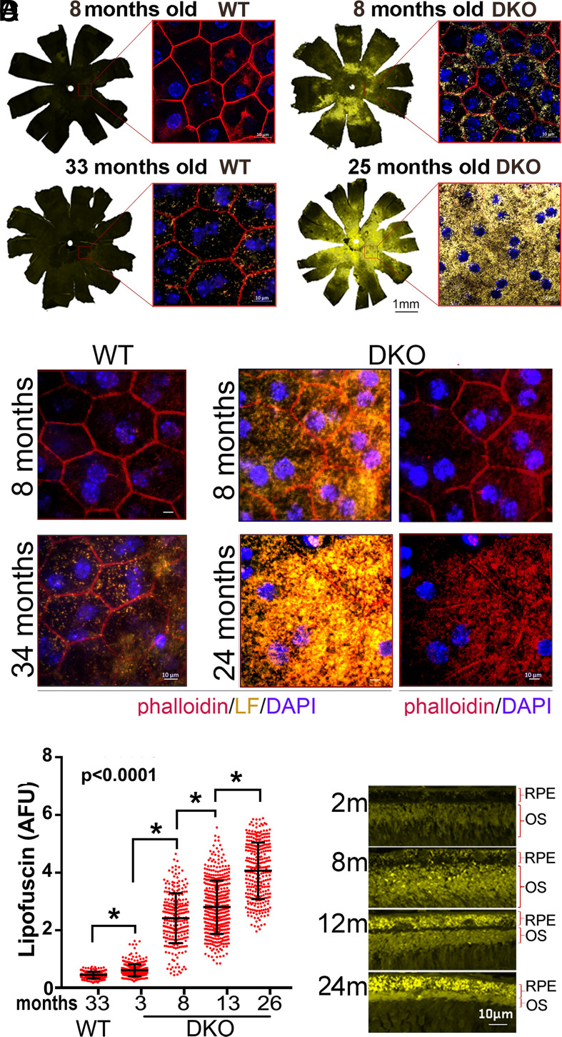
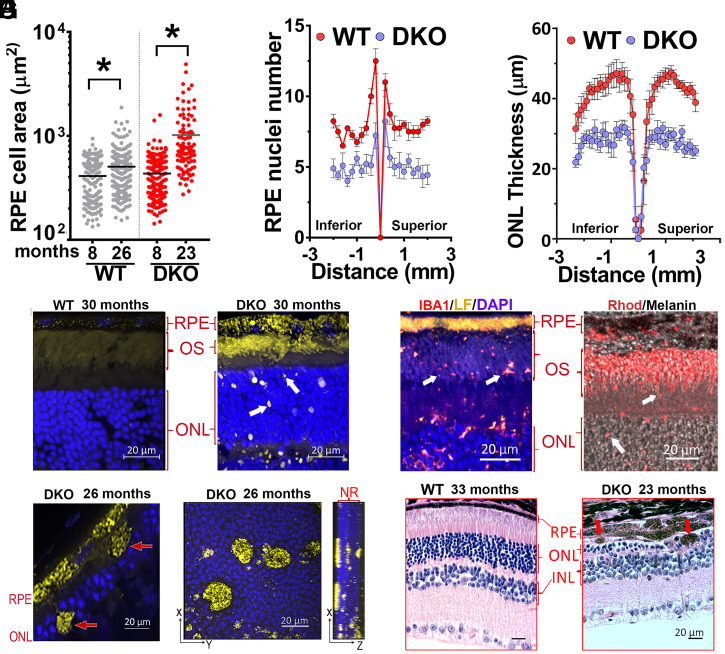
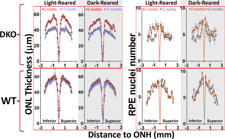
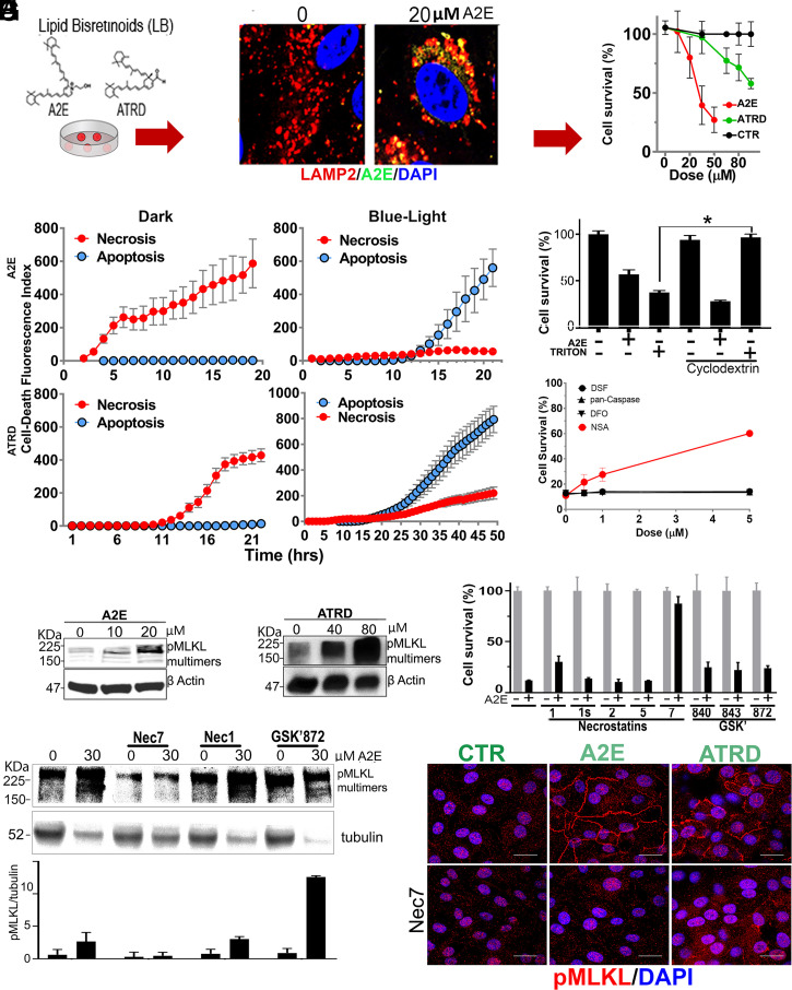
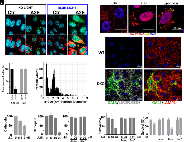
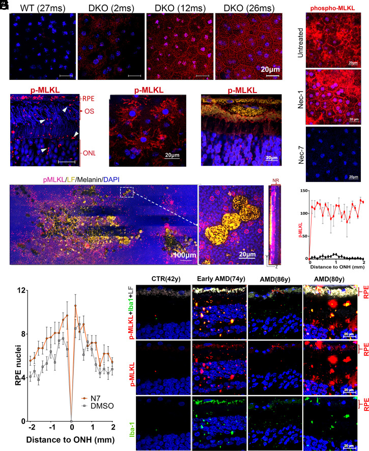
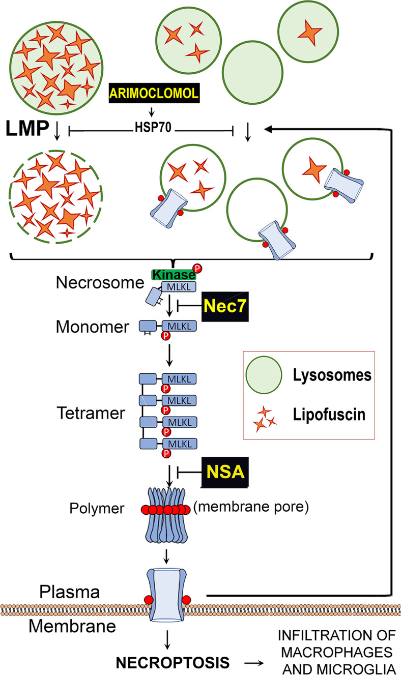
Lipofuscin granules enclose mixtures of cross-linked proteins and lipids in proportions that depend on the tissue analyzed. Retinal lipofuscin is unique in that it contains mostly lipids with very little proteins. However, retinal lipofuscin also presents biological and physicochemical characteristics indistinguishable from conventional granules, including indigestibility, tendency to cause lysosome swelling that results in rupture or defective functions, and ability to trigger NLRP3 inflammation, a symptom of low-level disruption of lysosomes. In addition, like conventional lipofuscins, it appears as an autofluorescent pigment, considered toxic waste, and a biomarker of aging. Ocular lipofuscin accumulates in the retinal pigment epithelium (RPE), whereby it interferes with the support of the neuroretina. RPE cell death is the primary cause of blindness in the most prevalent incurable genetic and age-related human disorders, Stargardt disease and age-related macular degeneration (AMD), respectively. Although retinal lipofuscin is directly linked to the cell death of the RPE in Stargardt, the extent to which it contributes to AMD is a matter of debate. Nonetheless, the number of AMD clinical trials that target lipofuscin formation speaks for the potential relevance for AMD as well. Here, we show that retinal lipofuscin triggers an atypical necroptotic cascade, amenable to pharmacological intervention. This pathway is distinct from canonic necroptosis and is instead dependent on the destabilization of lysosomes. We also provide evidence that necroptosis is activated in aged human retinas with AMD. Overall, this cytotoxicity mechanism may offer therapeutic targets and markers for genetic and age-related diseases associated with lipofuscin buildups.

Keywords: LMP; Lipofuscin; aging; lipid-bisretinoids; necroptosis.


Conflict of interest statement

Competing interest statement: A patent has been filed for targeting the LMP/necroptosis cascade.

Figures

Fig. 1.
Fig. 1.
(A) Llipofuscin content in RPE of WT and DKO mice at indicated ages. Fluorescence images of RPE flat mounts (10×) were stitched together to display the content in the whole eye. The orientation is as follows: dorsal–top; nasal–right. (Scale bar, 1 mm.) Inserts show high-magnification (63×) of central-retina RPE, with lipofuscin granules (yellow), ZO1 borders (red), and a nuclear Hoechst (blue). (Scale bars, 10 μm.) (B) 63× image of central retina’s RPE. Phalloidin are in red and lipofuscin granules are in yellow. Hexagonal RPE shape in both 8- and 34-mo WTs (Left two panels) contrasts with giant cells with lipofuscin in 8 and 24 mo DKOs. (Scale bars, 10 μm.) (C) Lipofuscin content per RPE, calculated from random fluorescent fields in flat-mounted RPE eyecups prestained with phalloidin. A 33-mo-old WT (n = 5) exhibited less lipofuscin/cell than 3-mo-old DKO (n = 4) (P < 0.01). The content of lipofuscin/cell was significantly different between 3- and 8- (n = 6); 8- and 13- (n = 4); and 13- and 26 (n = 5)-mo-old DKOs (mean ± SD, P < 0.0001). (D) Paraffin cross-section showing the progressive accumulation of lipofuscin in the RPE of DKO mice with age.
Fig. 2.
Fig. 2.
Degeneration in retinas with lipofuscin. (A) Individual and mean values of RPE’s sizes (micrometers2) in flat mounts from 8 (n = 5)- and 26 (n = 5)-mo-old WT (gray); 8 (n = 6)- and 23 (n = 5)-mo-old DKO (red) (P < 0.0001, unpaired t test). (B) RPE nuclei number in 26-mo-old DKO (blue) (n = 10) and WT (red) (n = 8) measured in paraffin cross-sections every 0.2 mm (P < 0.05, mean ± SEM, by multiple t tests). (C) ONL thicknesses in hematoxylin and eosin (H&E) stained cross-sections, along the vertical axis, in 26-mo-old DKO (blue) (n = 5) and WT (red) (n = 7) (P < 0.001, mean ± SEM, by ordinary two-way ANOVA). (D) Autofluorescence (yellow) in cryo-sections from 30-mo-old WT (n = 3) and DKO (n = 3) showing ∼1 to 3 μm lipofuscin debris (white arrows). (E, Left) ∼1 to 3 μm lipofuscin debris colocalize with Iba1 (white arrows) in 20-mo-old DKO retina. (E, Right) ∼1 to 3 μm lipofuscin debris (white arrows) are positive for rhodopsin (red) in 10-mo-old DKO. Bright-field image was overlapped to show RPE-melanin (black). (F) ∼5 to 10 μm RPE debris (arrows) in paraffin-section of 26-mo-old DKO. (G) Maximum projection of z-stacks from 26-mo-old DKO flat-mounted neuroretina. Noticeable were the large RPE debris inserted at different depths into the ONL. (H) H&E cross-sections. (Right) Damage in ONL overlying ∼10 μm RPE debris containing melanin. (Scale bars, 20 μm.)
Fig. 3.
Fig. 3.
Light-independent lipofuscin cytotoxicity contributes to degeneration in pigmented retinas. ONL thinning and nuclei RPE loss, between 2 and 12 mo of age, occurred at similar levels among light-cycled and dark-reared mice (P < 0.01, by two-way ANOVA). For ONL: 12 h-light-cycled mice, 2-mo DKO (n = 8); 12-mo DKO (n = 14); and 2- and 12-mo WT (n = 6 per group). For dark-reared: 2-mo DKO (n = 6); 12-mo DKO (n = 8); and 2- and 12-mo WT (n = 6 per group). For RPE nuclei number: 12 h-light-cycled mice, 2-mo DKO (n = 8); 12-mo DKO (n = 14); and 2- and 12-mo WT (n = 7 per group). For dark-reared: 2-mo DKO (n = 7); 12-mo DKO (n = 4); and 2-mo (n = 4) and 12-mo WT (n = 7) (P < 0.05, mean ± SEM, by multiple t test).
Fig. 4.
Fig. 4.
Mechanism of light-independent cytotoxicity. (A) Lipid-bisretinoids accumulation (green) into lamp-2 lysosomes (red) of ARPE-19 in cell-death assay. Viability was determined with Resazurin. (B) Real-time monitoring of apoptotic and necrotic death in ARPE19 cells in the dark or after blue-light exposure. For dark toxicity, cells received A2E (30 μM) or ATRD (80 μM) at time 0. For phototoxicity, experiments started after 10 min blue-light irradiation of cells preloaded with vehicle or sublethal doses of A2E (5 μM) or ATRD (20 μM) 24 h earlier. Cells were monitored at 30-min intervals for NUC405 (blue = caspase 3 activation) or DRAQ7 (red= plasma membrane leakage), respectively. (C) A2E (20 μM) lethal toxicity was not prevented by MβCD, while MβCD completely rescued cells from 500 μM Triton X-100 detergent cytotoxicity (P < 0.001, mean ± SEM, n = 3, two-tailed t test). (D) Inhibition of the main effector pathways of programed cell death: disulfiram for Gasdermin-D/pyroptosis; Deferoxamine (DFO) for ferroptosis; and NSA for MLKL/necroptosis (n = 3). (E and F) Dose-dependent phosphorylation and polymerization of MLKL by A2E and ATRD. (G) Protection from lipofuscin by Nec7 (mean ± SEM, P < 0.01, two-tailed t test, n = 3). (H) WB showing Nec7 but not Nec1 prevents MLKL phosphorylation/polymerization. (I) ARPE-19 monolayers with A2E or ATRD accumulations display phospho-MLKL in plasma membranes which was blocked by Nec7. (Scale bars, 20 μm.)
Fig. 5.
Fig. 5.
(A) Cellular ROS (red) were detected in mitochondria but not in lipofuscin granules (green). Only after blue-light exposure, ROS colocalized with lipofuscin. (B) A2E (MW = 592) passed through 3-μm but not 0.45-μm filters, suggesting that it forms aggregates. (C) Particle size measurement using TRPS shows that A2E formed aggregates larger than 500 nm in PBS. (D) Galectin-3 puncta assay in ARPE19 to evaluate LMP. Both the positive control LLOMe and A2E induced LMP. (E) Increased LMP in DKO retinas with lipofuscin. Galectin-3 puncta positive staining in lysosomes of cells in RPE eyecups from 20-mo-old DKO but not WT mice. (Scale bar, 10 μm.) (F) Loss of cathepsins activity in cells exposed to LLOMe doses or (G) A2E. (H) Loss of cathepsin activity can be prevented with arimoclomol or Nec7 in A2E loaded cells. (I) Arimoclomol or Nec7 promote survival to A2E accumulation. (J) The LMP inducer LLOMe promotes a necroptosis preventable with Nec7 and arimoclomol but not Nec1.
Fig. 6.
Fig. 6.
Necroptosis in retinas with lipofuscin. Central-RPEs, in flat-mounted DKO eyecups immune-stained with (A) anti-phospho-Ser345-MLKL (red) and DAPI (blue) (n = 4) (Scale bars, 20 μm.) (B) ∼1 to 3 μm lipofuscin debris (characterized in Fig. 2 as lipofuscin+, iba1+) were positive for phospho-MLKL (red) in demelanized paraffin cross-section from 20-mo-old DKO. (C) Microglia/macrophages attached to flat-mounted RPE eyecups from 20-mo-old DKO stained for phospho-MLKL. (D) Phospho-MLKL staining (red) of large RPE debris (yellow) shed into neuroretina. (E) Stitched panoramic of flat-mounted neuroretina containing clusters of large (∼10 μm) RPE fragments, described in Fig. 2 F and H. Very noticeable was the strong halo of phospho-MLKL (red) around these RPE inclusions. Right panel is the maximal projection of one of these lesions, obtained with 63× objective. It shows the spread of the phospho-MLKL signal in the ONL layer in contact with the invading RPE fragments. (F) Single intravitreal injection of Nec7 (DKO at 718 d-old, n = 4) but not Nec1 (DKO at 709 d-old, n = 4) eliminated phospho-MLKL staining in 20-mo-old DKO retinas (n = 4). (G) Phospho-MLKL expression in RPE flat mounts from 23-mo-old DKO, measured every 0.1-mm interval and plotted as a function of the distance from ONH in inferior hemiretina. Mock- (red line) and Nec7- (black line) treated eyes (**P < 0.01 by two-way ANOVA, n = 3). (H) Intraocular treatment with Nec7 (n = 10) prevented loss of RPE nuclei in DKOs compared to mock-treated eyes (n = 10) (mean ± SEM, P = 0.015, by t test, two samples equal variance). (I) Human retinas stained with anti-phospho-MLKL (red) and Iba1 (green). The staining was stronger in retinas with abundant lipofuscin and appeared in RPE, Iba1+ cells as well as in debris/fragments infiltrating neuroretina. Donors of 86 y and 80 y had advanced dry AMD diagnosis.
Fig. 7.
Fig. 7.
Working model of light-independent cytotoxicity elicited by the accumulation of lipofuscin. Lipofuscin forms solid crystals that, when in high amounts, damage the lysosomal membranes and cause LMP. The release of lysosomal enzymes triggers the formation of an atypical necrosome that phosphorylates MLKL, promoting its oligomerization and membranes translocation. Phospho-MLKL destabilizes the membrane of lysosomes promoting more LMP. When the levels of phospho-MLKL in plasma membrane become intolerable, the cell undergoes necroptosis.

Similar articles

Cited by

References

    1. Ng K.-P., et al. , Retinal pigment epithelium lipofuscin proteomics. Mol. Cell. Proteomics 7, 1397–1405 (2008). - PMC - PubMed
    1. Gómez-Sintes R., Ledesma M. D., Boya P., Lysosomal cell death mechanisms in aging. Ageing Res. Rev. 32, 150–168 (2016). - PubMed
    1. Ono K., Kim S. O., Han J., Susceptibility of lysosomes to rupture is a determinant for plasma membrane disruption in tumor necrosis factor alpha-induced cell death. Mol. Cell. Biol. 23, 665–676 (2003). - PMC - PubMed
    1. Brunk U., Brun A., The effect of aging on lysosomal permeability in nerve cells of the central nervous system. An enzyme histochemical study in rat. Histochemie 30, 315–324 (1972). - PubMed
    1. Sparrow J. R., Cai B., Jang Y. P., Zhou J., Nakanishi K., A2E, a Fluorophore of RPE Lipofuscin, Can Destabilize Membrane in Advances in Experimental Medicine and Biology, 2005, pp. 63–68. - PubMed

Publication types