. 2021 Nov 23;118(47):e2115435118.
doi: 10.1073/pnas.2115435118.

Development of a universal nanobody-binding Fab module for fiducial-assisted cryo-EM studies of membrane proteins

Affiliations

Development of a universal nanobody-binding Fab module for fiducial-assisted cryo-EM studies of membrane proteins

Joël S Bloch et al. Proc Natl Acad Sci U S A. .

Abstract

With conformation-specific nanobodies being used for a wide range of structural, biochemical, and cell biological applications, there is a demand for antigen-binding fragments (Fabs) that specifically and tightly bind these nanobodies without disturbing the nanobody-target protein interaction. Here, we describe the development of a synthetic Fab (termed NabFab) that binds the scaffold of an alpaca-derived nanobody with picomolar affinity. We demonstrate that upon complementary-determining region grafting onto this parent nanobody scaffold, nanobodies recognizing diverse target proteins and derived from llama or camel can cross-react with NabFab without loss of affinity. Using NabFab as a fiducial and size enhancer (50 kDa), we determined the high-resolution cryogenic electron microscopy (cryo-EM) structures of nanobody-bound VcNorM and ScaDMT, both small membrane proteins of ∼50 kDa. Using an additional anti-Fab nanobody further facilitated reliable initial three-dimensional structure determination from small cryo-EM test datasets. Given that NabFab is of synthetic origin, is humanized, and can be conveniently expressed in Escherichia coli in large amounts, it may be useful not only for structural biology but also for biomedical applications.

Keywords: antibody; cryogenic electron microscopy; membrane protein; nanobody; structure.


Conflict of interest statement

The authors declare no competing interests.

Figures

Fig. 1.
Fig. 1.
Universal binding of NabFab to the scaffold Nbs enabled by CDR grafting. (A) Schematic of NabFab application in Nb binding. The grafting of target-specific Nb CDRs onto the TC-Nb4 (33) scaffold, which contains the NabFab-binding epitope, enables rigid complex formation of target membrane protein, grafted Nb, NabFab, as well as an anti-Fab Nb (32) as symmetry breaker. Dashed boxes illustrate the rationale behind CDR grafting. On the example of the structure of NorM-Nb17_4 (ribbon representation), the TC-Nb4 scaffold (33) is shown in green and the grafted CDRs of NorM-Nb17 in red. On the right side, a sequence alignment of TC-Nb4 and NorM-Nb17 is shown in Kabat numbering (48). The scaffold and CDR regions that comprise the grafted NorM-Nb17_4 are highlighted with the same colors. Note that for NorM-Nb17_4, “CDR4” of TC-Nb4 was retained due to its similarity in sequence. (B) SPR sensograms of NabFab binding to TC-Nb4 (alpaca) and grafted Nbs BtuF-Nb9_4 (alpaca), Lyso-Nb_4 (camel), and RNAs-Nb_4 (camel).
Fig. 2.
Fig. 2.
Fiducial-assisted structure determination. (AD) Cryo-EM structure determination of VcNorM complex. (EH) Cryo-EM structure determination of ScaDMT complex. (A and E) Representative cryo-EM micrographs of complexes of detergent-solubilized membrane protein, grafted Nb, NabFab, and anti-Fab Nb. (B and F) Representative 2D classes as calculated in RELION 3.1 (43). (C and G) Processing of a small cryo-EM test dataset including initial model generation and 3D refinement as calculated in RELION 3.1 (43) and assignment of the correct handedness of the structure using structural knowledge of NabFab:anti-Fab Nb complex. (D and H) Unsharpened cryo-EM maps of VcNorM and ScaDMT complexes derived from the processing of full datasets [VcNorM in RELION 3.1 and ScaDMT in RELION 3.1 (43) and cryoSPARC V3.2 (49)]. VcNorM and ScaDMT are highlighted in light blue, grafted Nb CDRs in red, TCNb4 scaffold in green, NabFab HC in cyan and LC in black, and the anti-Fab Nb in yellow.
Fig. 3.
Fig. 3.
Structural analysis of NabFab binding to grafted Nbs of different origin. (A) Surface representation of complex of VcNorM (blue), NorM-Nb17_4 TC-Nb4 (33) scaffold green, NorM-Nb17 CDRs red), NabFab (HC cyan, LC black), and anti-Fab Nb (32) (yellow). (B) Zoomed-in view of NorM-Nb17_4:NabFab-binding interface with NorM-Nb17_4 in surface representation and NabFab in ribbon representation with interface residues in stick representation with oxygen atoms colored in red and nitrogen atoms in blue. (C) DMT-Nb17_4:NabFab-binding interface. (D) Lyso-Nb_4:NabFab-binding interface. (E) Representative section of EM map of NorM-Nb17_4:NabFab interface at 12 rmsd, carved to 2 Å. Ordered water molecules indicated by red crosses. (F) Representative section of EM map of DMT-Nb16_4:NabFab interface at 11 rmsd, carved to 2 Å. (G) Representative section of electron density of Lyso-Nb_4:NabFab interface at 1 rmsd, carved to 2 Å.
Fig. 4.
Fig. 4.
Hotspot analysis of NabFab-binding epitope on TC-Nb4 scaffold. (A) Detailed views of NorM-Nb17_4 residues at the NabFab-binding interface. Distances in angstrom are indicated with numbers and dotted lines. Note that R45 and Y91 are contributing to a cation–π ladder. (B) Alanine scanning of Nb residues at the interface to NabFab. Bars indicate the ratio of the KD for NabFab binding to the original TC-Nb4 scaffold and the respective surface mutants as derived from SPR measurements. Residues for which mutation to Ala reduced binding affinity more than 10-fold are highlighted in red. Note that Lyso-Nb_4 was used in this experiment.
Fig. 5.
Fig. 5.
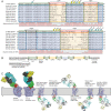
In silico CDR grafting and NabFab-binding analysis for potential structural targets. (A) Protein sequence alignment of TC-Nb4 (33), NorM-Nb17, DMT-Nb16 (38), and Lyso-Nb (36), as well as selected Nbs of known membrane protein–Nb complexes (–13, 50, 51) (PDB IDs: 3P0G, 4MQS, 6B73, 6IBB, and 6I6J), generated with Clustal Omega (52), in Kabat numbering (48). Green boxes indicate scaffold regions of TC-Nb4. Blue boxes indicate equivalent scaffold regions other Nbs. Red boxes indicate CDRs of TC-Nb4. Yellow boxes indicated equivalent CDR regions of other Nbs. A black box indicates “CDR4” of TC-Nb4. A gray box indicates the equivalent region in other Nbs. Note that “CDR4” is a nonvariable loop that in some cases interacts with the target protein of the respective Nb. Residues at the Nb-binding interface to NabFab are indicated with colored dots on top. Yellow dots indicate a strong contribution to NabFab binding. Cyan-colored dots indicate a weak contribution, as described in Fig. 3B. (B) Schematic of CDR-grafting strategy: Green boxes indicate regions of the TC-Nb4 scaffold that were retained during grafting. Yellow boxes indicate epitope-binding CDR regions of different Nbs that were grafted into the TC-Nb4 scaffold. A gray/blue box indicates “CDR4,” which can also be grafted. Flanking gray boxes indicate the secretion and purification tags. Note that the His-tags are not necessary for NabFab binding. (C) Structures of complexes of VcNorM and ScaDMT in surface representation as well as selected examples of in silico docked [PyMol (53)], putative complexes of selected membrane protein–Nb complexes in ribbon representation. Membrane proteins are colored in blue, original Nb CDRs in red, TC-Nb4-scaffold in green, NabFab HC in cyan, NabFab LC in gray, and anti-Fab Nb in yellow.

References

    1. Basu K., Green E. M., Cheng Y., Craik C. S., Why recombinant antibodies - benefits and applications. Curr. Opin. Biotechnol. 60, 153–158 (2019). - PMC - PubMed
    1. Chen G., Sidhu S. S., Nilvebrant J., Synthetic antibodies in infectious disease. Adv. Exp. Med. Biol. 1053, 79–98 (2017). - PubMed
    1. Adams J. J., Sidhu S. S., Synthetic antibody technologies. Curr. Opin. Struct. Biol. 24, 1–9 (2014). - PubMed
    1. Sidhu S. S., Kossiakoff A. A., Exploring and designing protein function with restricted diversity. Curr. Opin. Chem. Biol. 11, 347–354 (2007). - PubMed
    1. Muyldermans S., Nanobodies: Natural single-domain antibodies. Annu. Rev. Biochem. 82, 775–797 (2013). - PubMed

Publication types

Substances

LinkOut - more resources