

Nanometer-resolution in situ structure of the SARS-CoV-2 postfusion spike protein
- PMID: 34782481
- PMCID: PMC8640741
- DOI: 10.1073/pnas.2112703118
Nanometer-resolution in situ structure of the SARS-CoV-2 postfusion spike protein
Abstract
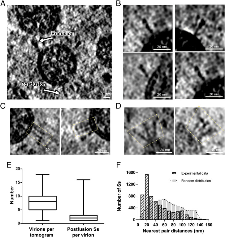
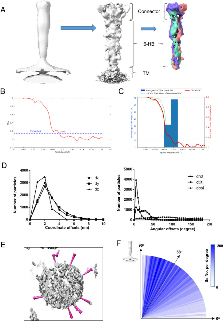
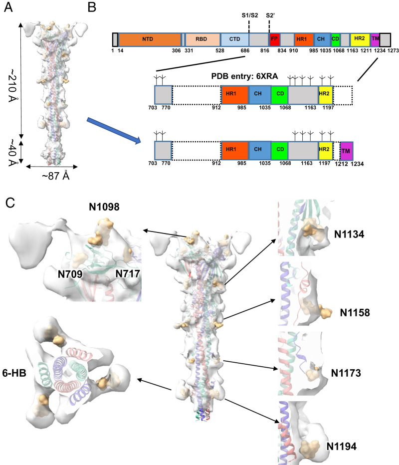
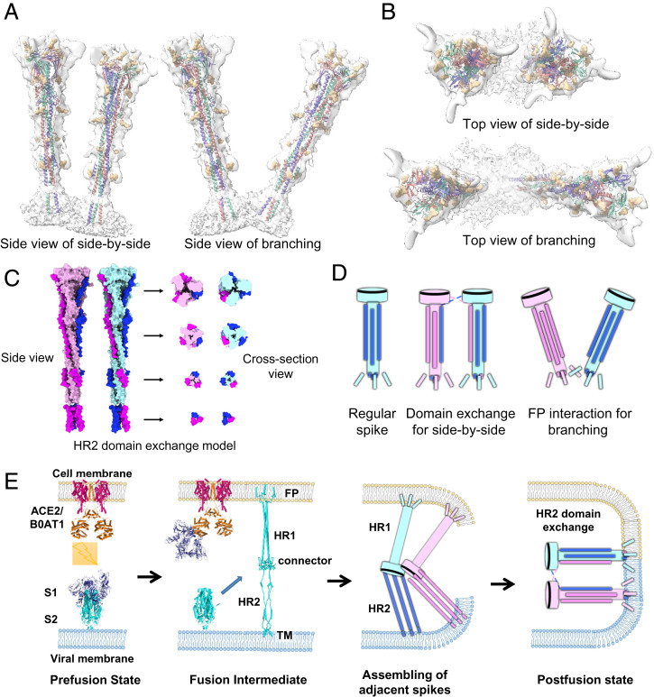
The spike protein of severe acute respiratory syndrome coronavirus 2 (SARS-CoV-2) mediates membrane fusion to allow entry of the viral genome into host cells. To understand its detailed entry mechanism and develop a specific entry inhibitor, in situ structural information on the SARS-CoV-2 spike protein in different states is urgent. Here, by using cryo-electron tomography, we observed both prefusion and postfusion spikes in β-propiolactone-inactivated SARS-CoV-2 virions and solved the in situ structure of the postfusion spike at nanometer resolution. Compared to previous reports, the six-helix bundle fusion core, the glycosylation sites, and the location of the transmembrane domain were clearly resolved. We observed oligomerization patterns of the spikes on the viral membrane, likely suggesting a mechanism of fusion pore formation.
Keywords: SARS-CoV-2; cryo-electron tomography; postfusion state; spike protein; subtomogram analysis.
Conflict of interest statement
The authors declare no competing interest.
Figures
Similar articles
-
The Architecture of Inactivated SARS-CoV-2 with Postfusion Spikes Revealed by Cryo-EM and Cryo-ET.Structure. 2020 Nov 3;28(11):1218-1224.e4. doi: 10.1016/j.str.2020.10.001. Epub 2020 Oct 15. Structure. 2020. PMID: 33058760 Free PMC article.
-
Unveiling the structural spectrum of SARS-CoV-2 fusion by in situ cryo-ET.Nat Commun. 2025 Jun 3;16(1):5150. doi: 10.1038/s41467-025-60406-z. Nat Commun. 2025. PMID: 40461447 Free PMC article.
-
Cryo-EM structure of the SARS coronavirus spike glycoprotein in complex with its host cell receptor ACE2.PLoS Pathog. 2018 Aug 13;14(8):e1007236. doi: 10.1371/journal.ppat.1007236. eCollection 2018 Aug. PLoS Pathog. 2018. PMID: 30102747 Free PMC article.
-
Structural basis of severe acute respiratory syndrome coronavirus 2 infection.Curr Opin HIV AIDS. 2021 Jan;16(1):74-81. doi: 10.1097/COH.0000000000000658. Curr Opin HIV AIDS. 2021. PMID: 33186231 Review.
-
Site-specific glycosylation of SARS-CoV-2: Big challenges in mass spectrometry analysis.Proteomics. 2022 Aug;22(15-16):e2100322. doi: 10.1002/pmic.202100322. Epub 2022 Jun 22. Proteomics. 2022. PMID: 35700310 Free PMC article. Review.
Cited by
-
Approaches to the Potential Therapy of COVID-19: A General Overview from the Medicinal Chemistry Perspective.Molecules. 2022 Jan 20;27(3):658. doi: 10.3390/molecules27030658. Molecules. 2022. PMID: 35163923 Free PMC article. Review.
-
A highly immunogenic live-attenuated vaccine candidate prevents SARS-CoV-2 infection and transmission in hamsters.Innovation (Camb). 2022 Mar 29;3(2):100221. doi: 10.1016/j.xinn.2022.100221. Epub 2022 Mar 2. Innovation (Camb). 2022. PMID: 35252935 Free PMC article.
-
Study of fusion peptide release for the spike protein of SARS-CoV-2.RSC Adv. 2023 Jun 6;13(25):16970-16983. doi: 10.1039/d3ra01764h. eCollection 2023 Jun 5. RSC Adv. 2023. PMID: 37288377 Free PMC article.
-
Broad Adaptability of Coronavirus Adhesion Revealed from the Complementary Surface Affinity of Membrane and Spikes.Adv Sci (Weinh). 2024 Nov;11(41):e2404186. doi: 10.1002/advs.202404186. Epub 2024 Sep 4. Adv Sci (Weinh). 2024. PMID: 39231361 Free PMC article.
-
Conserved role of spike S2 domain N-glycosylation across beta-coronavirus family.bioRxiv [Preprint]. 2024 Sep 5:2024.09.05.611372. doi: 10.1101/2024.09.05.611372. bioRxiv. 2024. Update in: Npj Viruses. 2025 Jan 25;3(1):4. doi: 10.1038/s44298-024-00085-7. PMID: 39282346 Free PMC article. Updated. Preprint.
References
-
- Ksiazek T. G., et al. , SARS Working Group, A novel coronavirus associated with severe acute respiratory syndrome. N. Engl. J. Med. 348, 1953–1966 (2003). - PubMed
-
- Zaki A. M., van Boheemen S., Bestebroer T. M., Osterhaus A. D., Fouchier R. A., Isolation of a novel coronavirus from a man with pneumonia in Saudi Arabia. N. Engl. J. Med. 367, 1814–1820 (2012). - PubMed
Publication types
MeSH terms
Substances
LinkOut - more resources
Full Text Sources
Miscellaneous
