

Promoting axon regeneration in the central nervous system by increasing PI3-kinase signaling
- PMID: 34782551
- PMCID: PMC8643051
- DOI: 10.4103/1673-5374.327324
Promoting axon regeneration in the central nervous system by increasing PI3-kinase signaling
Abstract
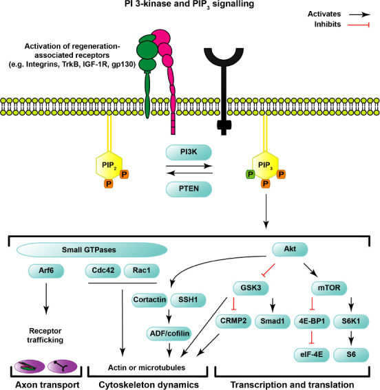
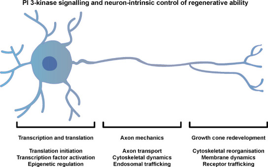
Much research has focused on the PI3-kinase and PTEN signaling pathway with the aim to stimulate repair of the injured central nervous system. Axons in the central nervous system fail to regenerate, meaning that injuries or diseases that cause loss of axonal connectivity have life-changing consequences. In 2008, genetic deletion of PTEN was identified as a means of stimulating robust regeneration in the optic nerve. PTEN is a phosphatase that opposes the actions of PI3-kinase, a family of enzymes that function to generate the membrane phospholipid PIP3 from PIP2 (phosphatidylinositol (3,4,5)-trisphosphate from phosphatidylinositol (4,5)-bisphosphate). Deletion of PTEN therefore allows elevated signaling downstream of PI3-kinase, and was initially demonstrated to promote axon regeneration by signaling through mTOR. More recently, additional mechanisms have been identified that contribute to the neuron-intrinsic control of regenerative ability. This review describes neuronal signaling pathways downstream of PI3-kinase and PIP3, and considers them in relation to both developmental and regenerative axon growth. We briefly discuss the key neuron-intrinsic mechanisms that govern regenerative ability, and describe how these are affected by signaling through PI3-kinase. We highlight the recent finding of a developmental decline in the generation of PIP3 as a key reason for regenerative failure, and summarize the studies that target an increase in signaling downstream of PI3-kinase to facilitate regeneration in the adult central nervous system. Finally, we discuss obstacles that remain to be overcome in order to generate a robust strategy for repairing the injured central nervous system through manipulation of PI3-kinase signaling.
Keywords: PI3-kinase; PI3K; PTEN; axon cytoskeleton; axon regeneration; axon transport; cell signaling; central nervous system; growth cone; neuroprotection; trafficking; transcription; translation.
Conflict of interest statement
None
Figures
References
-
- Anderson MA, O’Shea TM, Burda JE, Ao Y, Barlatey SL, Bernstein AM, Kim JH, James ND, Rogers A, Kato B, Wollenberg AL, Kawaguchi R, Coppola G, Wang C, Deming TJ, He Z, Courtine G, Sofroniew MV. Required growth facilitators propel axon regeneration across complete spinal cord injury. Nature. 2018;561:396–400. - PMC - PubMed
-
- Ashery U, Penner R, Spira ME. Acceleration of membrane recycling by axotomy of cultured aplysia neurons. Neuron. 1996;16:641–651. - PubMed
Publication types
Grants and funding
LinkOut - more resources
Full Text Sources
Research Materials
Miscellaneous
