Review
. 2022 Jun;17(6):1228-1239.
doi: 10.4103/1673-5374.327323.

SARS-CoV-2 involvement in central nervous system tissue damage

Affiliations
Review

SARS-CoV-2 involvement in central nervous system tissue damage

Muhammad Ali Haidar et al. Neural Regen Res. 2022 Jun.

Abstract

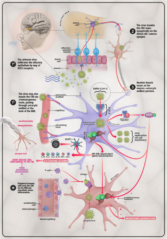
As the severe acute respiratory syndrome coronavirus 2 (SARS-CoV-2) continues to spread globally, it became evident that the SARS-CoV-2 virus infects multiple organs including the brain. Several clinical studies revealed that patients with COVID-19 infection experience an array of neurological signs ranging in severity from headaches to life-threatening strokes. Although the exact mechanism by which the SARS-CoV-2 virus directly impacts the brain is not fully understood, several theories have been suggested including direct and indirect pathways induced by the virus. One possible theory is the invasion of SARS-CoV-2 to the brain occurs either through the bloodstream or via the nerve endings which is considered to be the direct route. Such findings are based on studies reporting the presence of viral material in the cerebrospinal fluid and brain cells. Nevertheless, the indirect mechanisms, including blood-clotting abnormalities and prolonged activation of the immune system, can result in further tissue and organ damages seen during the course of the disease. This overview attempts to give a thorough insight into SARS-CoV-2 coronavirus neurological infection and highlights the possible mechanisms leading to the neurological manifestations observed in infected patients.

Keywords: CNS infection; COVID-19; autoantibodies; coagulopathy; encephalitis; neuroinflammation; renin-angiotensin system; viral encephalopathy.


Conflict of interest statement

None

Figures

Figure 1
Figure 1
Mechanisms of invasion and possible CNS effects of SARS-CoV-2. (1a) The virus first enters the body via the nasal passage and makes its way to the olfactory bulb by crossing the olfactory epithelium using ACE-2 receptors. From there the virus enters the CNS through (2) the mitral cell-neuron synapse. (3) The virus then infects astrocytes, ACE-2-expressing glial cells which can also be infected via (1b) astrocytic end-feet that surround endothelial cells at the level of the BBB. In addition to utilizing cell machinery to proliferate, SARS-CoV-2 provokes the downregulation of ACE-2 in host cells, which in turn leads to the over-activation of the RAAS system. This promotes the upregulation of the NF-κB pathway and the subsequent transcription of genes coding for pro-inflammatory mediators such as IL-1β, IL-6, IL-8, IL-10, TNF-α, and IFN-γ. This medley of cytokines, chemokines, and other pro-inflammatory molecules may develop into what is termed the cytokine storm, a life-threatening condition that has the potential to cause short- as well as long-term damage on the CNS in the form of stroke, auto-immune disease, or encephalopathy, etc. Disruption of the BBB and the ensuing infiltration of the CNS by peripheral immune cells (T-cells, macrophages, etc.), triggered by microglial antigen presentation, further excarbates the situation. The overall result is further aggravation of neuroinflammation and injury cascades that include, but are certainly not limited to, synaptic loss, demyelination, and excitotoxicity. ACE-2: Angiotensin-converting enzyme 2; BBB: blood-brain barrier; CNS: central nervous system; IFN: interferon; IL: interleukin; NF-κB: nuclear factor κB; RAAS: renin-angiotensin-aldosterone system; SARS-CoV-2: severe acute respiratory syndrome coronavirus 2; TNF-α: tumor necrosis factor-α.

Similar articles

Cited by

References

    1. Al Saiegh F, Ghosh R, Leibold A, Avery MB, Schmidt RF, Theofanis T, Mouchtouris N, Philipp L, Peiper SC, Wang ZX, Rincon F, Tjoumakaris SI, Jabbour P, Rosenwasser RH, Gooch MR. Status of SARS-CoV-2 in cerebrospinal fluid of patients with COVID-19 and stroke. J Neurol Neurosurg Psychiatry. 2020;91:846–848. - PubMed
    1. Alberti P, Beretta S, Piatti M, Karantzoulis A, Piatti ML, Santoro P, Vigano M, Giovannelli G, Pirro F, Montisano DA, Appollonio I, Ferrarese C. Guillain-Barre syndrome related to COVID-19 infection. Neurol Neuroimmunol Neuroinflamm. 2020;7:e741. - PMC - PubMed
    1. Amraie R, Napoleon MA, Yin W, Berrigan J, Suder E, Zhao G, Olejnik J, Gummuluru S, Muhlberger E, Chitalia V, Rahimi N. CD209L/L-SIGN and CD209/DC-SIGN act as receptors for SARS-CoV-2 and are differentially expressed in lung and kidney epithelial and endothelial cells. bioRxiv. 2020. doi:10.1101/2020.06.22.165803. Available from: https://www.biorxiv.org/content/10.1101/2020.06.22.165803v3 .
    1. Asadi-Pooya AA, Simani L, Shahisavandi M, Barzegar Z. COVID-19 de novo seizures and epilepsy: a systematic review. Neurol Sci. 2021;42:415–431. - PMC - PubMed
    1. Badenoch JB, Rengasamy ER, Watson CJ, Jansen K, Rooney AG. Persistent neuropsychiatric symptoms after COVID-19: a systematic review and meta-analysis. medRxiv. 2021 doi:10.1101/2021.04.30.21256413. - PMC - PubMed