. 2021 Nov 15;12(1):6586.
doi: 10.1038/s41467-021-26690-1.

Detrimental proarrhythmogenic interaction of Ca2+/calmodulin-dependent protein kinase II and NaV1.8 in heart failure

Affiliations

Detrimental proarrhythmogenic interaction of Ca2+/calmodulin-dependent protein kinase II and NaV1.8 in heart failure

Philipp Bengel et al. Nat Commun. .

Abstract

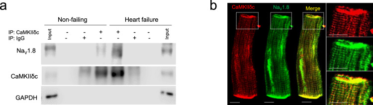
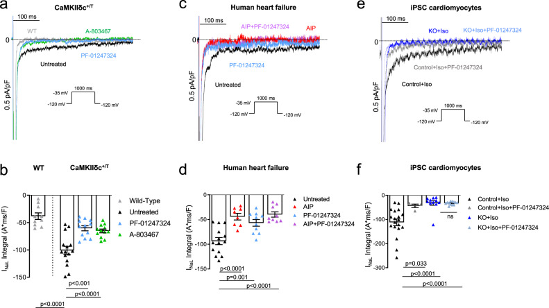
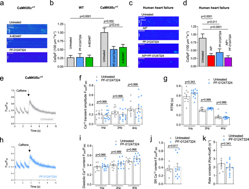
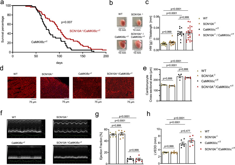
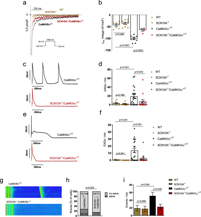
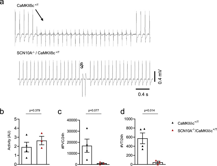
An interplay between Ca2+/calmodulin-dependent protein kinase IIδc (CaMKIIδc) and late Na+ current (INaL) is known to induce arrhythmias in the failing heart. Here, we elucidate the role of the sodium channel isoform NaV1.8 for CaMKIIδc-dependent proarrhythmia. In a CRISPR-Cas9-generated human iPSC-cardiomyocyte homozygous knock-out of NaV1.8, we demonstrate that NaV1.8 contributes to INaL formation. In addition, we reveal a direct interaction between NaV1.8 and CaMKIIδc in cardiomyocytes isolated from patients with heart failure (HF). Using specific blockers of NaV1.8 and CaMKIIδc, we show that NaV1.8-driven INaL is CaMKIIδc-dependent and that NaV1.8-inhibtion reduces diastolic SR-Ca2+ leak in human failing cardiomyocytes. Moreover, increased mortality of CaMKIIδc-overexpressing HF mice is reduced when a NaV1.8 knock-out is introduced. Cellular and in vivo experiments reveal reduced ventricular arrhythmias without changes in HF progression. Our work therefore identifies a proarrhythmic CaMKIIδc downstream target which may constitute a prognostic and antiarrhythmic strategy.


Conflict of interest statement

The authors declare no competing interests.

Figures

Fig. 1
Fig. 1. CaMKIIδc interacts with NaV1.8 in human myocardium and isolated cardiomyocytes.
a Co-immunoprecipitation of CaMKIIδc and NaV1.8 from left ventricular homogenates of human non-failing and failing hearts (NF: n = 7; HF: n = 7). b Co-localization of CaMKIIδc and NaV1.8 in human failing cardiomyocytes with immunofluorescence staining. Scale bar: 10 µm (staining was performed in cardiomyocytes isolated from five heart failure patients).
Fig. 2
Fig. 2. Reduced INaL upon NaV1.8 inhibition in human failing and mouse CaMKIIδc transgenic cardiomyocytes, and in SCN10A knockout iPSC-cardiomyocytes.
a Original traces of INaL in WT and CaMKIIδc+/T mouse ventricular cardiomyocytes elicited using the protocol shown in the inset. b Mean data ± SEM along with individual values shown in the graph plotting WT (n = 10 cells/5 mice) and CaMKIIδc+/T (untreated: n = 15 cells/7 mice; A-806467: n = 12 cells/7 mice; PF-01247324: n = 12 cells/7 mice). Probability vs. untreated (One-way ANOVA with post hoc Bonferroni’s correction). c Original traces of INaL from human failing ventricular cardiomyocytes elicited using the protocol shown in the inset. d Mean data ± SEM along with individual values shown in the graph plotting (untreated: n = 14 cells/8 patients; AIP: n = 8 cells/5 patients; PF-01247324: n = 10 cells/6 patients; AIP + PF-01247324 = 9 cells/5 patients). Probability vs. untreated (One-way ANOVA with post hoc Bonferroni’s correction). e Original traces of INaL from human ventricular SCN10A knockout iPSC-cardiomyocytes elicited using the protocol shown in the inset. f Mean data ± SEM along with individual values shown in the graph plotting (control + Iso: n = 19 cells/3 cardiac differentiations; control + Iso + PF: n = 4 cells/3 differentiations; SCN10A knockout (KO) + Iso: n = 11 cells/3 differentiations; KO + Iso + PF-01247324 = 12 cells/3 differentiations). Probability vs. control + Iso (One-way ANOVA with post hoc Bonferroni’s correction).
Fig. 3
Fig. 3. Effects NaV1.8 inhibition on intracellular Ca2+ handling.
a Representative line scan images of CaMKIIδc+/T ventricular cardiomyocytes. b CaSpF data shown as mean ± SEM for wildtype (WT) (untreated: n = 58 cells/4 mice; PF-01247324: n = 41 cells/4 mice; A-806467: n = 41 cells/4 mice) and CaMKIIδc+/T (untreated: n = 122 cells/8 mice; PF-01247324: n = 105 cells/7 mice; A-806467: n = 101 cells/8 mice). Data were analyzed by one-way ANOVA with post hoc Bonferroni’s correction. c Representative line scan images of human failing ventricular cardiomyocytes. d Data shown as mean ± SEM (untreated: n = 123 cells/14 patients; Autocamtide-2-related inhibitory peptide (AIP): n = 105 cells/15 patients; PF-01247324: n = 59 cells/10 patients; AIP + PF-01247324 = 89 cells/13 patients). Data were analyzed by one-way ANOVA with post hoc Bonferroni’s correction, Probability vs. untreated. e Representative Ca2+ transients stimulated at 1 Hz and caffeine-induced Ca2+ transients in ventricular cardiomyocytes from CaMKIIδc+/T under untreated conditions. f Mean data ± SEM show no effect of PF-01347324 treatment on Ca2+ transient amplitude at 1.0, 2.0, and 4.0 Hz stimulation (n = 13 cells/5 mice) compared to untreated (n = 10 cells/5 mice). g Ca2+-transient decay (90% of Ca2+-removal) RT90 was unchanged in PF-01347324-treated cells (n = 11 cells/5 mice) compared to untreated (n = 8 cells/5 mice). Data were presented as mean values ± SEM. h Representative Ca2+ transients stimulated at 1 Hz and caffeine-induced Ca2+ transients in ventricular cardiomyocytes from CaMKIIδc+/T treated with PF-01247324. i Diastolic Ca2+ after addition of PF-01347324 (n = 13 cells/5 mice) compared to untreated cells (n = 10 cells/5 mice) at different stimulation frequencies was unchanged (one-way ANOVA with post hoc Bonferroni’s correction, Fig. 3f, g, i). Data were presented as mean values ± SEM. j Mean and individual values ± SEM of caffeine-induced Ca2+ transients (untreated: n = 7 cells/4 mice, PF-01247324: n = 11 cells/5 mice) did not differ between the groups (Student’s t-test). k Ca2+-reuptake into the SR was not affected by inhibition of NaV1.8 (untreated: n = 7 cells/4 mice, PF-01247324: n = 11 cells/5 mice), analyzed by Student’s t-test. Data were presented as mean values ± SEM.
Fig. 4
Fig. 4. Knockout of the Scn10a (NaV1.8) gene in CaMKIIδc+/T mice improves survival.
a Survival curve of CaMKIIδc+/T and SCN10A−/−/CaMKIIδc+/T (43 vs. 64 animals, median survival 72 vs. 98 days, blinded analysis). Log-rank (Mantel–Cox test and Gehan–Breslow–Wilcoxon test (two-tailed analysis) were performed to calculate the survival percentage of mice. Probability vs CaMKIIδc+/T. b Hearts from WT, SCN10A−/−, CaMKIIδc+/T, and SCN10A−/−/CaMKIIδc+/T mice. c Ratio of heart weight to tibia length as a parameter of cardiac hypertrophy. CaMKIIδc+/T and SCN10A−/−/CaMKIIδc+/T showed a significant increase in this ratio compared to WT and SCN10A−/− mice. Data were analyzed by one-way ANOVA with post hoc Bonferroni’s correction. (N = hearts studied, WT = 14, SCN10A−/− = 16, CaMKIIδc+/T = 13, and SCN10A−/−/CaMKIIδc+/T = 23). Data were presented as mean values ± SEM. d Original histological wheat germ agglutinin staining from WT, SCN10A−/−, CaMKIIδc+/T, and SCN10A−/−/CaMKIIδc+/T mice. Scale bars = 75 µm. Stainings were produced from different sections and three different regions (basal, mid-ventricular, and apical) of each heart studied. e Cardiomyocyte cross-sectional-area (CSA) as a parameter for cellular hypertrophy. CaMKIIδc+/T and SCN10A−/−/CaMKIIδc+/T showed a significant increase in CSA compared to WT and SCN10A−/− mice. CSA in CaMKIIδc+/T and SCN10A−/−/CaMKIIδc+/T mice did not significantly differ. Data were analyzed by one-way ANOVA with post hoc Bonferroni’s correction. N = hearts studied (>300 cardiomyocytes were studied per heart, from different sections and different regions (basal, mid-ventricular, apical), WT = 5 hearts, SCN10A−/− = 5 hearts, CaMKIIδc+/T = 4 hearts, SCN10A−/−/CaMKIIδc+/T = 5 hearts. Data were presented as mean values ± SEM. f Original echocardiography recordings from WT, SCN10A−/−, CaMKIIδc+/T, and SCN10A−/−/CaMKIIδc+/T at M-mode in 12–13- week-old mice. g Echocardiography recordings revealed a decrease in left ventricular ejection fraction (EF) in CaMKIIδc+/T (six mice) and SCN10A−/−/CaMKIIδc+/T (six mice) compared to WT (seven mice) or SCN10A−/− (eight mice) (p < 0.0001(one-way ANOVA with post hoc Bonferroni’s correction). Data were presented as mean values ± SEM. h Echocardiography recordings revealed a significant increase in left ventricular end-diastolic diameter (LVEDD) in CaMKIIδc+/T (six mice) and SCN10A−/−/CaMKIIδc+/T (six mice) compared to WT (seven mice) or SCN10A−/− (eight mice) (p < 0.0001). LVEDD was not significantly different in WT vs SCN10A−/− or CaMKIIδc+/T vs SCN10A−/−/CaMKIIδc+/T (one-way ANOVA with post hoc Bonferroni’s correction). Data were presented as mean values ± SEM.
Fig. 5
Fig. 5. Knockout of Scn10a (NaV1.8) in CaMKIIδc+/T mice (SCN10A−/−/CaMKIIδc+/T) significantly reduces INaL and proarrhythmic triggers.
a Original traces of INaL in WT, SCN10A−/−, CaMKIIδc+/T, and SCN10A−/−/CaMKIIδc+/T mouse ventricular cardiomyocytes elicited using the protocol shown in the inset. b Mean data ± SEM along with individual values shown in the graph plotting (WT: n = 7 cells/4 mice, SCN10A−/− n = 10 cells/5 mice, CaMKIIδc+/T: n = 9 cells/5 mice; SCN10A−/−/CaMKIIδc+/T: n = 4 cells/4 mice), there was a significantly reduced INaL in SCN10A−/−/CaMKIIδc+/T cardiomyocytes compared to CaMKIIδc+/T. Data were analyzed by one-way ANOVA with post hoc Bonferroni’s correction. c Original traces of action potentials showing triggered action potentials originating from delayed afterdepolarizations (DADs) in CaMKIIδc+/T and SCN10A−/−/CaMKIIδc+/T cardiomyocytes. d Graph of mean data ± SEM along with individual values showing DADs per minute in WT (n = 10 cells/5 mice), SCN10A−/− (n = 12 cells/5 mice), CaMKIIδc+/T (n = 21 cells/5 mice) and SCN10A−/−/CaMKIIδc+/T (n = 15 cells/5 mice) cardiomyocytes. There were significantly less events of afterdepolarizations in SCN10A−/−/CaMKIIδc+/T compared to CaMKIIδc+/T cardiomyocytes. Data were analyzed by one-way ANOVA with the post hoc two-stage step-up method of Benjamini, Krieger, and Yekutieli. e Original traces of action potential showing early afterdepolarizations (EADs) in CaMKIIδc+/T and SCN10A−/−/CaMKIIδc+/T cardiomyocytes. f Graph of mean data ± SEM along with individual values showing EADs per minute in WT (n = 10 cells/5 mice), SCN10A−/− (n = 12 cells/5 mice), CaMKIIδc+/T (n = 21 cells/5 mice) and SCN10A−/−/CaMKIIδc+/T (n = 16 cells/5 mice) cardiomyocytes. There were significantly less events of afterdepolarizations in SCN10A−/−/CaMKIIδc+/T compared to CaMKIIδc+/T cardiomyocytes. Data were analyzed by one-way ANOVA with the post hoc two-stage step-up method of Benjamini, Krieger, and Yekutieli. g Original confocal line scans images of CaMKIIδc+/T and SCN10A−/−/CaMKIIδc+/T cardiomyocytes showing diastolic Ca2+ waves. h Percentage of cells exhibiting waves was significantly less in SCN10A−/−/CaMKIIδc+/T (n = 74 cells/7 mice) compared to CaMKIIδc+/T (n = 104 cells/9 mice). Data were analyzed by Chi-square test, two-tailed analysis. i Significantly decreased number of Ca2+ waves per minute in SCN10A−/−/CaMKIIδc+/T compared to CaMKIIδc+/T. Data were analyzed by one-way ANOVA with post hoc Bonferroni’s correction. Cells/mice studied, WT: n = 48 cells/5 mice, SCN10A−/−: n = 52 cells/5 mice, CaMKIIδc+/T: n = 104 cells/9 mice; SCN10A−/−/CaMKIIδc+/T: n = 74 cells/7 mice. Data were presented as mean values ± SEM.
Fig. 6
Fig. 6. SCN10A−/−/CaMKIIδc+/T exhibit less in vivo arrhythmias compared to CaMKIIδc+/T mice.
a Original ECG traces from telemetry recordings of 10-week-old CaMKIIδc+/T and SCN10A−/−/CaMKIIδc+/T mice showing ventricular arrhythmias. b Unchanged activity levels in SCN10A−/−/CaMKIIδc+/T compared to CaMKIIδc+/T (CaMKIIδc+/T (four mice); SCN10A−/−/CaMKIIδc+/T (three mice), four individual recordings each). Data were presented as mean values ± SEM, analyzed by unpaired two-tailed Student’s t-test. c Mean values of premature ventricular contractions (PVCs) in SCN10A−/−/CaMKIIδc+/T (p = 0.08, Unpaired two-tailed Student’s t-test), CaMKIIδc+/T (four mice); SCN10A−/−/CaMKIIδc+/T (three mice), four individual recordings each. Data were presented as mean values ± SEM. d Reduction of ventricular tachycardia (VT) incidence in SCN10A−/−/CaMKIIδc+/T (p < 0.05, Unpaired two-tailed Student’s t-test), CaMKIIδc+/T (four mice); SCN10A−/−/CaMKIIδc+/T (three mice), four individual recordings each. Data were presented as mean values ± SEM.

Similar articles

Cited by

References

    1. Despa S, Islam MA, Weber CR, Pogwizd SM, Bers DM. Intracellular Na(+) concentration is elevated in heart failure but Na/K pump function is unchanged. Circulation. 2002;105:2543–2548. - PubMed
    1. Maltsev VA, et al. Novel, ultraslow inactivating sodium current in human ventricular cardiomyocytes. Circulation. 1998;98:2545–2552. - PubMed
    1. Maltsev VA, Silverman N, Sabbah HN, Undrovinas AI. Chronic heart failure slows late sodium current in human and canine ventricular myocytes: implications for repolarization variability. Eur. J. Heart Fail. 2007;9:219–227. - PMC - PubMed
    1. Song Y, Shryock JC, Belardinelli L. An increase of late sodium current induces delayed afterdepolarizations and sustained triggered activity in atrial myocytes. AJP Hear. Circ. Physiol. 2008;294:H2031–H2039. - PubMed
    1. Valdivia CR, et al. Increased late sodium current in myocytes from a canine heart failure model and from failing human heart. J. Mol. Cell. Cardiol. 2005;38:475–483. - PubMed

Publication types

Substances