. 2021 Nov 16;7(1):110.
doi: 10.1038/s41421-021-00327-5.

Bi-allelic variants in human WDR63 cause male infertility via abnormal inner dynein arms assembly

Affiliations

Bi-allelic variants in human WDR63 cause male infertility via abnormal inner dynein arms assembly

Shuai Lu et al. Cell Discov. .

Abstract

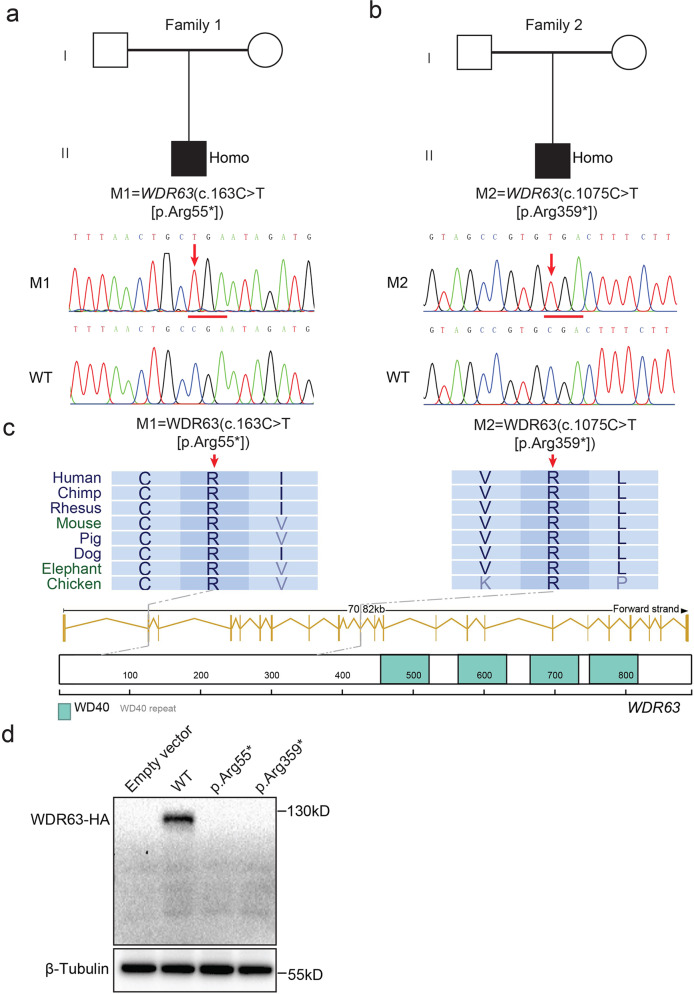
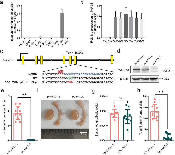
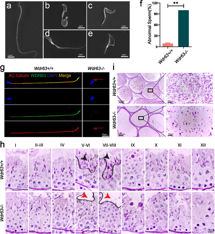
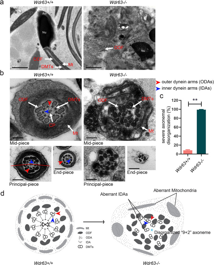
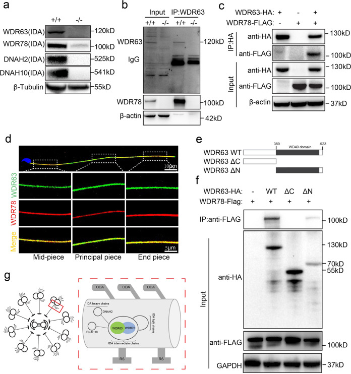
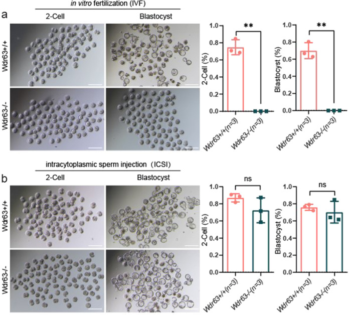
Inner dynein arm (IDA), composed of a series of protein complex, is necessary to cilia and flagella bend formation and beating. Previous studies indicated that defects of IDA protein complex result in multiple morphological abnormalities of the sperm flagellum (MMAF) and male infertility. However, the genetic causes and molecular mechanisms in the IDAs need further exploration. Here we identified two loss-of-function variants of WDR63 in both MMAF and non-obstructive azoospermia (NOA) affected cohorts. WDR63 encodes an IDA-associated protein that is dominantly expressed in testis. We next generated Wdr63-knockout (Wdr63-KO) mice through the CRISPR-Cas9 technology. Remarkably, Wdr63-KO induced decreased sperm number, abnormal flagellar morphology and male infertility. In addition, transmission electron microscopy assay showed severely disorganized "9 + 2" axoneme and absent inner dynein arms in the spermatozoa from Wdr63-KO male mice. Mechanistically, we found that WDR63 interacted with WDR78 mainly via WD40-repeat domain and is necessary for IDA assembly. Furthermore, WDR63-associated male infertility in human and mice could be overcome by intracytoplasmic sperm injection (ICSI) treatment. In conclusion, the present study demonstrates that bi-allelic variants of WDR63 cause male infertility via abnormal inner dynein arms assembly and flagella formation and can be used as a genetic diagnostic indicator for infertility males.


Conflict of interest statement

The authors declare no competing interests.

Figures

Fig. 1
Fig. 1. Identification of bi-allelic variants of WDR63 in patients with MMAF.
a, b Pedigree of family 1 affected by bi-allelic WDR63 variant that was identified by WES from MMAF-affected men (a). Pedigree of family 2 affected by bi-allelic WDR63 variant that were identified by Sanger sequencing from NOA-affected men (b). Filled black filled squares indicate infertile men in this family. Sanger sequencing results are shown under the pedigrees. The mutated positions are indicated by red arrows. c Schematic representation of the domains of WDR63 and locations of WDR63 variants identified in this study. Sequence alignment shows conservation of the mutated residues across different species according to UCSC genome browser. The green boxes indicate Trp-Asp (WD40) repeat domains as described by the UniProt server. d WDR63 variants cause the degradation of WDR63 protein. Full-length wild-type and mutant WDR63 cDNA constructs were overexpressed in HEK293T cells followed by immunoblotting analysis. Abbreviations: M1, mutation 1; M2, mutation 2; Homo, homozygous; WD40, Trp-Asp repeat.
Fig. 2
Fig. 2. WDR63 protein is highly expressed in late stages of spermatogenesis and Wdr63-null males are infertile.
a Real-time q-PCR for Wdr63 transcripts in various 8-weeks mouse tissues, and Gapdh gene is used as a control. Error bars, SEM (n = 3). b mRNA levels of Wdr63 in mouse testis at the indicated time points. Error bars, SEM (n = 3). c Schematic diagram of CRISPR/Cas9 strategy for the generation of Wdr63 KO mice. The sgRNA was designed to target exon 10 of the Wdr63, a nonsense mutation (GCG-TGA) plus a 2-bp deletion was obtained. d Western blot analysis of WDR63 protein in wild-type (Wdr63+/+), heterozygous (Wdr63+/−) and homozygous (Wdr63−/−) mice with a mouse polyclonal antibody raised against WDR63. β-actin was used as a loading control. e Fertility analysis for adult Wdr63+/+ and Wdr63−/−. f, g The morphology and testis weight of testes and epididymis from Wdr63+/+ and Wdr63−/− males at 8-week-old. Scale bars, 0.5 cm. h Total sperm number was dramatically decreased in Wdr63−/− males compared to that of Wdr63+/+ males. Abbreviations: W, week; M, million. For e to h, n = 10 and the bars represent means ± SEM. The statistical analysis was carried out using One-way ANOVA test, ** denotes P < 0.01; ns, not significant.
Fig. 3
Fig. 3. Wdr63-KO males showed typical MMAF phenotypes.
a SEM shows a spermatozoon with normal morphology from the epididymis of Wdr63+/+ males. Scale bars, 10 μm. be SEM shows the spermatozoon of Wdr63−/− males presented with MMAF phenotypes, such as short and coiled flagella, flagella of irregular caliber, and other malformations diagnosed as MMAF. Scale bars, 5 μm. f Spermatocytograms showing the number of abnormal sperms in WT and homozygous mutant males. g Immunofluorescent staining of WDR63 (green), AC-Tubulin (red) and DAPI (blue) on the spermatozoa collected from Wdr63+/+ and Wdr63−/− males. WDR63 localized at entire flagella in WT male mice. Scale bars, 10 μm. h Periodic acid–Schiff (PAS) staining of testis sections from adult Wdr63+/+ and Wdr63−/− mice. Black arrows indicated normal spermatozoon flagella in stage VII–VIII seminiferous tubules of Wdr63+/+ male mice, whereas red arrows indicated the absence of elongated tails in Wdr63−/− male mice. Scale bars, 10 μm. i HE staining of the cauda epididymis from male mice. Decreased sperm quantity was observed in the epididymis from Wdr63−/− male mice, when compared with that of Wdr63+/+ male mice. Scale bars, 10 μm. Abbreviations: PL, preleptotene; P, pachytene; RS, round spermatids; ES, elongated spermatids. For f, n = 3 and the bars represent means ± SEM. The statistical analysis was carried out using one-way ANOVA test, ** denotes P < 0.01.
Fig. 4
Fig. 4. Ultrastructure analyses for Wdr63-KO male mice show a severely disorganized axoneme disorganization arrangement.
a Longitudinal sections of spermatozoon mid-piece in adult male mice. Wdr63+/+ male mice sperm had a symmetrical mid-piece with smooth axoneme surrounding with a regularly arranged mitochondrial sheath. In contrast, Wdr63−/− male mice showed a seriously disorganized ODF and deficient of axoneme and mitochondrial sheath. Scale bars, 2 μm. b, c Cross-sections of spermatozoon flagella the mid-piece, principal piece, and end-piece in adult male mice. Wdr63+/+ male mice show the typical “9 + 2” microtubule structure, whereas nearly all spermatozoon of Wdr63−/− male mice show severe disorganized “9 + 2” axoneme. Scale bars, 200 nm. d Schematic of spermatozoon flagella cross-sections ultrastructure in Wdr63+/+ and Wdr63−/− male mice. Abbreviations: Nu, nucleus; Acr, acrosome; DMT, peripheral microtubule doublets; ODF, outer dense fibers; CP, central pair of microtubules; Mt, mitochondrial sheath; ODA, outer dynein arms (red arrows); IDA, inner dynein arms (blue arrows). For c, n = 3 and the bars represent means ± SEM. The statistical analysis was carried out using One-way ANOVA test, ** denotes P < 0.01.
Fig. 5
Fig. 5. WDR63 associates with WDR78 and their interaction is necessary for IDA assembly.
a Western blot analysis of WDR63, WDR78, DNAH2 and DNAH10 in testes of Wdr63+/+ and Wdr63−/− male mice. b Immunoprecipitation with testicular extracts using anti-WDR63 antibodies, Mass spectrometric analysis identified WDR78 is the potential associated proteins. Western blot analysis and confirmed that WDR63 interacts with WDR78. c HA-WDR63 and/or FLAG-WDR78 expression constructs were transfected into HEK293T cells. After 48 h, the cells were collected for immunoprecipitation analysis with indicated anti-HA and anti-FLAG antibodies. d Immunofluorescence analysis indicated that WDR63 co-localized with WDR78 in normal spermatozoon. e Schematic diagram of mouse wild type WDR63 (NP_766452.2) and truncated constructs. WD40 repeats domains are marked in a gray box (398–785 aa, UniProt ID: B2RY71). WDR63 ΔC, deletion of the C terminal domain of WDR63 (398–923 aa); WDR63 ΔN, deletion of the N terminal domain of WDR63 (1–397 aa). f HA-WDR63 WT, HA-WDR63 ΔC and HA-WDR63 ΔN were co-transfected with FLAG-WDR78 into HEK293T cells, respectively. After 48 h, the cells were collected for immunoprecipitation analysis with the indicated antibodies. g Schematic of the role of WDR63 interacts with WDR78 in IDAs structure.
Fig. 6
Fig. 6. WDR63-associated male infertility could be rescue by ICSI but not IVF.
a Representative two-cell embryos and blastocysts from in vitro fertilization. Scale bar, 200 μm. Compared with WT male mice, the rates of 2-cell and blastocyst as lower as zero in Wdr63 KO male mice. b Representative two-cell embryos and blastocysts from intracytoplasmic sperm injection. The 2-cell and blastocyst were successfully obtained upon ICSI using the spermatozoa from Wdr63 KO male mice. Scale bar, 200 μm. For a and b, n = 3 and the bars represent means ± SEM. The statistical analysis was carried out using One-way ANOVA test, ** denotes P < 0.01; ns, not significant.

Similar articles

Cited by

References

    1. Zhu Z, et al. Spermatogenesis is normal in Tex33 knockout mice. Peer J. 2020;8:e9629. - PMC - PubMed
    1. Agarwal A, Mulgund A, Hamada A, Chyatte MR. A unique view on male infertility around the globe. Reprod. Biol. Endocrinol. 2015;13:37. - PMC - PubMed
    1. Tuttelmann F, Ruckert C, Ropke A. Disorders of spermatogenesis: perspectives for novel genetic diagnostics after 20 years of unchanged routine. Med. Genet. 2018;30:12–20. - PMC - PubMed
    1. Feng T, et al. Eef2k is not required for fertility in male mice. Transl. Androl. Urol. 2021;10:1988–1999. - PMC - PubMed
    1. Toure A, et al. The genetic architecture of morphological abnormalities of the sperm tail. Hum. Genet. 2021;140:21–42. - PubMed