. 2021 Nov 15;11(1):22224.
doi: 10.1038/s41598-021-01622-7.

The diagnostic value of contrast-enhanced 2D mammography in everyday clinical use

Affiliations

The diagnostic value of contrast-enhanced 2D mammography in everyday clinical use

L M F H Neeter et al. Sci Rep. .

Abstract

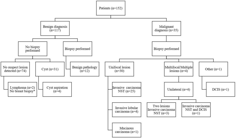
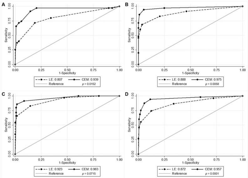
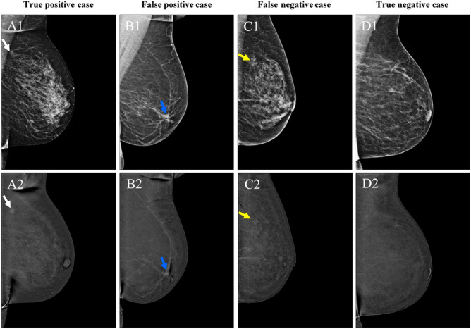
Contrast-enhanced mammography (CEM) has shown to be superior to full-field digital mammography (FFDM), but current results are dominated by studies performed on systems by one vendor. Information on diagnostic accuracy of other CEM systems is limited. Therefore, we aimed to evaluate the diagnostic performance of CEM on an alternative vendor's system. We included all patients who underwent CEM in one hospital in 2019, except those with missing data or in whom CEM was used as response monitoring tool. Three experienced breast radiologists scored the low-energy images using the BI-RADS classification. Next, the complete CEM exams were scored similarly. Histopathological results or a minimum of one year follow-up were used as reference standard. Diagnostic performance and AUC were calculated and compared between low-energy images and the complete CEM examination, for all readers independently as well as combined. Breast cancer was diagnosed in 23.0% of the patients (35/152). Compared to low-energy images, overall CEM sensitivity increased from 74.3 to 87.6% (p < 0.0001), specificity from 87.8 to 94.6% (p = 0.0146). AUC increased from 0.872 to 0.957 (p = 0.0001). Performing CEM on the system tested, showed that, similar to earlier studies mainly performed on another vendor's systems, both sensitivity and specificity improved when compared to FFDM.


Conflict of interest statement

ML received several research grant and speaker’s fees from GE Healthcare, Hologic, Bayer and Guerbet. JW received institutional grants and speaker’s fees from AGFA, Bayer Healthcare, Bard Medical, GE Healthcare, Optimed, Philips Healthcare, Siemens Healthineers. The other authors declare no competing interests.

Figures

Figure 1
Figure 1
Patient diagnostic flowchart. *Axillary biopsy revealed lymphoma, no suspect lesion on CEM. NST No special type, DCIS Ductal carcinoma in situ, CEM Contrast-enhanced mammography.
Figure 2
Figure 2
ROC curves and corresponding AUC values for LE and CEM, and p values. (A) Reader 1; (B) Reader 2; (C) Reader 3; (D) All readers combined. ROC Receiver operating characteristics, AUC Area under the curve, CI Confidence interval, LE Low-energy, CEM Contrast-enhanced mammography.
Figure 3
Figure 3
Examples of a true positive (A), false positive (B), false negative case (C), and true negative case (D). Top row represents the low-energy images, bottom row represents the recombined images. A1 shows an ill-defined, round mass anterior to the pectoral muscle (white arrow). The mass shows enhancement (A2). In B1, an ill-defined mass can be observed in the retro areolar zone with spiculated margins, also showing enhancement on the recombined images (B2, blue arrow). In C1 and C2, none of the radiologists classified this case as malignant, although a cancer was present as a subtle, ill-defined focal asymmetry at the site of the yellow arrow. The distortion did not show any enhancement. Finally, D1 and D2 show a negative case (no abnormalities or focal enhancement visible in the breast). However, this patient suffered from an axillary lymphoma and was therefore classified as ‘negative’, as no breast cancer was detected. Histopathological results from A, B and C were invasive carcinoma of no special type, fibroglandular tissue, and invasive lobular carcinoma, respectively.

References

    1. GE Healthcare. GE Healthcare announces FDA 510(k) clearance of SenoBright Contrast Enhanced Spectral Mammography (CESM) for breast cancer diagnosis. https://www.ge.com/news/press-releases/ge-healthcare-announces-fda-510k-... (2011).
    1. Jochelson MS, Lobbes MBI. Contrast-enhanced mammography: State of the art. Radiology. 2021;299:36–48. doi: 10.1148/radiol.2021201948. - DOI - PMC - PubMed
    1. Sorin V, et al. Contrast-enhanced spectral mammography in women with intermediate breast cancer risk and dense breasts. AJR. Am. J. Roentgenol. 2018;211:W267–W274. doi: 10.2214/AJR.17.19355. - DOI - PubMed
    1. Sung JS, et al. Performance of dual-energy contrast-enhanced digital mammography for screening women at increased risk of breast cancer. Radiology. 2019;293:81–88. doi: 10.1148/radiol.2019182660. - DOI - PMC - PubMed
    1. Zhu X, et al. Diagnostic value of contrast-enhanced spectral mammography for screening breast cancer: Systematic review and meta-analysis. Clin. Breast Cancer. 2018;18:e985–e995. doi: 10.1016/j.clbc.2018.06.003. - DOI - PubMed

Publication types

MeSH terms