

Chromosome clustering in mitosis by the nuclear protein Ki-67
- PMID: 34783345
- PMCID: PMC8786303
- DOI: 10.1042/BST20210717
Chromosome clustering in mitosis by the nuclear protein Ki-67
Abstract
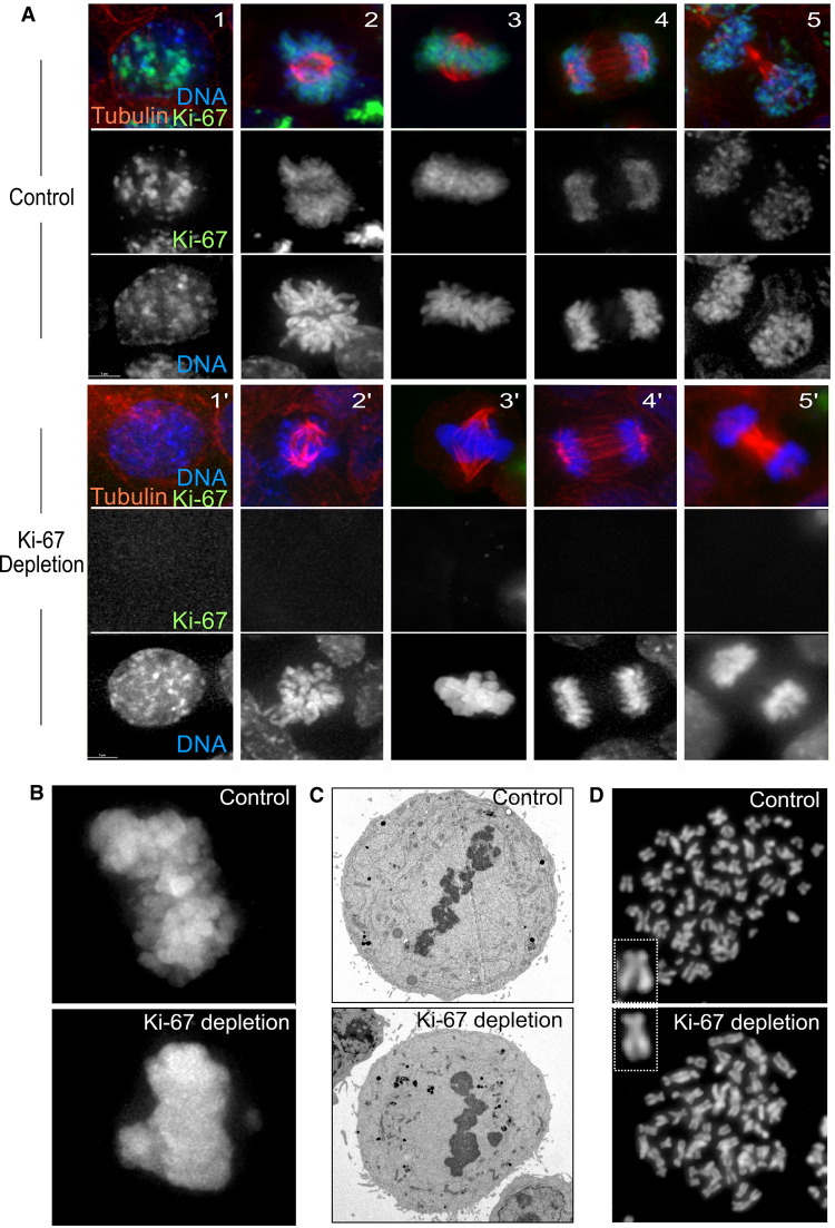
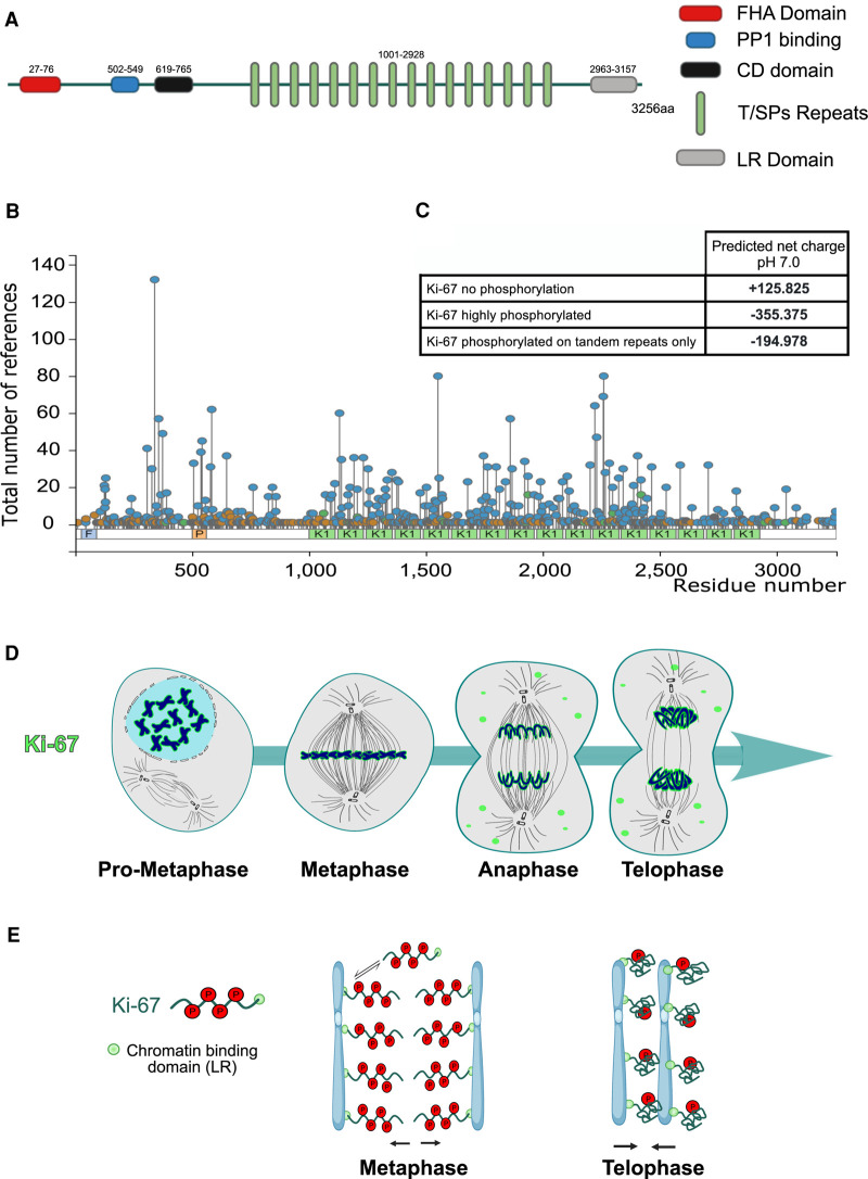
Ki-67 is highly expressed in proliferating cells, a characteristic that made the protein a very important proliferation marker widely used in the clinic. However, the molecular functions and properties of Ki-67 remained quite obscure for a long time. Only recently important discoveries have shed some light on its function and shown that Ki-67 has a major role in the formation of mitotic chromosome periphery compartment, it is associated with protein phosphatase one (PP1) and regulates chromatin function in interphase and mitosis. In this review, we discuss the role of Ki-67 during cell division. Specifically, we focus on the importance of Ki-67 in chromosome individualisation at mitotic entry (prometaphase) and its contribution to chromosome clustering and nuclear remodelling during mitotic exit.
Keywords: Ki-67; chromosomes; mitosis.
© 2021 The Author(s).
Conflict of interest statement
The authors declare that there are no competing interests associated with the manuscript.
Figures
Similar articles
-
A liquid-like coat mediates chromosome clustering during mitotic exit.Mol Cell. 2024 Sep 5;84(17):3254-3270.e9. doi: 10.1016/j.molcel.2024.07.022. Epub 2024 Aug 16. Mol Cell. 2024. PMID: 39153474
-
Ki-67 is a PP1-interacting protein that organises the mitotic chromosome periphery.Elife. 2014 May 27;3:e01641. doi: 10.7554/eLife.01641. Elife. 2014. PMID: 24867636 Free PMC article.
-
Ki-67 acts as a biological surfactant to disperse mitotic chromosomes.Nature. 2016 Jul 14;535(7611):308-12. doi: 10.1038/nature18610. Epub 2016 Jun 29. Nature. 2016. PMID: 27362226 Free PMC article.
-
The intrinsically disorderly story of Ki-67.Open Biol. 2021 Aug;11(8):210120. doi: 10.1098/rsob.210120. Epub 2021 Aug 11. Open Biol. 2021. PMID: 34375547 Free PMC article. Review.
-
Ki-67 and the Chromosome Periphery Compartment in Mitosis.Trends Cell Biol. 2017 Dec;27(12):906-916. doi: 10.1016/j.tcb.2017.08.001. Epub 2017 Aug 23. Trends Cell Biol. 2017. PMID: 28838621 Review.
Cited by
-
Dephosphorylation in nuclear reassembly after mitosis.Front Cell Dev Biol. 2022 Oct 4;10:1012768. doi: 10.3389/fcell.2022.1012768. eCollection 2022. Front Cell Dev Biol. 2022. PMID: 36268509 Free PMC article. Review.
-
Ki-67 is necessary during DNA replication for fork protection and genome stability.Genome Biol. 2024 Apr 22;25(1):105. doi: 10.1186/s13059-024-03243-5. Genome Biol. 2024. PMID: 38649976 Free PMC article.
-
Prognostic Biomarkers of Cell Proliferation in Colorectal Cancer (CRC): From Immunohistochemistry to Molecular Biology Techniques.Cancers (Basel). 2023 Sep 15;15(18):4570. doi: 10.3390/cancers15184570. Cancers (Basel). 2023. PMID: 37760539 Free PMC article. Review.
-
Between Centralization and Fragmentation: The Past, Present, and Future of Phage Collections.Phage (New Rochelle). 2024 Mar 18;5(1):22-29. doi: 10.1089/phage.2023.0043. eCollection 2024 Mar. Phage (New Rochelle). 2024. PMID: 40114810 Free PMC article. Review.
-
Ki-67 and CDK1 control the dynamic association of nuclear lipids with mitotic chromosomes.J Lipid Res. 2025 Jan;66(1):100731. doi: 10.1016/j.jlr.2024.100731. Epub 2024 Dec 18. J Lipid Res. 2025. PMID: 39706365 Free PMC article.
References
Publication types
MeSH terms
Substances
Grants and funding
LinkOut - more resources
Full Text Sources