Review
. 2021 Dec 2;13(46):19306-19323.
doi: 10.1039/d0nr03872e.

Silver chalcogenide nanoparticles: a review of their biomedical applications

Affiliations
Review

Silver chalcogenide nanoparticles: a review of their biomedical applications

Lenitza M Nieves et al. Nanoscale. .

Abstract

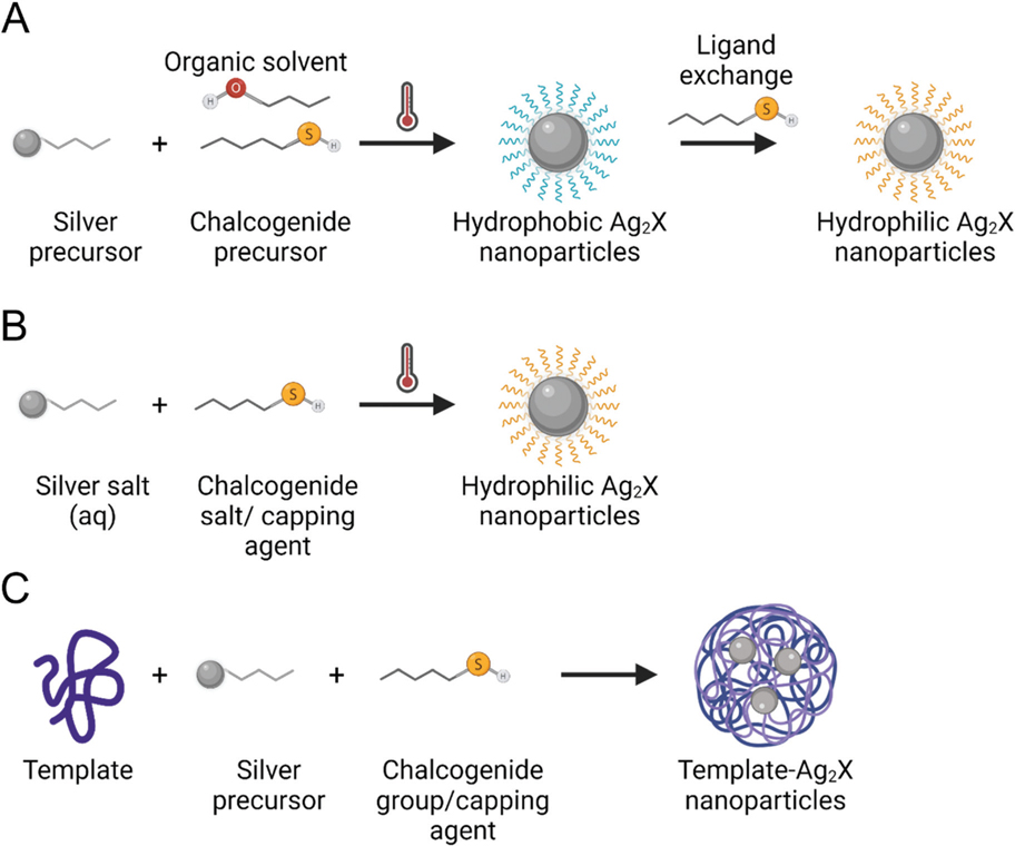
Silver chalcogenide (Ag2X, where X = S, Se, or Te) nanoparticles have been extensively investigated for their applications in electronics but have only recently been explored for biomedical applications. In the past 10 years, Ag2X, primarily silver sulfides at first, have become of great importance as quantum dots, since they not only possess excellent deep tissue imaging properties in the near-infrared regions I and II, but also have low toxicities. Their appealing properties have led to numerous recent developments of Ag2X for biomedical applications. Furthermore, Ag2X have been discovered in the past 2-3 years to be potent X-ray contrast agents, adding to the numerous biomedical uses of these nanoparticles. In this review, we discuss the most recent advances in silver chalcogenide nanoparticle use in areas such as bio-imaging, theranostics, and biosensors. Moreover, we examine the advances in synthetic approaches for these nanoparticles, which include aqueous and organic syntheses routes. Finally, we discuss the advantages and current limitations in the use of silver chalcogenides for different biomedical applications and their potential for advancement and expansions in use.


Conflict of interest statement

Conflicts of interest

ADAM and DPC are named as inventors on a patent application concerning the use of silver chalcogenides as X-ray contrast agents, as well as both holding stock in a company, Daimroc Imaging, whose goal is to commercially develop such nanoparticles.

Figures

Fig. 1
Fig. 1
Examples of synthetic approaches used for the development of Ag2X nanoparticles in the biomedical field. Representations of (A) organic synthesis followed by ligand substitution, (B) aqueous synthesis, and (C) template-based synthesis.
Fig. 2
Fig. 2
(A) Schematic illustration of glucose functionalized-Ag2Se QDs synthesis. (B) Absorbance (left) - fluorescence (right) spectra of Ag2Se QDs (black) and glucose-Ag2Se QDs (red). (C) TEM micrograph of glucose-Ag2Se QDs. (D) Laser confocal scanning imaging of MCF-7 cells incubated with glucose-Ag2Se QDs or controls (Ag2Se QDs and PBS) for 4 h at a concentration of 500 μg ml−1. (E) NIR in vivo images of mice at different time points post-intravenous injection of glucose-Ag2Se QDs. (F) Ex vivo images of mice organs after intravenous injection of glucose-Ag2Se QDs: (1) heart, (2) liver, (3) spleen, (4) lungs, (5) kidneys, (6) brain, (7) large intestine, (8) small intestine, (9) stomach and (10) urinary bladder. This figure has been adapted from ref. with permission from The Royal Society of Chemistry, copyright 2019.
Fig. 3
Fig. 3
(A) Schematic illustration of the nanobioprobe preparation and application. PQDs: NIR II fluorescent assembly of Ag2Te QDs and PLGA; CVs: cell membrane-derived vesicles; CPQDs: CVs-camouflaged PQDs; FL: fluorescence. (B) TEM micrograph of CPQDs. (C) In vivo NIR-II fluorescence imaging of 4T1 tumor-bearing mice injected with PQDs, and CPQDs over 48 hours. The white and green dashed circles indicate tumor and normal tissue areas, respectively. (D) Ex vivo NIR-II fluorescence imaging of the tumors and major organs collected from representative mice at 48 hours post-injection. This figure has been adapted from ref. with permission from Wiley, copyright 2019.
Fig. 4
Fig. 4
(A) Schematic depiction of the synthesis of Ag2Te nanoparticles. (B) Transmission electron micrograph of Ag2Te NPs. (C-E) Effect of Ag2Te (black), Ag2S (dark gray), and Ag (light gray) nanoparticles at different concentrations on the viability of (C) HepG2, (D) MDA-MB-231, and (E) J774A.1 cells. (F) DEM phantom imaging showing the contrast produced by the different solutions at a concentration of 10 mg mL−1 of the element of interest. Ag denotes AgNO3, Te denotes sodium telluride, and I denotes iopamidol. (G) Quantification of the contrast-to-noise ratio from the different solutions imaged in DEM. (H) CT contrast in the tumor (yellow circle) of breast cancer tumor-bearing mice prior to and after intravenous injection with the different silver chalcogenides at different time points. (I) Quantification of the change in CT attenuation in the tumor from the different silver chalcogenide nanoparticles in the tumor. This figure has been adapted from ref. with permission from The Royal Society of Chemistry, copyright 2021.
Fig. 5
Fig. 5
(A) Schematic illustration of the all-in-one nanoparticles (AION) and its components. (B) Transmission electron micrograph of AION. Scale bar is 10 nm. (C) Transmission electron micrograph of AION showing the inclusion of Ag2S-NP (red circle) and IO-NP (yellow circle). Scale bar is 10 nm. (D) In vivo tumor DEM images pre-injection and 120 minutes post-injection with AION. (E) In vivo CT images pre-injection and 24 hours post-injection with AION. (F) MR images (including R2 map overlay) of tumor-bearing mice pre-injection and 24 hours post-injection with AION. Red circles show the tumor. (G) In vivo NIR fluorescence imaging pre-injection and 24 hours post-injection with AION. Black circles represent the tumor. This figure has been adapted from ref. with permission from The Royal Society of Chemistry, copyright 2018.
Fig. 6
Fig. 6
(A) Schematic illustration of theranostic Ag2S nanodots synthesized through controlled growth in albumin nanocages. (B) In vivo PA imaging in 4T1-tumor-bearing mice treated with Ag2S nanodots at a dose of 50.0 μmol kg−1 Ag over 24 h post-injection. (C) Tumor growth profiles of 4T1-tumor-bearing mice treated with Ag2S nanodots with or without 5 min laser irradiation (785 nm, 1.5 W cm−2). (D) Photograph of tumors extracted from the mice at 30 days post-irradiation. This figure has been adapted from ref. with permission from American Chemical Society, copyright 2017.
Fig. 7
Fig. 7
(A) Schematic illustration of UCNPs-QDs synthesis, components, and application for photodynamic therapy when combined with a photosensitizer (RB). (B) TEM of UCNP-QDs. (C) Viability of HeLa cells treated with UCQR, UCRs + NIR, or UCQRs + NIR. (D) Schematic illustration of in vivo antitumor treatment and in vivo fluorescence imaging of UCQRs at different time points after intravenous injection. (E) Relative tumor volume from mice after various treatments. (F) Representative photographs of excised tumors after treatment with PBS (i), NIR (ii), UCQRs (iii), UCR + NIR (iv), and UCQRs + NIR (v). This figure has been adapted from ref. with permission from American Chemical Society, copyright 2019.
Fig. 8
Fig. 8
(A) Schematic of the thrombin sensor synthesis and mechanism of action. (B) Transmission electron micrograph of as-prepared GSH-Ag2Se QDs. (C) UCL spectra of the biosensor in a 100-fold diluted serum sample under different thrombin concentrations. (D) Linear relationship between F/F0 and the concentration of thrombin within the range of 0.1–125 nM. This figure has been adapted from ref. with permission from The Royal Society of Chemistry, copyright 2020.

References

    1. Bouché M, Hsu JC, Dong YC, Kim J, Taing K and Cormode DP, Bioconjugate Chem, 2019, 31, 303–314. - PMC - PubMed
    1. Hsu JC, Nieves LM, Betzer O, Sadan T, Noël PB, Popovtzer R and Cormode DP, Wiley Interdiscip. Rev.: Nanomed. Nanobiotechnol, 2020,12, e1642. - PMC - PubMed
    1. Upputuri PK and Pramanik M, Wiley Interdiscip. Rev.: Nanomed. Nanobiotechnol, 2020,12, e1618. - PubMed
    1. Han X, Xu K, Taratula O and Farsad K, Nanoscale, 2019, 11, 799–819. - PMC - PubMed
    1. Yang S and Li Y, Wiley Interdiscip. Rev.: Nanomed. Nanobiotechnol, 2020, 12, el603. - PubMed

LinkOut - more resources