. 2021 Nov 16;19(11):e3001424.
doi: 10.1371/journal.pbio.3001424. eCollection 2021 Nov.

Systematic exploration of Escherichia coli phage-host interactions with the BASEL phage collection

Affiliations

Systematic exploration of Escherichia coli phage-host interactions with the BASEL phage collection

Enea Maffei et al. PLoS Biol. .

Abstract

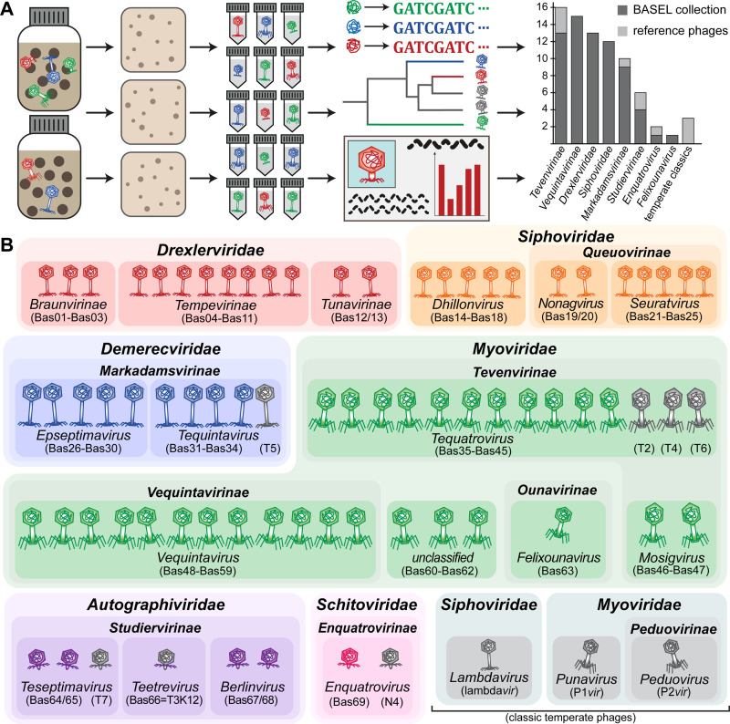
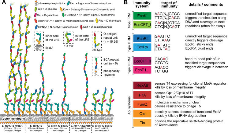
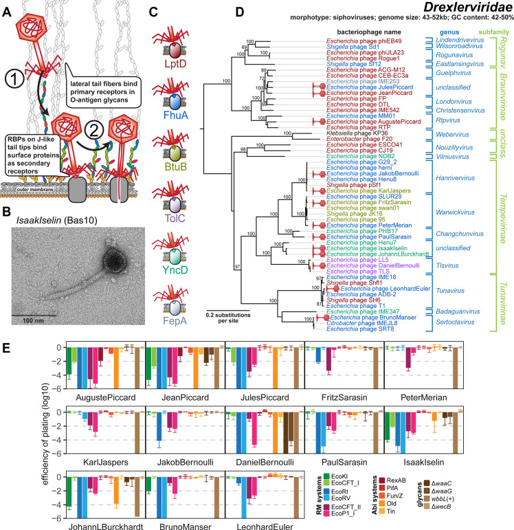
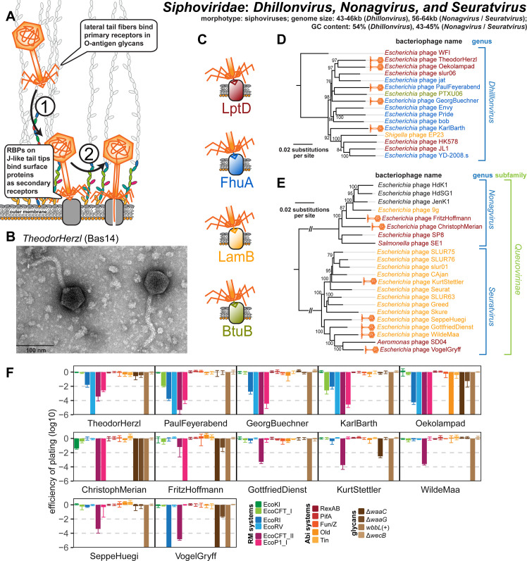
Bacteriophages, the viruses infecting bacteria, hold great potential for the treatment of multidrug-resistant bacterial infections and other applications due to their unparalleled diversity and recent breakthroughs in their genetic engineering. However, fundamental knowledge of the molecular mechanisms underlying phage-host interactions is mostly confined to a few traditional model systems and did not keep pace with the recent massive expansion of the field. The true potential of molecular biology encoded by these viruses has therefore remained largely untapped, and phages for therapy or other applications are often still selected empirically. We therefore sought to promote a systematic exploration of phage-host interactions by composing a well-assorted library of 68 newly isolated phages infecting the model organism Escherichia coli that we share with the community as the BASEL (BActeriophage SElection for your Laboratory) collection. This collection is largely representative of natural E. coli phage diversity and was intensively characterized phenotypically and genomically alongside 10 well-studied traditional model phages. We experimentally determined essential host receptors of all phages, quantified their sensitivity to 11 defense systems across different layers of bacterial immunity, and matched these results to the phages' host range across a panel of pathogenic enterobacterial strains. Clear patterns in the distribution of phage phenotypes and genomic features highlighted systematic differences in the potency of different immunity systems and suggested the molecular basis of receptor specificity in several phage groups. Our results also indicate strong trade-offs between fitness traits like broad host recognition and resistance to bacterial immunity that might drive the divergent adaptation of different phage groups to specific ecological niches. We envision that the BASEL collection will inspire future work exploring the biology of bacteriophages and their hosts by facilitating the discovery of underlying molecular mechanisms as the basis for an effective translation into biotechnology or therapeutic applications.


Conflict of interest statement

The authors have declared that no competing interests exist.

Figures

Fig 1
Fig 1. Overview of the BASEL collection.
(A) Illustration of the workflow of bacteriophage isolation, characterization, and selection that resulted in the BASEL collection (details in Materials and methods; the bar diagram includes phi92-like phages in Vequintavirinae for simplicity). (B) Taxonomic overview of the bacteriophages included in the BASEL collection and their unique Bas## identifiers. Newly isolated phages are colored by current taxonomic classification, while reference phages are shown in gray. BASEL, BActeriophage SElection for your Laboratory.
Fig 2
Fig 2. Overview of E. coli surface glycan variants and the immunity systems used in this study.
(A) The surface glycans of different E. coli K-12 MG1655 variants are shown schematically (details in running text and Materials and methods). Note that the E. coli K-12 MG1655 laboratory wild type does not merely display the K12-type core LPS (classical rough LPS phenotype) but also the most proximal D-glucose of the O16-type O-antigen. (B) Key features of the 6 RM systems (each 2 of type I, type II, and type III) and the 5 Abi systems used for the phenotyping of this study are summarized schematically. Recognition sites of RM systems have either been determined experimentally or were predicted in REBASE (red nucleotides: methylation sites; dotted lines: cleavage sites) [–47,129]. The Abi systems have been characterized to very different extent but constitute the most well-understood representatives of these immunity systems of E. coli [10,48]. Abi, abortive infection; ECA, enterobacterial common antigen; LPS, lipopolysaccharide; RM, restriction–modification.
Fig 3
Fig 3. Overview of Drexlerviridae phages.
(A) Schematic illustration of host recognition by Drexlerviridae. (B) Representative TEM micrograph of phage IsaakIselin (Bas10). (C) Color code of terminal receptor specificity. (D) Maximum-Likelihood phylogeny of Drexlerviridae based on several core genes with bootstrap support of branches shown if >70/100. Newly isolated phages of the BASEL collection are highlighted by red phage icons, and the determined or proposed terminal receptor specificity is highlighted at the phage names using the color code highlighted in (C). The phylogeny was rooted based on a representative phylogeny including Dhillonvirus sequences as outgroup (S1A Fig). (E) The results of quantitative phenotyping experiments with Drexlerviridae phages regarding sensitivity to altered surface glycans and bacterial immunity systems are presented as EOP. Data points and error bars represent average and standard deviation of at least 3 independent experiments. Raw data and calculations are available in S1 Data. BASEL, BActeriophage SElection for your Laboratory; EOP, efficiency of plating; RBP, receptor-binding protein; TEM, transmission electron microscopy.
Fig 4
Fig 4. Shared bona fide RBP loci of small siphoviruses mediate specificity to terminal receptors including LptD.
(A) The 7 identified receptors of small siphoviruses are shown with a color code that is also used to annotate demonstrated or predicted receptor specificity in the phylogenies of Figs 3D, 5D, and 5E. (B) We show representative bona fide RBP loci that seem to encode the receptor specificity of the small siphoviruses studied in this work (same color code as in (A)). Note that the loci linked to each receptor are very similar while the genetic arrangement differs considerably between loci linked to different terminal host receptors (see also S1B Fig). (C) Whole-genome sequencing of bacterial mutants exhibiting spontaneous resistance to 7 small siphoviruses with no previously known receptor revealed different mutations or small deletions in the essential gene lptD that encodes the LptD LPS export channel. Top agar assays with 2 representative mutants in comparison to the ancestral E. coli K-12 BW25113 strain were performed with serial 10-fold dilutions of 12 different phages (undiluted high-titer stocks at the bottom and increasingly diluted samples toward the top). Both mutants display complete resistance to the 7 small siphoviruses of diverse genera within Drexlerviridae and Siphoviridae families that share the same bona fide RBP module (S1B Fig) while no other phage of the BASEL collection was affected. We excluded indirect effects, e.g., via changes in the LPS composition in the lptD mutants, by confirming that 5 LPS-targeting phages of diverse families (see below) showed full infectivity on all strains. (D) The amino acid sequence alignment of wild-type LptD with the 2 mutants highlighted in (A) shows that resistance to LptD-targeting phages is linked to small deletions in or adjacent to regions encoding extracellular loops as defined in previous work [150], suggesting that they abolish the RBP–receptor interaction. BASEL, BActeriophage SElection for your Laboratory; LPS, lipopolysaccharide; RBP, receptor-binding protein.
Fig 5
Fig 5. Overview of Siphoviridae genera Dhillonvirus, Nonagvirus, and Seuratvirus.
(A) Schematic illustration of host recognition by small siphoviruses. (B) Representative TEM micrograph of phage TheodorHerzl (Bas14). (C) Color code of terminal receptor specificity (same as in Figs 3 and 4). (D) Maximum-Likelihood phylogeny of the Dhillonvirus genus based on a whole-genome alignment with bootstrap support of branches shown if >70/100. Newly isolated phages of the BASEL collection are highlighted by orange phage icons and the determined or proposed terminal receptor specificity is highlighted at the phage names using the color code highlighted in (C). The phylogeny was rooted between phage WFI and all others based on a representative phylogeny including Drexlerviridae sequences as outgroup (S1A Fig). (E) Maximum-Likelihood phylogeny of the Nonagvirus and Seuratvirus genera based on a whole-genome alignment with bootstrap support of branches shown if >70/100. Newly isolated phages of the BASEL collection are highlighted by orange phage icons, and the determined or proposed terminal receptor specificity is highlighted at the phage names using the color code highlighted in (C) (see S2B Fig for the 3 phages without coloring). The phylogeny was rooted between the 2 genera that belong both to the Queuovirinae subfamily of Siphoviridae. (F) The results of quantitative phenotyping experiments with Dhillonvirus, Nonagvirus, and Seuratvirus phages regarding sensitivity to altered surface glycans and bacterial immunity systems are presented as EOP. Data points and error bars represent average and standard deviation of at least 3 independent experiments. Raw data and calculations are available in S1 Data. Abi, abortive infection; BASEL, BActeriophage SElection for your Laboratory; EOP, efficiency of plating; RBP, receptor-binding protein; RM, restriction–modification; TEM, transmission electron microscopy.
Fig 6
Fig 6. Overview of Demerecviridae subfamily Markadamsvirinae.
(A) Schematic illustration of host recognition by T5-like siphoviruses. (B) Representative TEM micrograph of phage GreteKellenberger (Bas26). (C) Maximum-Likelihood phylogeny of the Markadamsvirinae subfamily of Demerecviridae based on several core genes with bootstrap support of branches shown if >70/100. Phages of the BASEL collection are highlighted by little phage icons, and the determined or proposed terminal receptor specificity is highlighted at the phage names using the color code highlighted in (D). The phylogeny was rooted between the Epseptimavirus and Tequintavirus genera. (D) Color code of terminal receptor specificity (same as in Figs 3–5). (E) The results of quantitative phenotyping experiments with Markadamsvirinae phages regarding sensitivity to altered surface glycans and bacterial immunity systems are presented as EOP. Data points and error bars represent average and standard deviation of at least 3 independent experiments. Raw data and calculations are available in S1 Data. Abi, abortive infection; BASEL, BActeriophage SElection for your Laboratory; EOP, efficiency of plating; RBP, receptor-binding protein; RM, restriction–modification; TEM, transmission electron microscopy.
Fig 7
Fig 7. Overview of the Myoviridae subfamily Tevenvirinae.
(A) Schematic illustration of host recognition by T-even myoviruses. (B) Representative TEM micrograph of phage WilhelmHis (Bas35). (C) Maximum-Likelihood phylogeny of the Tevenvirinae subfamily of Myoviridae based on a curated whole-genome alignment with bootstrap support of branches shown if >70/100. The phylogeny was rooted between the Tequatrovirus and Mosigvirus genera. Phages of the BASEL collection are highlighted by little phage icons and experimentally determined primary receptor specificity is highlighted at the phage names using the color code highlighted in (D). Primary receptor specificity of T-even phages depends on RBPs expressed either as a C-terminal extension of the distal half fiber (T4 and other OmpC-targeting phages) or as separate small fiber tip adhesins [65], but sequence analyses of the latter remained ambiguous. We therefore only annotated experimentally determined primary receptors (see also S4A Fig) [51,65]. (D) Color code of primary receptor specificity. (E) The results of quantitative phenotyping experiments with Tevenvirinae phages regarding sensitivity to altered surface glycans and bacterial immunity systems are presented as EOP. Data points and error bars represent average and standard deviation of at least 3 independent experiments. Raw data and calculations are available in S1 Data. Abi, abortive infection; BASEL, BActeriophage SElection for your Laboratory; EOP, efficiency of plating; LPS, lipopolysaccharide; RBP, receptor-binding protein; RM, restriction–modification; TEM, transmission electron microscopy.
Fig 8
Fig 8. Overview of the Myoviridae subfamily Vequintavirinae and relatives.
(A) Schematic illustration of host recognition by Vequintavirinae and related myoviruses. (B) Representative TEM micrograph of phage HeinrichReichert (Bas58). (C) Maximum-Likelihood phylogeny of the Vequintavirinae subfamily of Myoviridae and relatives based on a curated whole-genome alignment with bootstrap support of branches shown if >70/100. The phylogeny was rooted between the Vequintavirinae sensu stricto and the 2 closely related, formally unclassified groups at the bottom. Newly isolated phages of the BASEL collection are highlighted by green phage icons. (D) The results of quantitative phenotyping experiments with Vequintavirinae and their phi92-like relatives regarding sensitivity to altered surface glycans and bacterial immunity systems are presented as EOP. Data points and error bars represent average and standard deviation of at least 3 independent experiments. Raw data and calculations are available in S1 Data. Abi, abortive infection; BASEL, BActeriophage SElection for your Laboratory; ECA, enterobacterial common antigen; EOP, efficiency of plating; LPS, lipopolysaccharide; RM, restriction–modification; TEM, transmission electron microscopy.
Fig 9
Fig 9. Overview of Autographiviridae subfamily Studiervirinae and Schitoviridae subfamily Enquatrovirinae.
(A) Schematic illustration of host recognition by the studied Autographiviridae and Schitoviridae phages. (B) Representative TEM micrograph of T7-like Autographiviridae phage JacobBurckhardt (Bas65). (C) Representative TEM micrograph of Enquatrovirus AlfredRasser (Bas69). (D) Maximum-Likelihood phylogeny of the Studiervirinae subfamily of Autographiviridae based on several core genes with bootstrap support of branches shown if >70/100. The phylogeny was midpoint-rooted between the clade formed by Teseptimavirus and Teetrevirus and the other genera. Phages of the BASEL collection are highlighted by little phage icons. (E) Maximum-Likelihood phylogeny of the Enquatrovirus genus and related groups of Enquatrovirinae and other Schitoviridae based on several core genes with bootstrap support of branches shown if >70/100. The phylogeny was midpoint-rooted between the distantly related Jwalphavirus and Inbricusvirus genera and the other shown Schitoviridae. Phages of the BASEL collection are highlighted by little phage icons. (F) The results of quantitative phenotyping experiments with Autographiviridae and Schitoviridae phages regarding sensitivity to altered surface glycans and bacterial immunity systems are presented as EOP. Data points and error bars represent average and standard deviation of at least 3 independent experiments. Raw data and calculations are available in S1 Data. Abi, abortive infection; BASEL, BActeriophage SElection for your Laboratory; ECA, enterobacterial common antigen; EOP, efficiency of plating; LPS, lipopolysaccharide; RM, restriction–modification; TEM, transmission electron microscopy.
Fig 10
Fig 10. Overview of Myoviridae: Ounavirinae and classic temperate phages.
(A) Schematic illustration of host recognition by Ounavirinae: Felixounavirus phages. Note that the illustration shows short tail fibers simply in analogy to Tevenvirinae or Vequintavirinae (Figs 7A and 8A), but any role for such structures has not been explored for Felix O1 and relatives. (B) Representative TEM micrograph of phage JohannRWettstein (Bas63). (C) Maximum-Likelihood phylogeny of the Ounavirinae subfamily of Myoviridae based on several core genes with bootstrap support of branches shown if >70/100. The phylogeny was midpoint-rooted between Kolesnikvirus and the other genera. Our new isolate JohannRWettstein is highlighted by a green phage icon. (D) The results of quantitative phenotyping experiments with JohannRWettstein regarding sensitivity to altered surface glycans and bacterial immunity systems are presented as EOP. (E, F) Schematic illustration of host recognition by classic temperate phages lambda, P1, and P2. Note the absence of lateral tail fibers due to a mutation in lambda PaPa laboratory strains [93]. (G) The results of quantitative phenotyping experiments with the 3 classic temperate phages regarding sensitivity to altered surface glycans and bacterial immunity systems are presented as EOP. Data points and error bars in (D) and (G) represent average and standard deviation of at least 3 independent experiments. Raw data and calculations are available in S1 Data. Abi, abortive infection; EOP, efficiency of plating; LPS, lipopolysaccharide; RBP, receptor-binding protein; RM, restriction–modification; TEM, transmission electron microscopy.
Fig 11
Fig 11. Surface glycans of the enterobacterial strains used in this work.
The known surface glycans of different enterobacterial strains used in this work were drawn in comparison to highlight the diversity and barrier function of their O-antigen as well as the slight differences between LPS core types (see Materials and methods for details on how the illustration was composed). ECA, enterobacterial common antigen; LPS, lipopolysaccharide.
Fig 12
Fig 12. Host range of phages in the BASEL collection.
(A) Schematic overview of qualitative spot test assays with high-titer phage stocks (>109 pfu/ml; left) and quantitative EOP assays using serial dilutions of phage stocks (right) to determine the lysis host range and the plaquing host range of bacteriophages, respectively (see Materials and methods for details of the methodology). The graph at the bottom highlights how the results of the 4 fictional phages in this example would be plotted (with colorful bars at the top indicating the lysis host range and a bar diagrams for the EOP at the bottom that shows the plaquing host range). (B-H) The lysis host range and the plaquing host range of all phages in the BASEL collection across different enterobacteria were determined and visualized as shown in (A). Data points and error bars of the EOP plots represent average and standard deviation of at least 3 independent experiments. Raw data and calculations are available in S1 Data. BASEL, BActeriophage SElection for your Laboratory; EOP, efficiency of plating.

Similar articles

Cited by

References

    1. Mahmoudabadi G, Phillips R. A comprehensive and quantitative exploration of thousands of viral genomes. elife. 2018;7. doi: 10.7554/eLife.31955 ; PubMed Central PMCID: PMC5908442. - DOI - PMC - PubMed
    1. Dion MB, Oechslin F, Moineau S. Phage diversity, genomics and phylogeny. Nat Rev Microbiol. 2020;18(3):125–38. Epub 2020/02/06. doi: 10.1038/s41579-019-0311-5 . - DOI - PubMed
    1. Cobian Güemes AG, Youle M, Cantu VA, Felts B, Nulton J, Rohwer F. Viruses as Winners in the Game of Life. Annu Rev Virol. 2016;3(1):197–214. Epub 2016/10/16. doi: 10.1146/annurev-virology-100114-054952 . - DOI - PubMed
    1. Keen EC. A century of phage research: bacteriophages and the shaping of modern biology. BioEssays. 2015;37(1):6–9. doi: 10.1002/bies.201400152 ; PubMed Central PMCID: PMC4418462. - DOI - PMC - PubMed
    1. Demerec M, Fano U. Bacteriophage-Resistant Mutants in Escherichia Coli. Genetics. 1945;30(2):119–36. Epub 1945/03/01. doi: 10.1093/genetics/30.2.119 ; PubMed Central PMCID: PMC1209279. - DOI - PMC - PubMed

Publication types

LinkOut - more resources