. 2021 Nov;11(11):210117.
doi: 10.1098/rsob.210117. Epub 2021 Nov 17.

A large panel of chicken cells are invaded in vivo by Salmonella Typhimurium even when depleted of all known invasion factors

Affiliations

A large panel of chicken cells are invaded in vivo by Salmonella Typhimurium even when depleted of all known invasion factors

S M Roche et al. Open Biol. 2021 Nov.

Abstract

Poultry are the main source of human infection by Salmonella. As infected poultry are asymptomatic, identifying infected poultry farms is difficult, thus controlling animal infections is of primary importance. As cell tropism is known to govern disease, our aim was therefore to identify infected host-cell types in the organs of chicks known to be involved in Salmonella infection and investigate the role of the three known invasion factors in this process (T3SS-1, Rck and PagN). Chicks were inoculated with wild-type or isogenic fluorescent Salmonella Typhimurium mutants via the intracoelomic route. Our results show that liver, spleen, gall bladder and aortic vessels could be foci of infection, and that phagocytic and non-phagocytic cells, including immune, epithelial and endothelial cells, are invaded in vivo in each organ. Moreover, a mutant defective for the T3SS-1, Rck and PagN remained able to colonize organs like the wild-type strain and invaded non-phagocytic cells in each organ studied. As the infection of the gall bladder had not previously been described in chicks, invasion of gall bladder cells was confirmed by immunohistochemistry and infection was shown to last several weeks after inoculation. Altogether, for the first time these findings provide insights into cell tropism of Salmonella in relevant organs involved in Salmonella infection in chicks and also demonstrate that the known invasion factors are not required for entry into these cell types.

Keywords: Salmonella; gall bladder; host–pathogen interaction; invasion; poultry.


Conflict of interest statement

We declare we have no competing interests.

Figures

Figure 1.
Figure 1.
Level of different S. Typhimurium strains in organs of chicks after intracoelomic inoculation. Five-day-old chicks were inoculated in the coelomic cavity with around 6 × 10⁷ CFU per chick with S. Typhimurium 14028 turboFP650 wild-type strain (WT; blue diamond), ΔinvA::kan mutant strain (ΔinvA; T3SS-1 defective; green diamond) or the ΔinvA::kan ΔpagN::cm Δrck mutant strain (3Δ; T3SS-1, Rck, PagN defective; yellow diamond). Two days post infection, spleens, livers, aortic vessels and gall bladders were removed aseptically from each animal for quantification of bacterial load. Results are expressed as number of bacteria per gram of organ (log CFU per gram of organ). The medians are represented by a red dash. A Kruskal–Wallis test was conducted, followed by Dunn's multiple comparisons test (GraphPad Software). Significance was *p < 0.05 and **p < 0.01.
Figure 2.
Figure 2.
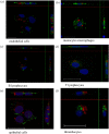
Intracellular localization of Salmonella in cells purified from in vivo infected organs. Five-day-old chicks were inoculated in the coelomic cavity with around 6 × 10⁷ CFU per chick with S. Typhimurium 14028 turboFP650 wild-type strain, ΔinvA::kan mutant strain or the ΔinvA::kan ΔpagN::cm Δrck mutant strain. Two days post infection, animals were sacrificed and the different organs removed. Cells were then isolated from organs, they were sorted using a high-speed cell sorter, MoFlo Astrios EQ and deposited on glass coverslips after cytospin at 200 r.p.m. for 10 min. Next, cells were fixed in formaldehyde. Nucleus staining was performed with Dapi (blue). The bacteria are in red (turboFP650), whereas cells are identified in green due to FITC or Alexa Fluor 488 conjugated antibodies. Cells were observed under a SP8 confocal laser-scanning microscope equipped with a 100× oil immersion objective (Leica). Z-stacks were re-sliced horizontally and vertically to obtain the projections of perpendicular views from three-dimensional images, allowing a view of all bacteria in the cells, using Las AF lite 2.6.3 build 8173 software (Leica). White dashes represent 20 µm. (a) Represents endothelial cells from the aortic vessels, infected with the 3Δ strain. Picture size 32.54 × 38.45 µm. (b) Represents monocytes–macrophages from the liver, infected with the ΔinvA strain. Picture size 116.25 × 116.25 µm. (c) Represents B lymphocytes, infected with the wild-type strain. Picture size 58.13 × 58.13 µm. (d) Represents T lymphocytes, infected with the wild-type strain. Picture size 39.88 × 39.88 µm. (e) Represents epithelial cells in the gall bladder, infected with the 3Δ strain. Picture size 37.80 × 37.80 µm. (f) Represents thrombocytes in the aortic vessels, infected with the 3Δ strain. Picture size: 116.25 × 116.25 µm.
Figure 3.
Figure 3.
Percentage of identified and Salmonella infected cells in spleen 5-day-old chicks were inoculated in the coelomic cavity with around 6 × 10⁷ CFU per chick with S. Typhimurium 14028 turboFP650 wild-type strain (WT; blue diamond), ΔinvA::kan mutant strain (ΔinvA; T3SS-1 defective; green diamond) or the ΔinvA::kan ΔpagN::cm Δrck mutant strain (3Δ; T3SS-1, Rck, PagN defective; yellow diamond). Two days post infection, animals were sacrificed and the different organs removed. Cells from uninfected animals of the same age were used as a control. After labelling with the corresponding antibodies, the percentages of macrophages–monocytes, B and T lymphocytes, thrombocytes and epithelial and endothelial cells were quantified by flow cytometry. The percentage of labelled cells (a) and the percentage of labelled infected cells (b) are represented. All negative responses were scored at 0.001%. The medians are represented by a red dash. Asymptotic two-sample Fisher–Pitman permutation tests (one-way test) were performed (R software). Significance was *p < 0.05.
Figure 4.
Figure 4.
Percentage of identified and Salmonella infected cells in liver. Five-day-old chicks were inoculated in the coelomic cavity with around 6 × 10⁷ CFU per chick with S. Typhimurium 14028 turboFP650 wild-type strain (WT; blue diamond), ΔinvA::kan mutant strain (ΔinvA; T3SS-1 defective; green diamond) or the ΔinvA::kan ΔpagN::cm Δrck mutant strain (3Δ; T3SS-1, Rck, PagN defective; yellow diamond). Two days post infection, animals were sacrificed and the different organs removed. Cells from uninfected animals of the same age were used as a control. After labelling with the corresponding antibodies, the percentages of macrophages–monocytes, B and T lymphocytes, thrombocytes and epithelial and endothelial cells were quantified through flow cytometry. The percentage of labelled cells (a) and the percentage of labelled infected cells (b) are represented. All negative responses were scored at 0.001%. The medians are represented by a red dash. Asymptotic two-sample Fisher–Pitman permutation tests (one-way test) were performed (R software). Significance was *p < 0.05.
Figure 5.
Figure 5.
Percentage of identified and Salmonella infected cells in aortic vessels. Five-day-old chicks were inoculated in the coelomic cavity with around 6.107 CFU per chick with S. Typhimurium 14028 turboFP650 wild-type strain (WT; blue diamond), ΔinvA::kan mutant strain (ΔinvA; T3SS-1 defective; green diamond) or the ΔinvA::kan ΔpagN::cm Δrck mutant strain (3Δ; T3SS-1, Rck, PagN defective; yellow diamond). Two days post infection, animals were sacrificed and the different organs removed. Cells from uninfected animals of the same age were used as a control. After labelling with the corresponding antibodies, the percentages of macrophages–monocytes, B and T lymphocytes, thrombocytes and epithelial and endothelial cells were quantified by flow cytometry. The percentage of labelled cells (a) and the percentage of labelled infected cells (b) are represented. All negative responses were scored at 0.001%. The medians are represented by a red dash. Asymptotic two-sample Fisher–Pitman permutation tests (one-way test) were performed (R software). Significance was *p < 0.05.
Figure 6.
Figure 6.
Percentage of identified and Salmonella infected cells in gall bladder. Five-day-old chicks were inoculated in the coelomic cavity with around 6.107 CFU per chick with S. Typhimurium 14028 turboFP650 wild-type strain (WT; blue diamond), ΔinvA::kan mutant strain (ΔinvA; T3SS-1 defective; green diamond) or the ΔinvA::kan ΔpagN::cm Δrck mutant strain (3Δ; T3SS-1, Rck, PagN defective; yellow diamond). Two days post infection, animals were sacrificed and the different organs removed. Cells from uninfected animals of the same age were used as a control. After labelling with the corresponding antibodies, the percentages of macrophages–monocytes, B and T lymphocytes, thrombocytes and epithelial and endothelial cells were quantified by flow cytometry. The percentage of labelled cells (a) and the percentage of labelled infected cells (b) are represented. All negative responses were scored at 0.001%. The medians are represented by a red dash. Asymptotic two-sample Fisher–Pitman permutation tests (one-way test) were performed (R software). Significance was *p < 0.05.
Figure 7.
Figure 7.
Immunohistochemistry of chick gall bladder infected with S. Typhimurium wild-type stain or with a mutant deleted of the three known invasion factors 5-day-old chicks were inoculated in the coelomic cavity with around 6 × 10⁷ CFU per chick with S. Typhimurium 14028 turboFP650 wild-type strain (WT) or the ΔinvA::kan ΔpagN::cm Δrck mutant strain (3Δ). Two days post infection, animals were sacrificed. Gall bladders were removed and fixed in 4% buffered paraformaldehyde at 4°C for 24 h. Tissues were processed using routine methods, paraffin embedded, cut in sections (thickness, 5 µm) and stained with diaminobenzidine for IHC with HRP detection. The primary antibody was a rabbit anti-Salmonella O:4,5 lipopolysaccharide marker. Tissues were examined and photographed with a light microscope Eclipse 80i, Nikon with DXM 1200C digital camera (Nikon Instruments, Europe, Amsterdam, The Netherlands) and NIS-Elements D Microscope Imaging Software. Tissues were counterstained in blue with Harris's haematoxylin and Salmonella were stained in brown with HRP detection. Representative pictures are presented. Bacteria are seen (→) within the epithelium (e) and the mucosa (ma). Sections of a gall bladder of (a) an uninfected chick, (b,d,e) a chick infected by the wild-type strain and (c,f) a chick infected by the 3Δ mutant are represented. Scale bar, 100 μm in (ac) and 20 μm in (df).
Figure 8.
Figure 8.
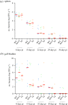
Persistence of S. Typhimurium in the spleen and in the gall bladder after intracoelomic inoculation. Five-day-old chicks were inoculated in the coelomic cavity with around 3 × 10⁷ CFU per chick with S. Typhimurium 14028 turboFP650 wild-type (WT; blue diamond), ΔinvA::kan mutant strain (ΔinvA; T3SS-1 defective; green diamond) or the ΔinvA::kan ΔpagN::cm Δrck mutant strain (3Δ; T3SS-1, Rck, PagN defective; yellow diamond). Each week, seven animals were sacrificed and their spleens and gall bladders removed. The kinetics of spleen (a) and gall bladder (b) colonization were monitored each week for a period of 36 days. Results are expressed as number of bacteria (log CFU per gram of organ). The medians are represented by a red dash. A Kruskal–Wallis test was conducted, followed by a Dunn's multiple comparisons test (GraphPad Software). Significance was *p < 0.05.

Similar articles

Cited by

References

    1. WHO. 2017. WHO estimates of the global burden of foodborne diseases. See https://apps.who.int/iris/handle/10665/199350.
    1. ECDC Ea. 2019. The European Union summary report on trends and sources of zoonoses, zoonotic agents and food-borne outbreaks in 2016. EFSA J. 15, 5077. (10.2903/j.efsa.2010.1496) - DOI - PMC - PubMed
    1. Ferrari RG, Rosario DKA, Cunha-Neto A, Mano SB, Figueiredo EES, Conte-Junior CA. 2019. Worldwide epidemiology of Salmonella serovars in animal-based foods: a meta-analysis. Appl. Environ. Microbiol. 85, e00591-19. (10.1128/AEM.00591-19) - DOI - PMC - PubMed
    1. Rabinowitz PM, Conti LA. 2009. Human-clinical-medicine: clinical approaches to zoonoses, toxicants and other shared health risks. 1st edn. Maryland Heights, MD: Saunders.
    1. Knight-Jones TJ, Mylrea GE, Kahn S. 2010. Animal production food safety: priority pathogens for standard setting by the World Organisation for Animal Health. Rev. Sci. Technol. 29, 523-535. (10.20506/rst.29.3.1994) - DOI - PubMed

Publication types

Substances