. 2021 Nov 16;23(1):287.
doi: 10.1186/s13075-021-02663-z.

Expansion of myeloid-derived suppressor cells contributes to metabolic osteoarthritis through subchondral bone remodeling

Affiliations

Expansion of myeloid-derived suppressor cells contributes to metabolic osteoarthritis through subchondral bone remodeling

Lixia Zhang et al. Arthritis Res Ther. .

Abstract

Background: Osteoarthritis (OA) subsequent to acute joint injury accounts for a significant proportion of all arthropathies. Myeloid-derived suppressor cells (MDSCs) are a heterogeneous population of myeloid progenitor cells classically known for potent immune-suppressive activity; however, MDSCs can also differentiate into osteoclasts. In addition, this population is known to be expanded during metabolic disease. The objective of this study was to determine the role of MDSCs in the context of OA pathophysiology.

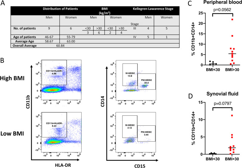
Methods: In this study, we examined the differentiation and functional capacity of MDSCs to become osteoclasts in vitro and in vivo using mouse models of OA and in MDSC quantitation in humans with OA pathology relative to obesity status.

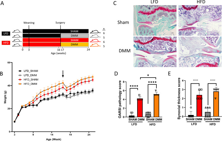
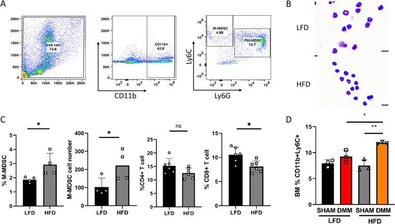
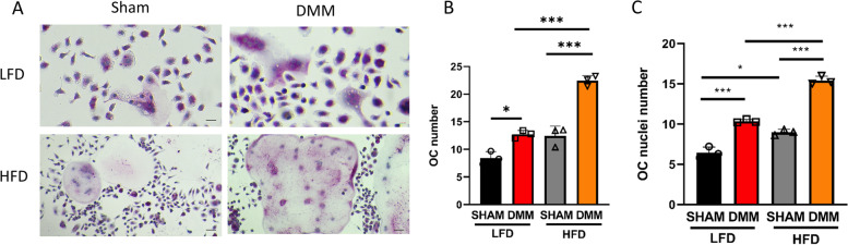
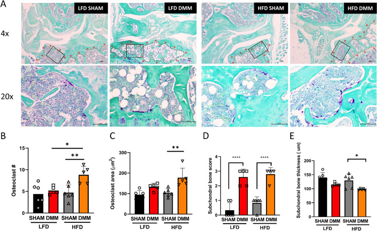
Results: We observed that MDSCs are expanded in mice and humans during obesity. MDSCs were expanded in peripheral blood of OA subjects relative to body mass index and in mice fed a high-fat diet (HFD) compared to mice fed a low-fat diet (LFD). In mice, monocytic MDSC (M-MDSC) was expanded in diet-induced obesity (DIO) with a further expansion after destabilization of the medial meniscus (DMM) surgery to induce post-traumatic OA (PTOA) (compared to sham-operated controls). M-MDSCs from DIO mice had a greater capacity to form osteoclasts in culture with increased subchondral bone osteoclast number. In humans, we observed an expansion of M-MDSCs in peripheral blood and synovial fluid of obese subjects compared to lean subjects with OA.

Conclusion: These data suggest that MDSCs are reprogrammed in metabolic disease, with the potential to contribute towards OA progression and severity.

Keywords: Myeloid-derived suppressor cells; Obesity; Osteoarthritis; Osteoclasts; Subchondral bone.


Conflict of interest statement

The authors declare that they have no competing interests.

Figures

Fig. 1
Fig. 1
High-fat diet promotes post-traumatic osteoarthritis in mice. A Overview of the experimental design. Mice were fed either a low-fat diet (LFD) or a high-fat diet (HFD) for 12 weeks and sham or destabilization of the medial meniscus (DMM) surgery was performed at weeks 16–17. Sham group (n = 6), DMM group (n = 5). B Changes in weight over time. Arrow represents the time of the surgery. HFD increases body weight compared to LFD. C Representative histopathological section from mouse knee joints stained with fast green and Safranin-O. D Glasson Modified OARSI pathology score measuring osteoarthritic damage (n = 6 in the sham group, n = 5 in the DMM group). E Synovial thickness score (n = 6 in the sham group, n = 5 in the DMM group). Scale bar = 100μM. **P < 0.01, ****P < 0.0001. Two-way ANOVA Tukey’s multiple comparison test
Fig. 2
Fig. 2
High-fat diet increases monocytic-MDSC expansion during PTOA. A Gating strategy used to define the M-MDSC subpopulation (CD11b+Ly6C+Ly6G cells) in mouse bone marrow. Following the initial FS/SC discrimination, the gate was set on CD11b+ cells. After exclusion of doublets (not shown), live CD11b+ cells were gated and Ly6C+ and Ly6G+ populations. B Cytospin images of M-MDSC cells from the bone marrow from LFD and HFD mice (at 16 weeks). Scale bar denotes 10 microns. C HFD increases the BM M-MDSC population in mice (n = 4 in each group) by total number and percentage with concomitant suppression of CD8+ and CD4+ T-cells compared to LFD mice (n = 6 in each group, at 16 weeks). D HFD increases the BM M-MDSC population in DMM mice (n = 3 in each group). *P < 0.05, **P < 0.01, ***P < 0.001, ****P < 0.0001 by two-way ANOVA Tukey’s multiple comparison test
Fig. 3
Fig. 3
High-fat diet (HFD) increases osteoclastogenesis in DMM mice. A Representative images of isolated bone marrow M-MDSC post-M-CSF and RANKL stimulation stained with tartrate-resistant acid phosphatase (TRAP; n = 3 in each group). B Osteoclast number (n = 3 in each group), number of osteoclasts per field of view, each enumeration utilized 5 fields of view/slide at 20×. C Osteoclast nuclei number per osteoclast (n = 3 in each group). *P < 0.05, **P < 0.01, ***P < 0.001, ****P < 0.0001 by two-way ANOVA Tukey’s multiple comparison test
Fig. 4
Fig. 4
High-fat diet (HFD) alters subchondral bone parameters with increased osteoclast formation. A Representative histopathological section from mouse subchondral bone stained with tartrate-resistant acid phosphatase (TRAP). B HFD increases osteoclast number in DMM. C Osteoclast area (μm2). D Subchondral bone score. E Subchondral bone thickness. n = 6 in the sham group, n = 5 in the DMM group; scale bar = 100μM. *P < 0.05, ****P < 0.0001 two-way ANOVA Tukey’s multiple comparison test
Fig. 5
Fig. 5
MDSCs are expanded in obese patients with post-traumatic osteoarthritis. A Demographic of the patient population. B Gating strategy used to define the M-MDSC subpopulation (CD11b+ HLADR CD14+ CD15 cells). The M-MDSC population is expanded in C peripheral blood and D synovial fluid of obese (BMI > 30; N = 10) compared to non-obese (BMI < 30; N = 5) patients diagnosed with post-traumatic osteoarthritis. Two-tailed unpaired Student t-test

References

    1. Felson DT, Lawrence RC, Dieppe PA, Hirsch R, Helmick CG, Jordan JM, Kington RS, Lane NE, Nevitt MC, Zhang Y, Sowers M, McAlindon T, Spector TD, Poole AR, Yanovski SZ, Ateshian G, Sharma L, Buckwalter JA, Brandt KD, Fries JF. Osteoarthritis: new insights. Part 1: the disease and its risk factors. Ann Intern Med. 2000;133(8):635–646. doi: 10.7326/0003-4819-133-8-200010170-00016. - DOI - PubMed
    1. Lories RJ, Luyten FP. The bone-cartilage unit in osteoarthritis. Nat Rev Rheumatol. 2011;7(1):43–49. doi: 10.1038/nrrheum.2010.197. - DOI - PubMed
    1. Houard X, Goldring MB, Berenbaum F. Homeostatic mechanisms in articular cartilage and role of inflammation in osteoarthritis. Curr Rheumatol Rep. 2013;15(11):375. doi: 10.1007/s11926-013-0375-6. - DOI - PMC - PubMed
    1. Wojdasiewicz P, Poniatowski LA, Szukiewicz D. The role of inflammatory and anti-inflammatory cytokines in the pathogenesis of osteoarthritis. Mediators Inflamm. 2014;2014:561459. doi: 10.1155/2014/561459. - DOI - PMC - PubMed
    1. Orlowsky EW, Kraus VB. The role of innate immunity in osteoarthritis: when our first line of defense goes on the offensive. J Rheumatol. 2015;42(3):363–371. doi: 10.3899/jrheum.140382. - DOI - PMC - PubMed

Publication types

LinkOut - more resources