

Basaloid follicular hamartoma associated with follicular mucinosis and inflammation
- PMID: 34785065
- PMCID: PMC8799845
- DOI: 10.1016/j.abd.2020.10.012
Basaloid follicular hamartoma associated with follicular mucinosis and inflammation
Abstract
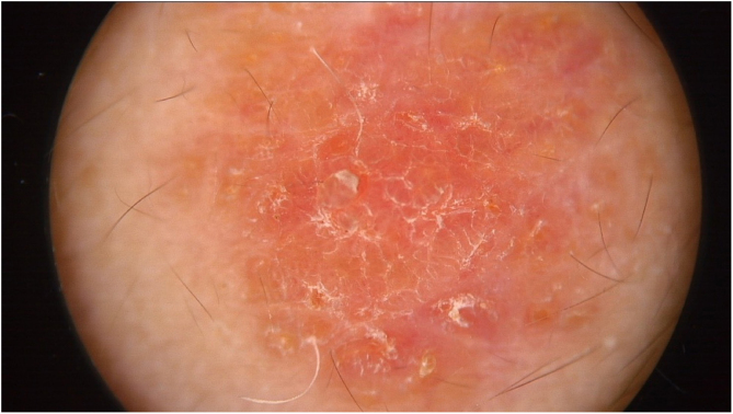
Basaloid follicular hamartoma is a benign, superficial malformation of hair follicles that can be mistaken both clinical and histopathologically for basal cell carcinoma. Basaloid follicular hamartoma has been linked to a mutation in the PTCH-1 gene, which is part of the same pathway involved in Gorlin-Goltz syndrome. Here we present a 9-year-old patient with an asymptomatic congenital lesion on the forehead, which increased in size over the years. Histopathology showed a basaloid follicular hamartoma associated with follicular mucinosis and inflammation. Gorlin-Goltz syndrome was ruled out by clinical examination.
Keywords: Carcinoma, basal cell; Genes, tumor suppressor; Hamartoma.
Copyright © 2021 Sociedade Brasileira de Dermatologia. Published by Elsevier España, S.L.U. All rights reserved.
Figures


Similar articles
-
Generalized basaloid follicular hamartoma syndrome versus Gorlin syndrome: A diagnostic challenge.Pediatr Dermatol. 2018 Nov;35(6):e396-e397. doi: 10.1111/pde.13614. Epub 2018 Aug 28. Pediatr Dermatol. 2018. PMID: 30152544
-
Basaloid follicular hamartoma.Arch Pathol Lab Med. 2010 Aug;134(8):1215-9. doi: 10.5858/2008-0620-RS.1. Arch Pathol Lab Med. 2010. PMID: 20670146 Review.
-
Basaloid follicular hamartoma syndrome: acquired sporadic variant with hypothyroidism, hypohidrosis and alopecia, a rare case.J Pak Med Assoc. 2023 Jun;73(6):1308-1310. doi: 10.47391/JPMA.6560. J Pak Med Assoc. 2023. PMID: 37427638
-
Cutaneous inflammation as a marker of malignant transformation in a patient with linear unilateral basaloid follicular hamartoma.Indian J Dermatol Venereol Leprol. 2019 May-Jun;85(3):287-290. doi: 10.4103/ijdvl.IJDVL_927_16. Indian J Dermatol Venereol Leprol. 2019. PMID: 30246704
-
Familial basaloid follicular hamartoma: lesional characterization and review of the literature.Am J Dermatopathol. 2003 Apr;25(2):130-7. doi: 10.1097/00000372-200304000-00006. Am J Dermatopathol. 2003. PMID: 12652194 Review.
Cited by
-
Multiple basal cell carcinomas within a Blaschkoid basaloid follicular hamartoma: A case report.JAAD Case Rep. 2025 Jul 28;64:30-32. doi: 10.1016/j.jdcr.2025.07.006. eCollection 2025 Oct. JAAD Case Rep. 2025. PMID: 40918551 Free PMC article. No abstract available.
References
-
- Mills O., Thomas L.B. Basaloid Follicular Hamartoma. Arch Pathol Lab Med. 2010;134:1215–1219. - PubMed
-
- Cabrera H.N., Giovanna P.D., García S., Nadur K.V. Hamartoma folicular basaloide linear unilateral con lesiones cutáneas y mucosas. Dermatol Argent. 2013;19:100–105.
-
- Boccaletti V., Accorsi P., Pinelli L., Ungari M., Giordano L., Neri I., et al. Congenital Systematized Basaloid Follicular Hamartoma with Microphthalmia and Hemimegalencephaly. Pediatr Dermatol. 2011;28:555–560. - PubMed
-
- Choi E., Liau M., Huang J., Tan K.B., Aw D. Basaloid follicular hamartoma: clinical, dermoscopic, and histopathological characteristics of case. Dermatol Online J. 2017;23:13030. - PubMed
-
- Saxena A., Shapiro M., Kasper D.A., Fitzpatrick J.E., Mellette Junior J.R. Basaloid follicular hamartoma: a cautionary tale and review of the literature. Dermatol Surg. 2007;33:1130–1135. - PubMed
Publication types
MeSH terms
LinkOut - more resources
Full Text Sources
Medical
