

Resting-state functional connectivity of the amygdala subregions in unmedicated patients with obsessive-compulsive disorder before and after cognitive behavioural therapy
- PMID: 34785511
- PMCID: PMC8598242
- DOI: 10.1503/jpn.210084
Resting-state functional connectivity of the amygdala subregions in unmedicated patients with obsessive-compulsive disorder before and after cognitive behavioural therapy
Abstract
Background: Cognitive behavioural therapy (CBT) is considered an effective first-line treatment for obsessive-compulsive disorder (OCD). However, the neural basis of CBT for OCD has not yet been elucidated. The role of the amygdala in OCD and its functional coupling with the cerebral cortex have received increasing attention, and may provide new understanding of the neural basis of CBT for OCD.
Methods: We acquired baseline resting-state functional MRI (fMRI) scans from 45 unmedicated patients with OCD and 40 healthy controls; we then acquired another wave of resting-state fMRI scans from the patients with OCD after 12 weeks of CBT. We performed seed-based resting-state functional connectivity analyses of the amygdala subregions to examine changes in patients with OCD as a result of CBT.
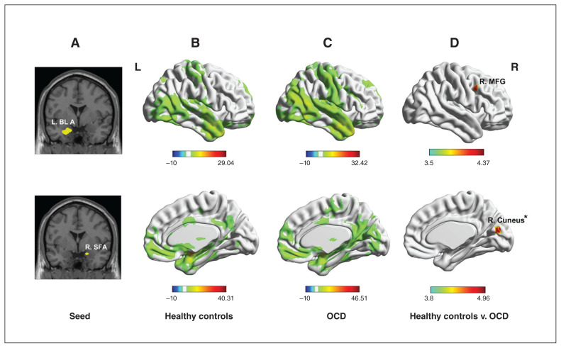
Results: Compared to healthy controls, patients with OCD showed significantly increased resting-state functional connectivity at baseline between the left basolateral amygdala and the right middle frontal gyrus, and between the superficial amygdala and the right cuneus. In patients with OCD who responded to CBT, we found decreased resting-state functional connectivity after CBT between the amygdala subregions and the visual association cortices and increased resting-state functional connectivity between the amygdala subregions and the right inferior parietal lobe. Furthermore, these changes in resting-state functional connectivity were positively associated with changes in scores on the compulsion or obsession subscales of the Yale-Brown Obsessive-Compulsive Scale.
Limitations: Because of the lack of a second scan for healthy controls after 12 weeks, our results may have been confounded by other variables.
Conclusion: Our findings yield insights into the pathophysiology of OCD; they also reveal the potential neural changes elicited by CBT, and thus have implications for guiding effective treatment strategies with CBT for OCD.
© 2021 CMA Joule Inc. or its licensors.
Conflict of interest statement
Competing interests: None declared.
Figures


Similar articles
-
Resting-state functional connectivity of amygdala subregions predicts treatment outcome for cognitive behavioral therapy in obsessive-compulsive disorder at a 4-month follow-up.Psychiatry Res. 2024 May;335:115876. doi: 10.1016/j.psychres.2024.115876. Epub 2024 Mar 28. Psychiatry Res. 2024. PMID: 38564923
-
Resting-state functional connectivity of amygdala subregions across different symptom subtypes of obsessive-compulsive disorder patients.Neuroimage Clin. 2024;43:103644. doi: 10.1016/j.nicl.2024.103644. Epub 2024 Jul 18. Neuroimage Clin. 2024. PMID: 39042954 Free PMC article.
-
Basolateral amygdala-ventromedial prefrontal cortex connectivity predicts cognitive behavioural therapy outcome in adults with obsessive-compulsive disorder.J Psychiatry Neurosci. 2017 Nov;42(6):378-385. doi: 10.1503/jpn.160215. J Psychiatry Neurosci. 2017. PMID: 28632120 Free PMC article.
-
Predictors of response to cognitive behavioural therapy (CBT) for individuals with obsessive-compulsive disorder (OCD): a systematic review.Behav Cogn Psychother. 2023 Jul;51(4):302-319. doi: 10.1017/S1352465823000103. Epub 2023 Apr 4. Behav Cogn Psychother. 2023. PMID: 37013903
-
Task fMRI studies investigating inhibitory control in patients with obsessive-compulsive disorder and eating disorders: A comparative meta-analysis.World J Biol Psychiatry. 2024 Jan-Feb;25(1):26-42. doi: 10.1080/15622975.2023.2251057. Epub 2023 Aug 28. World J Biol Psychiatry. 2024. PMID: 37640027 Review.
Cited by
-
Left Amygdala Functional Connectivity Decreased after Fear of Negative Events was Disregarded in Obsessive-Compulsive Disorder.Neurosci Insights. 2022 Jul 23;17:26331055221114823. doi: 10.1177/26331055221114823. eCollection 2022. Neurosci Insights. 2022. PMID: 36081984 Free PMC article.
-
Effects of cognitive behavioural therapy and exposure-response prevention on brain activation in obsessive-compulsive disorder patients: systematic review and meta-analysis.Eur Arch Psychiatry Clin Neurosci. 2025 Aug;275(5):1491-1507. doi: 10.1007/s00406-024-01852-6. Epub 2024 Jun 27. Eur Arch Psychiatry Clin Neurosci. 2025. PMID: 38935215
-
Neurobiological correlates of CBT response in OCD through the analysis of resting state networks.Int J Clin Health Psychol. 2025 Apr-Jun;25(2):100585. doi: 10.1016/j.ijchp.2025.100585. Epub 2025 May 12. Int J Clin Health Psychol. 2025. PMID: 40476043 Free PMC article.
-
Right Prefrontal Cortical Thickness Is Associated With Response to Cognitive-Behavioral Therapy in Children With Obsessive-Compulsive Disorder.J Am Acad Child Adolesc Psychiatry. 2023 Apr;62(4):403-414. doi: 10.1016/j.jaac.2022.07.865. Epub 2022 Dec 13. J Am Acad Child Adolesc Psychiatry. 2023. PMID: 36526161 Free PMC article.
-
Exploring functional connectivity in large-scale brain networks in obsessive-compulsive disorder: a systematic review of EEG and fMRI studies.Cereb Cortex. 2024 Aug 1;34(8):bhae327. doi: 10.1093/cercor/bhae327. Cereb Cortex. 2024. PMID: 39152672 Free PMC article.
References
-
- Olatunji BO, Davis ML, Powers MB, et al. . Cognitive-behavioral therapy for obsessive-compulsive disorder: a meta-analysis of treatment outcome and moderators. J Psychiatr Res 2013;47:33–41. - PubMed
-
- Sander D, Grafman J, Zalla T. The human amygdala: an evolved system for relevance detection. Rev Neurosci 2003;14:303–16. - PubMed
-
- Wendt J, Weike AI, Lotze M, et al. . The functional connectivity between amygdala and extrastriate visual cortex activity during emotional picture processing depends on stimulus novelty. Biol Psychol 2011;86:203–9. - PubMed
Publication types
MeSH terms
LinkOut - more resources
Full Text Sources
Medical