. 2021 Nov 24;8(6):ENEURO.0363-21.2021.
doi: 10.1523/ENEURO.0363-21.2021. Print 2021 Nov-Dec.

Off-Target Expression of Cre-Dependent Adeno-Associated Viruses in Wild-Type C57BL/6J Mice

Affiliations

Off-Target Expression of Cre-Dependent Adeno-Associated Viruses in Wild-Type C57BL/6J Mice

Justin J Botterill et al. eNeuro. .

Abstract

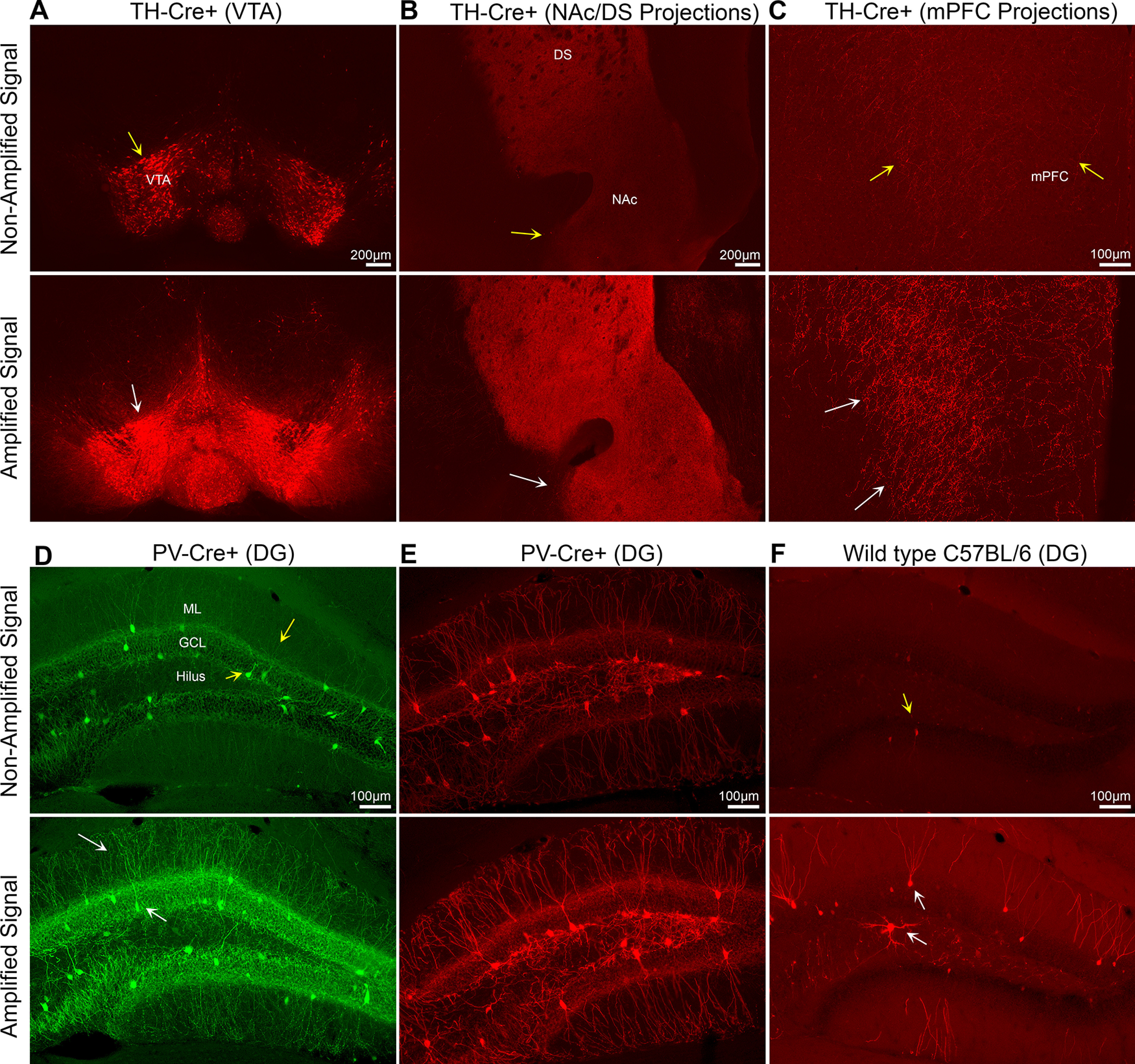
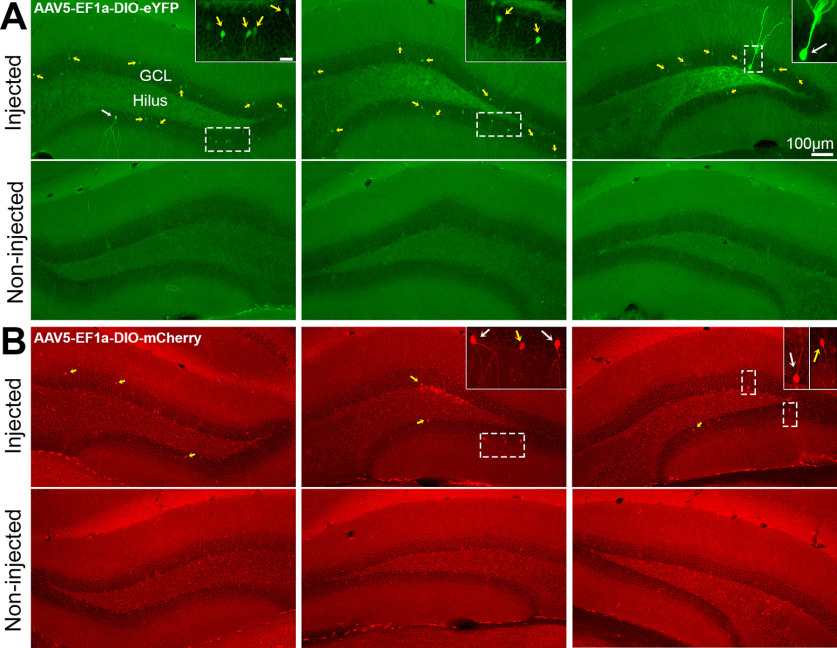
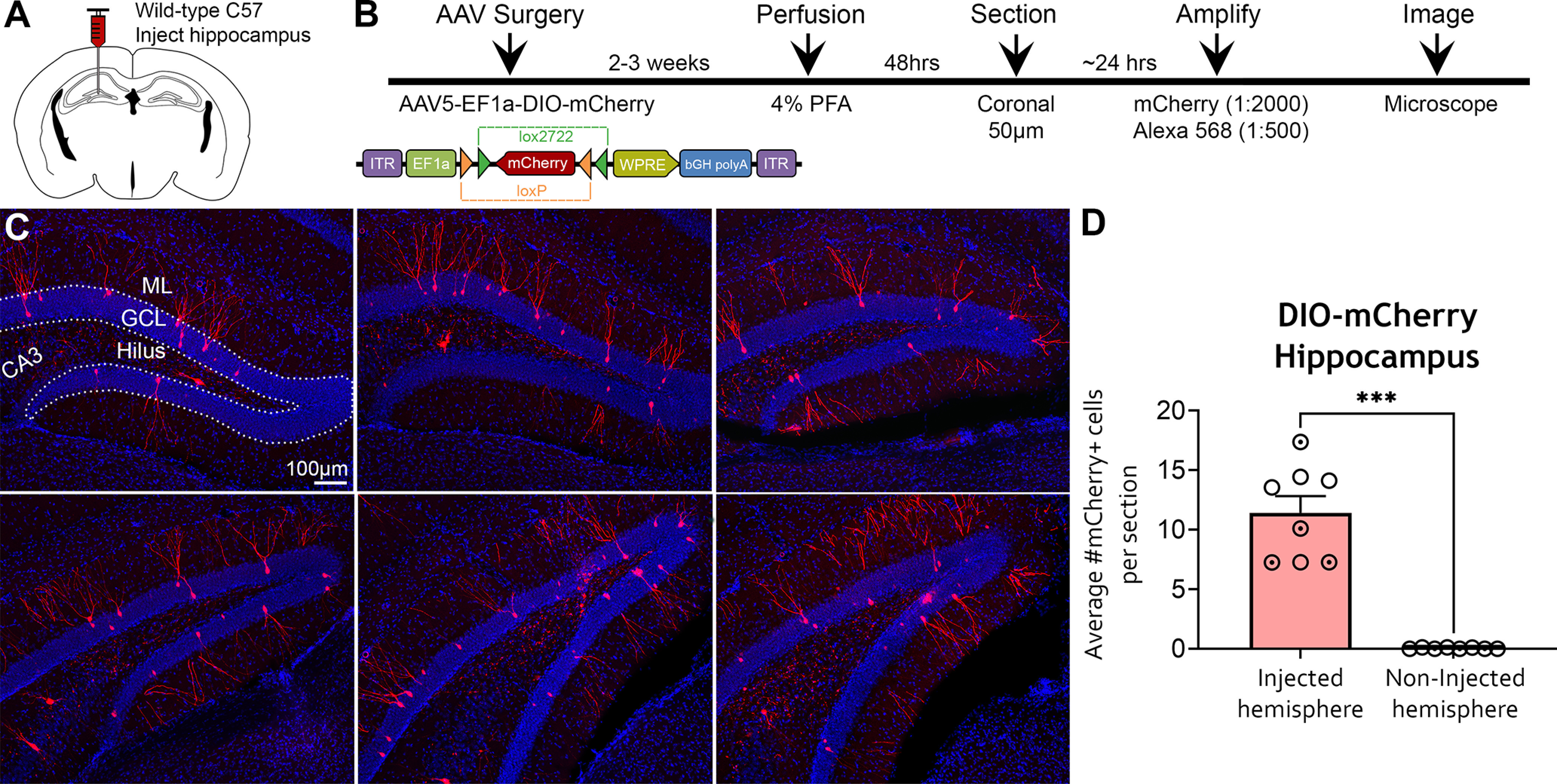
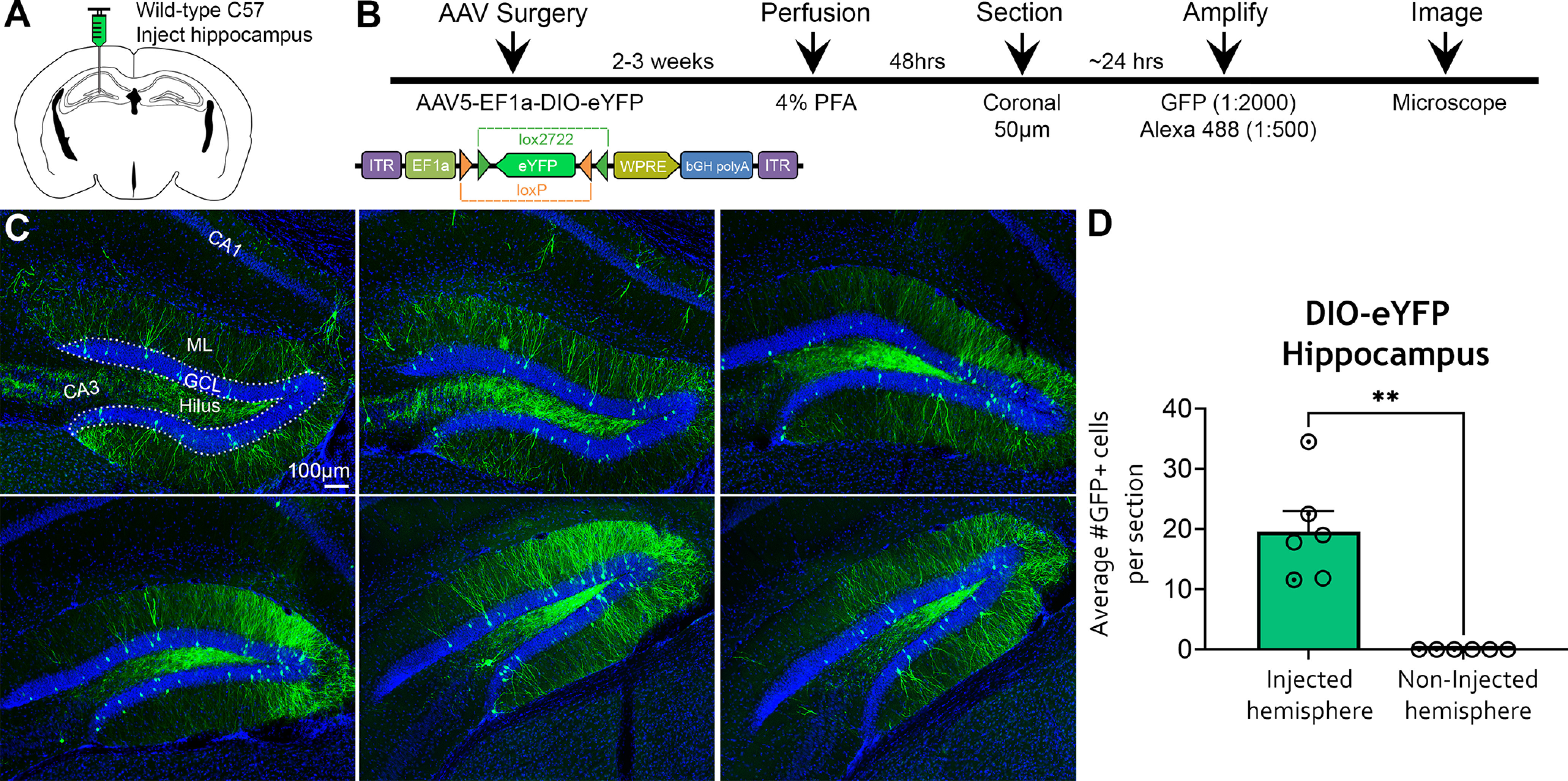
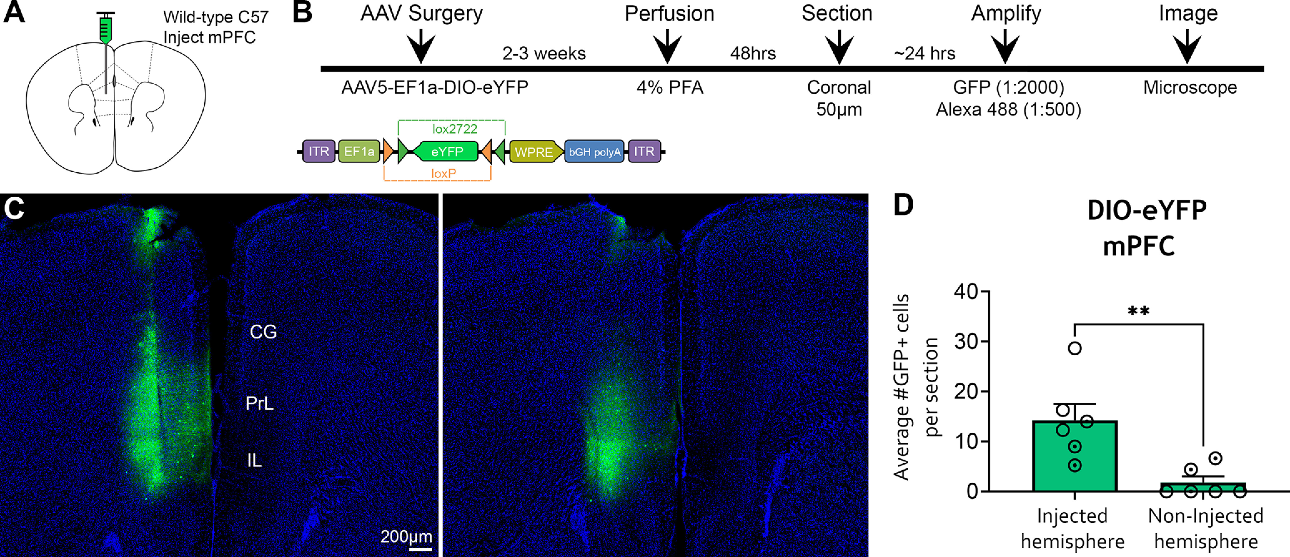
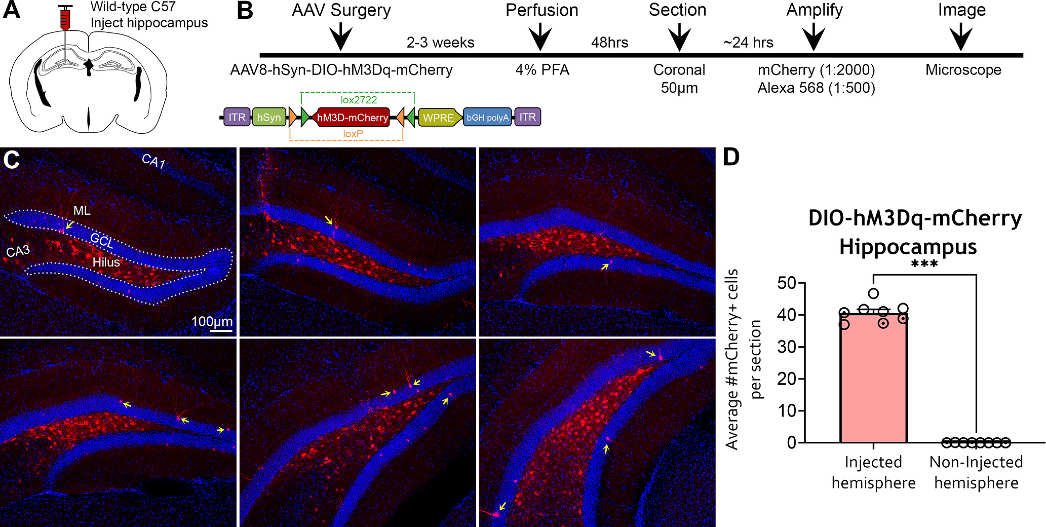
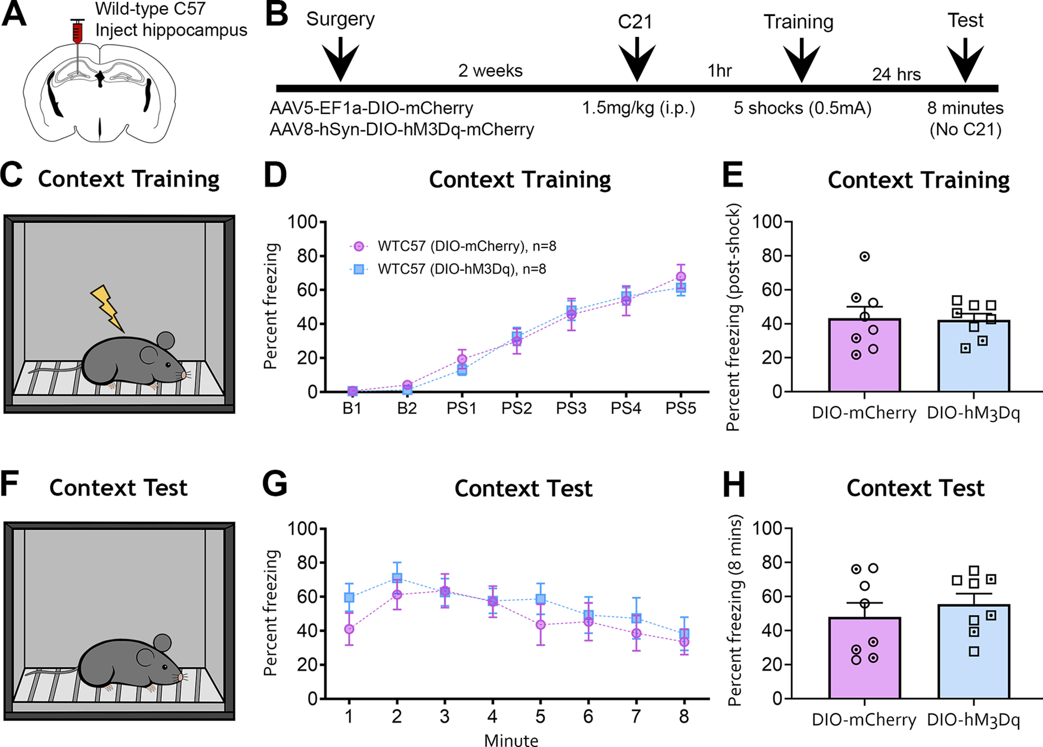
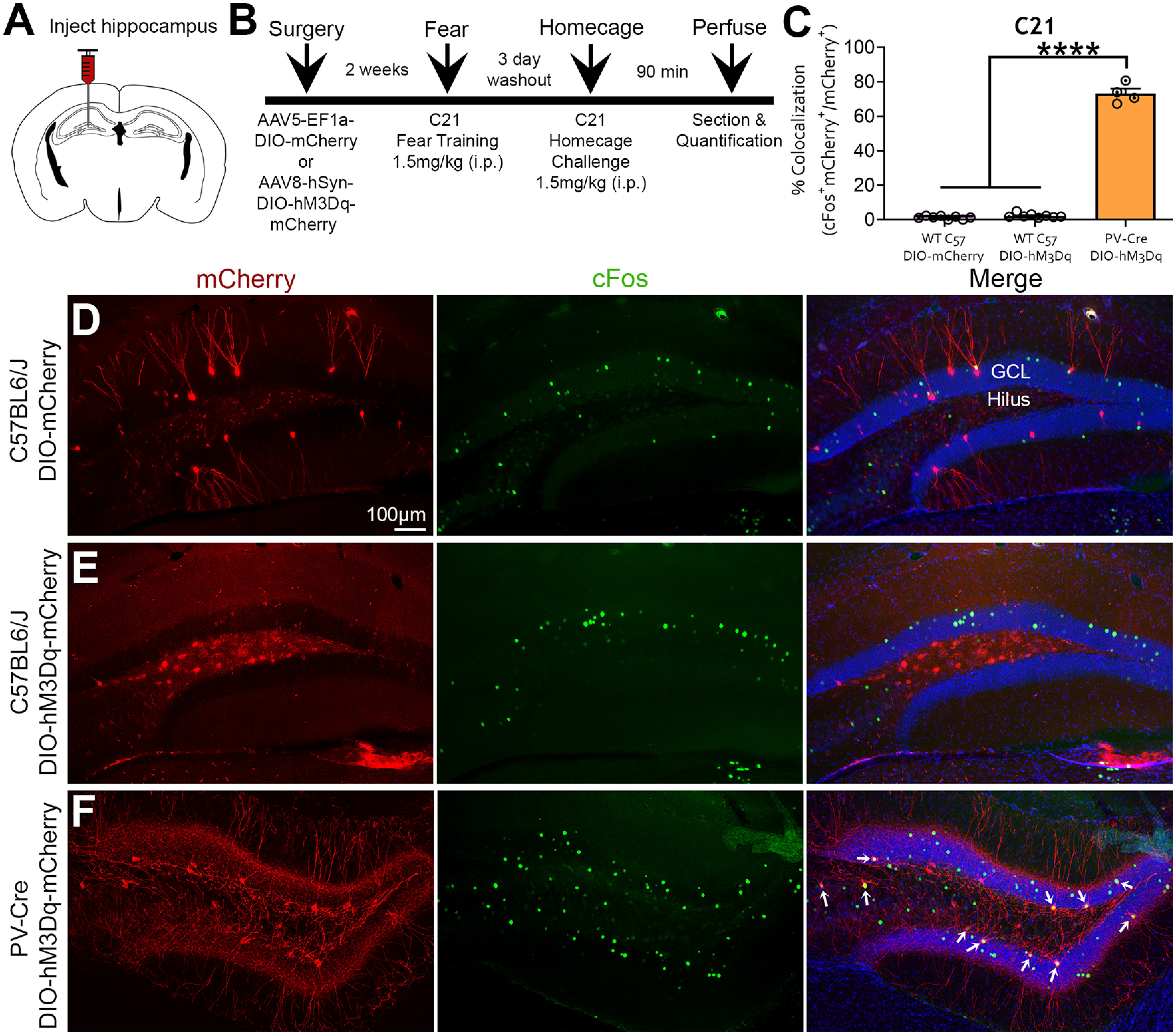
Adeno-associated viruses (AAVs) are a commonly used tool in neuroscience to efficiently label, trace, and/or manipulate neuronal populations. Highly specific targeting can be achieved through recombinase-dependent AAVs in combination with transgenic rodent lines that express Cre-recombinase in specific cell types. Visualization of viral expression is typically achieved through fluorescent reporter proteins (e.g., GFP or mCherry) packaged within the AAV genome. Although nonamplified fluorescence is usually sufficient to observe viral expression, immunohistochemical amplification of the fluorescent reporter is routinely used to improve viral visualization. In the present study, Cre-dependent AAVs were injected into the neocortex of wild-type C57BL/6J mice. While we observed weak but consistent nonamplified off-target double inverted open reading frame (DIO) expression in C57BL/6J mice, antibody amplification of the GFP or mCherry reporter revealed notable Cre-independent viral expression. Off-target expression of DIO constructs in wild-type C57BL/6J mice occurred independent of vendor, AAV serotype, or promoter. We also evaluated whether Cre-independent expression had functional effects via designer receptors exclusively activated by designer drugs (DREADDs). The DREADD agonist C21 (compound 21) had no effect on contextual fear conditioning or c-Fos expression in DIO-hM3Dq-mCherry+ cells of C57BL/6J mice. Together, our results indicate that DIO constructs have off-target expression in wild-type subjects. Our findings are particularly important for the design of experiments featuring sensitive systems and/or quantitative measurements that could be negatively impacted by off-target expression.

Keywords: Cre/loxP; DREADDs; Immunofluorescence; antibody amplification; c-Fos; double inverted open reading frame; fear conditioning.


Figures

Figure 1.
Figure 1.
Antibody amplification of Cre-dependent viral expression. A, Representative images from a TH-Cre mouse injected in the VTA with AAV5-EF1a-DIO-mCherry show a similar pattern of expression between nonamplified and amplified fluorescence (yellow and white arrows). B, Long-range VTA–NAc/DS projections are easier to visualize following mCherry amplification (yellow vs white arrow). C, Similarly, nonamplified fluorescence of VTA to mPFC projections was generally weak (yellow arrows), and the fluorescence signal was significantly improved following mCherry amplification (white arrows). D, E, Representative images from PV-Cre mice injected with AAV5-EF1a-DIO-EYFP (D) or AAV5-EF1a-DIO-mCherry (E). The nonamplified fluorescence signal was similar between eYFP and mCherry constructs. Moreover, fluorescence signal amplification is similar to the nonamplified signal (yellow arrows) but is brighter and easier to visualize (white arrows), especially the dendrites in the ML. F, Representative images from a C57BL/6J mouse injected with AAV5-EF1a-DIO-mCherry show minimal nonamplified fluorescence (yellow arrow). Remarkably, amplification of adjacent sections from the same mouse revealed mCherry expression within the DG (white arrows). Scale bars: Panels AB: 200 μm; Panels CF: 100 μm.
Figure 2.
Figure 2.
Nonamplified fluorescence of DIO constructs in WT C57BL/6J mice. A, B, Representative photomicrographs of nonamplified fluorescence signal in C57BL/6J mice injected with AAV5-EF1a-DIO-eYFP (A) or AAV5-EF1a-DIO-mCherry (B). Nonamplified immunofluorescence was generally weak and primarily restricted to the soma (yellow arrows; see insets) of the injected hemisphere only. We hypothesize that the weak nonamplified immunofluorescence in these cells is significantly enhanced after antibody amplification. In addition, a very small number of cells with bright immunofluorescence throughout the cell body and its processes were observed (white arrows; see insets). Scale bars: 10× objective, 100 μm; 20× objective, insets, 25 μm.
Figure 3.
Figure 3.
Amplified expression of DIO-mCherry in the hippocampus of WT C57BL/6J mice. A, B, Experimental design and timeline. AAV5-EF1a-DIO-mCherry was injected into the anterior and posterior hippocampi of C57BL/6J mice (n =8) and perfused 2–3 weeks later. Brains were sectioned in the coronal plane, and viral signal was amplified with rabbit anti-mCherry and goat anti-rabbit 568 antibodies. C, Representative immunofluorescence of mCherry throughout the relatively dorsal (top) and caudal (bottom) DG. Expression of mCherry was primarily observed in the GCL and dendrites extending into the ML (putative dentate GCs). The amplified mCherry signal also resulted in labeling of mossy fibers and cells in the hilus. D, Quantification of mCherry+ cells indicated that somatic expression was restricted to the injected hemisphere. Female (clear circles) and male (dotted circles) data points are identified, but no sex differences were found. ***p <0.001. Scale bar, 100 μm. Data for this figure are shown in Extended Data Figure 3-1.
Figure 4.
Figure 4.
Amplified expression of DIO-eYFP in the hippocampus of WT C57BL/6J mice. A, B, Experimental design and timeline. AAV5-EF1a-DIO-eYFP was injected into the anterior and posterior hippocampi of C57BL/6J mice (n =6) and perfused 2–3 weeks later. The eYFP signal was amplified with chicken anti-GFP and goat anti-chicken 488 antibodies. C, Representative immunofluorescence of GFP throughout the DG. GFP expression was observed primarily in the DG, characterized by robust labeling of putative GCs within the GCL and their dendrites. The hilus also showed bright GFP signal, with expression in mossy fibers and hilar cells. D, Quantification of GFP+ cells revealed that somatic expression was restricted to the injected hemisphere. Female (clear circles) and male (dotted circles) data points are identified, but no sex differences were found. **p <0.005. Scale bar, 100 μm. Data for this figure are shown in Extended Data Figures 4-1 and 4-2.
Figure 5.
Figure 5.
Amplified expression of DIO-eYFP in the mPFC of WT C57BL/6J mice. A, B, Experimental design and timeline. AAV5-EF1a-DIO-eYFP was injected into left mPFC of C57BL/6J mice (n =6), and mice were perfused 2–3 weeks later. Viral signal was amplified with chicken anti-GFP and goat anti-chicken 488 antibodies. C, Representative GFP immunofluorescence in the mPFC of two sections from the same mouse. D, Quantification of GFP+ cells in the mPFC showed that expression was primarily restricted to the injected hemisphere, but two mice had sparse expression of GFP+ cells in the noninjected hemisphere, presumably resulting from viral spread because of the close proximity of the left and right mPFC. Female (clear circles) and male (dotted circles) data points are identified, but no sex differences were found. CG, cingulate gyrus; PrL, Prelimbic cortex; IL, infralimbic cortex. **p <0.005. Scale bar, 200 μm.
Figure 6.
Figure 6.
Amplified expression of DIO-hM3Dq-mCherry in the hippocampi of WT C57BL/6J mice. A, B, Experimental design and timeline. AAV8-hSyn-DIO-hM3Dq-mCherry was injected into the anterior and posterior hippocampus of C57BL/6J mice (n =8), and mice were perfused 2–3 weeks later. The viral signal was amplified with rabbit anti-mCherry and goat anti-rabbit 568 antibodies and was visualized on an epifluorescence microscope. C, Representative mCherry immunofluorescence in relatively dorsal (top) and caudal (bottom) sections of the DG. Amplified mCherry expression appeared primarily within hilar cells and a sparse number of GCs (yellow arrows). D, Quantification of mCherry+ cells revealed that expression was restricted to the injected hippocampus. Female (clear circles) and male (dotted circles) data points are identified, but no sex differences were found. ***p <0.001. Scale bar, 100 μm. Data for this figure are shown in Extended Data Figures 6-1 and 6-2.
Figure 7.
Figure 7.
The hM3Dq agonist C21 does not affect fear behavior in C57BL/6J mice injected with DIO-mCherry or DIO-hM3Dq-mCherry in the DG. A, B, Experimental design and timeline. Adult C57BL/6J mice underwent surgery to receive intrahippocampal injections of AAV-EF1a-DIO-mCherry or AAV-hSyn-DIO-hM3Dq-mCherry. After a 2 week recovery period, mice were injected with the hM3Dq agonist C21 1 h before contextual fear training. C, Mice were then placed in a fear-conditioning chamber. Baseline activity was assessed over 2 min, followed by five footshocks (0.5 mA) spaced 1 min apart. D, Minute-by-minute analysis of the training session revealed that freezing behavior did not differ between EF1a-DIO-mCherry or hSyn-DIO-hM3Dq-mCherry groups. E, The average postshock freezing did not differ between the EF1a-DIO-mCherry and hSyn-DIO-hM3Dq-mCherry groups. F, Mice were returned to the same operant chamber 24 h later to test contextual fear memory. Notably, C21 was not administered a second time before the contextual memory test. G, Minute-by-minute analysis revealed that conditioned freezing did not differ between the EF1a-DIO-mCherry or hSyn-DIO-hM3Dq-mCherry groups. H, Average freezing during the memory test did not differ between groups. Female (clear points) and male (dotted points) data points are identified, but no sex differences were found.
Figure 8.
Figure 8.
mCherry and c-Fos immunofluorescence following C21 home-cage challenge. A, B, Experimental design and timeline. Mice underwent surgery for AAV injection and were allowed 2 weeks for recovery. Mice underwent behavioral testing and were then given a 3 d washout period. Mice were then injected with C21 (1.5 mg/kg) in their home cage and were euthanized 90 min later to evaluate the immediate early gene c-Fos. C, The percentage of colocalization of c-Fos+ and mCherry+ cells following C21 challenge was significantly lower in C57BL/6J mice injected with DIO-mCherry (7 c-Fos+mCherry+/497 mCherry+ cells = 1.41%) or DIO-hM3Dq-mCherry (23 c-Fos+mCherry+/1062 mCherry+ cells = 2.17%) compared with PV-Cre+ mice injected with DIO-hM3Dq-mCherry (267 c-Fos+mCherry/367 mCherry+ cells = 72.75%). D–F, Representative images show that C57BL/6J mice lacked the clear elevation of c-Fos (green) in mCherry+ cells seen in PV-Cre+ mice (yellow; white arrows). ****p <0.0001. Scale bar, 100 μm.

Similar articles

Cited by

References

    1. Alexander GM, Brown LY, Farris S, Lustberg D, Pantazis C, Gloss B, Plummer NW, Jensen P, Dudek SM (2018) CA2 neuronal activity controls hippocampal low gamma and ripple oscillations. Elife 7:e38052. 10.7554/eLife.38052 - DOI - PMC - PubMed
    1. Alkaabi KM, Yafea A, Ashraf SS (2005) Effect of pH on thermal- and chemical-induced denaturation of GFP. Appl Biochem Biotechnol 126:149–156. 10.1385/abab:126:2:149 - DOI - PubMed
    1. Arruda-Carvalho M, Sakaguchi M, Akers KG, Josselyn SA, Frankland PW (2011) Posttraining ablation of adult-generated neurons degrades previously acquired memories. J Neurosci 31:15113–15127. 10.1523/JNEUROSCI.3432-11.2011 - DOI - PMC - PubMed
    1. Atasoy D, Aponte Y, Su HH, Sternson SM (2008) A FLEX switch targets Channelrhodopsin-2 to multiple cell types for imaging and long-range circuit mapping. J Neurosci 28:7025–7030. 10.1523/JNEUROSCI.1954-08.2008 - DOI - PMC - PubMed
    1. Barzel A, Paulk NK, Shi Y, Huang Y, Chu K, Zhang F, Valdmanis PN, Spector LP, Porteus MH, Gaensler KM, Kay MA (2015) Promoterless gene targeting without nucleases ameliorates haemophilia B in mice. Nature 517:360–364. 10.1038/nature13864 - DOI - PMC - PubMed

Publication types

LinkOut - more resources