. 2021 Nov 16;12(1):6612.
doi: 10.1038/s41467-021-26884-7.

Organ-specific genome diversity of replication-competent SARS-CoV-2

Affiliations

Organ-specific genome diversity of replication-competent SARS-CoV-2

Jolien Van Cleemput et al. Nat Commun. .

Erratum in

  • Author Correction: Organ-specific genome diversity of replication-competent SARS-CoV-2.
    Van Cleemput J, van Snippenberg W, Lambrechts L, Dendooven A, D'Onofrio V, Couck L, Trypsteen W, Vanrusselt J, Theuns S, Vereecke N, van den Bosch TPP, Lammens M, Driessen A, Achten R, Bracke KR, Van den Broeck W, Von der Thüsen J, Nauwynck H, Van Dorpe J, Gerlo S, Maes P, Cox J, Vandekerckhove L. Van Cleemput J, et al. Nat Commun. 2022 Oct 21;13(1):6247. doi: 10.1038/s41467-022-33970-x. Nat Commun. 2022. PMID: 36271014 Free PMC article. No abstract available.

Abstract

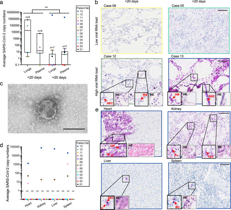
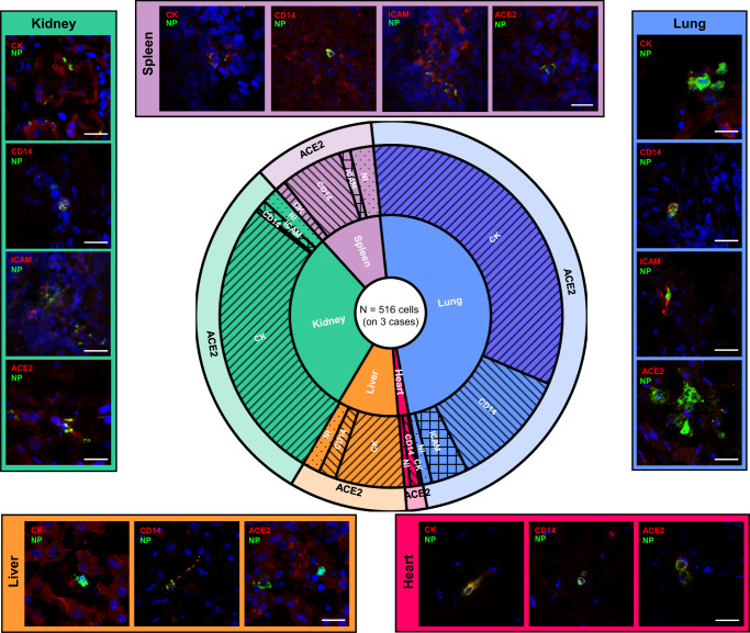
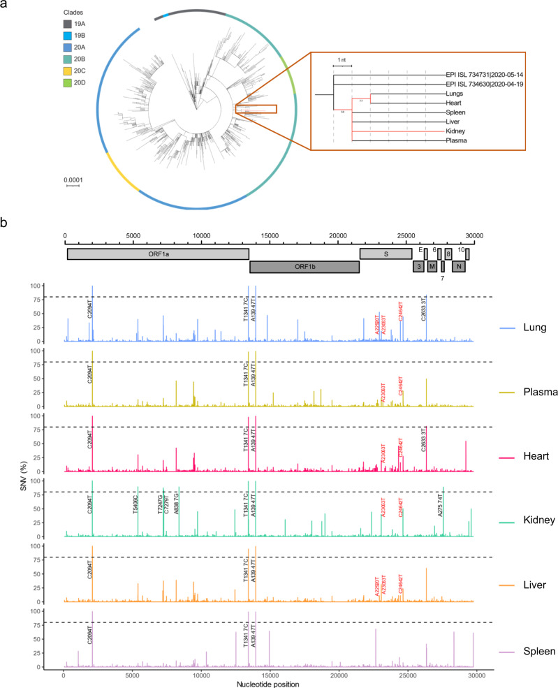
Severe acute respiratory syndrome coronavirus 2 (SARS-CoV-2) infection is not always confined to the respiratory system, as it impacts people on a broad clinical spectrum from asymptomatic to severe systemic manifestations resulting in death. Further, accumulation of intra-host single nucleotide variants during prolonged SARS-CoV-2 infection may lead to emergence of variants of concern (VOCs). Still, information on virus infectivity and intra-host evolution across organs is sparse. We report a detailed virological analysis of thirteen postmortem coronavirus disease 2019 (COVID-19) cases that provides proof of viremia and presence of replication-competent SARS-CoV-2 in extrapulmonary organs of immunocompromised patients, including heart, kidney, liver, and spleen (NCT04366882). In parallel, we identify organ-specific SARS-CoV-2 genome diversity and mutations of concern N501Y, T1027I, and Y453F, while the patient had died long before reported emergence of VOCs. These mutations appear in multiple organs and replicate in Vero E6 cells, highlighting their infectivity. Finally, we show two stages of fatal disease evolution based on disease duration and viral loads in lungs and plasma. Our results provide insights about the pathogenesis and intra-host evolution of SARS-CoV-2 and show that COVID-19 treatment and hygiene measures need to be tailored to specific needs of immunocompromised patients, even when respiratory symptoms cease.


Conflict of interest statement

The authors declare no competing interests.

Figures

Fig. 1
Fig. 1. Detailed virological analysis of 13 postmortem COVID-19 cases.
a SARS-CoV-2 RNA loads, as measured with RT-qPCR, on a total of 40 ng RNA in the lungs or 1 mL of plasma of different cases (different colors) with short- or long-lived disease (<20 or >20 days after onset of symptoms, respectively). Central tendencies for SARS-CoV-2 copies are illustrated as a boxplot where the band indicates the median, the box indicates the first and third quartiles, and the whiskers indicate the minimum and maximum of all of the data. The significance of two-way ANOVA (with categorical variables duration and anatomical compartment) between the mean SARS-CoV-2 copy numbers of long and short duration is indicated on top (**P = 0.002; F = 12.589; df = 23). Source data are provided as a Source Data file. b SARS-CoV-2 nucleocapsid (NP) staining (in purple; large images and 2X-enlarged smaller images delineated in black) and corresponding hematoxylin-eosin (HE) staining (2X-enlarged smaller images delineated in gray) of paraffin-embedded sections of lung tissue of four different cases with low (<5 copies/40 ng RNA) or high (>5 copies/40 ng RNA) viral RNA loads and short- or long-lived disease. Arrows show specific SARS-CoV-2 NP-positive cells or hyaline membranes (HM). Picture linings correspond to the respective case colors shown in the legend of Fig. 1a. Lower magnification IHC and HE images are shown in Fig. S3. All scale bars represent 100 µm. AEC alveolar epithelial cell, BEC bronchiolar epithelial cell, HM hyaline membrane, Mɸ macrophage. c Transmission electron microscopic image of a SARS-CoV-2 particle from plasma-derived viral progeny on Vero E6 cells. Controls are shown in Fig. S4c. Scale bar indicates 100 nm. d SARS-CoV-2 RNA loads on a total of 40 ng RNA, as measured with RT-qPCR, in selected extrapulmonary organs. Source data are provided as a Source Data file. e SARS-CoV-2 NP staining (in purple; large images and 2X-enlarged smaller images delineated in black) and corresponding hematoxylin-eosin (HE) staining (2X-enlarged smaller images delineated in gray) of paraffin-embedded sections of extrapulmonary tissues of the case 13. Arrowheads indicate SARS-CoV-2 NP-positive cells. Picture delineations correspond to the respective case colors shown in the legend of Fig. 1d. Lower magnification IHC and HE images are shown in Fig. S5. CM cardiomyocyte, IF interstitial fibroblast, SEC sinusoidal endothelial cell, H hepatocyte, MC myeloid cell, Po podocyte, TEC tubular epithelial cell. Scale bars represent 100 µm.
Fig. 2
Fig. 2. Delineation of SARS-CoV-2 NP-positive cells and total viral burden from postmortem biopsies, by tissue type.
A total of 516 SARS-CoV-2 nucleocapsid protein (NP)-positive cells were evaluated for marker expression (123 for ACE2, 133 for cytokeratin [CK], 136 for CD14, and 120 for ICAM) across different organs of three cases with viral dissemination (case 06, 07, and 13). The first level of the sunburst chart represents the distribution of SARS-CoV-2 NP-positive cells across different organs (blue = lung, red = heart, brown = liver, green = kidney, purple = spleen). The second level of the sunburst chart depicts the percentage of cell types positive for SARS-CoV-2 NP per organ (forward slash = cytokeratin [CK], backward slash = CD14, crosshatch = ICAM, dotted = not identified [NI]), and the third level shows co-localization data with ACE2 (long and short form). Source data are provided as a Source Data file. Representative confocal images are grouped per organ (different colors) at the outer edges. Scale bars represent 25 µm.
Fig. 3
Fig. 3. Tissue-specific SARS-CoV-2 evolution in an immune-suppressed individual with profound viral spread.
a Left: A circular maximum-likelihood phylogenetic tree rooted against the Wuhan-Hu-1 reference sequence, including SARS-CoV-2 consensus genomes from case 13 (in red) and public Belgian genomes from GISAID sampled between January 2020 and June 2020 (see also Table S6). The scale is proportional to the number of substitutions per site. Right: A detailed sub-tree highlighting case 13, displaying the underlying relation between the different anatomical compartments. Bootstrap values above 50 are shown. GISAID references are given in Table S6. b SARS-CoV-2 genome variations as compared to clade 20B consensus genome listed per anatomical compartment (different colors). Nucleotide positions and single nucleotide variation (SNV) frequencies are indicated on the X-axis and the Y-axis, respectively. SNVs with frequencies above 80% are annotated in black. SNVs with frequencies above 10% that are associated with variants of concern (VOCs) are annotated in red. A complete list of SNVs with allele frequencies per anatomical compartment can be found in Tables 1 and S3.

References

    1. da Silva PG, Mesquita JR, de São José Nascimento M, Ferreira VAM. Viral, host and environmental factors that favor anthropozoonotic spillover of coronaviruses: an opinionated review, focusing on SARS-CoV, MERS-CoV and SARS-CoV-2. Sci. Total Environ. 2021;750:141483. doi: 10.1016/j.scitotenv.2020.141483. - DOI - PMC - PubMed
    1. Phan T. Genetic diversity and evolution of SARS-CoV-2. Infect. Genet. Evol. 2020;81:104260. doi: 10.1016/j.meegid.2020.104260. - DOI - PMC - PubMed
    1. Andersen KG, Rambaut A, Lipkin WI, Holmes EC, Garry RF. The proximal origin of SARS-CoV-2. Nat. Med. 2020;26:450–452. doi: 10.1038/s41591-020-0820-9. - DOI - PMC - PubMed
    1. Robba C, Battaglini D, Pelosi P, Rocco PRM. Multiple organ dysfunction in SARS-CoV-2: MODS-CoV-2. Expert Rev. Respir. Med. 2020;14:865–868. doi: 10.1080/17476348.2020.1778470. - DOI - PMC - PubMed
    1. Karamitros T, et al. SARS-CoV-2 exhibits intra-host genomic plasticity and low-frequency polymorphic quasispecies. J. Clin. Virol. 2020;131:104585–104585. doi: 10.1016/j.jcv.2020.104585. - DOI - PMC - PubMed

Publication types

MeSH terms

Associated data