. 2021 Nov 16;11(1):22370.
doi: 10.1038/s41598-021-01853-8.

Repulsive guidance molecule acts in axon branching in Caenorhabditis elegans

Affiliations

Repulsive guidance molecule acts in axon branching in Caenorhabditis elegans

Kaname Tsutsui et al. Sci Rep. .

Abstract

Repulsive guidance molecules (RGMs) are evolutionarily conserved proteins implicated in repulsive axon guidance. Here we report the function of the Caenorhabditis elegans ortholog DRAG-1 in axon branching. The axons of hermaphrodite-specific neurons (HSNs) extend dorsal branches at the region abutting the vulval muscles. The drag-1 mutants exhibited defects in HSN axon branching in addition to a small body size phenotype. DRAG-1 expression in the hypodermal cells was required for the branching of the axons. Although DRAG-1 is normally expressed in the ventral hypodermis excepting the vulval region, its ectopic expression in vulval precursor cells was sufficient to induce the branching. The C-terminal glycosylphosphatidylinositol anchor of DRAG-1 was important for its function, suggesting that DRAG-1 should be anchored to the cell surface. Genetic analyses suggested that the membrane receptor UNC-40 acts in the same pathway with DRAG-1 in HSN branching. We propose that DRAG-1 expressed in the ventral hypodermis signals via the UNC-40 receptor expressed in HSNs to elicit branching activity of HSN axons.


Conflict of interest statement

The authors declare no competing interests.

Figures

Figure 1
Figure 1
Gene structure and mutant phenotype of drag-1 animals. (a) Structure of drag-1 and mutation sites of the tk81 and tm3773 alleles. The exon and intron regions were determined by sequencing cDNA generated from isolated drag-1 mRNA. Black, yellow, blue, and magenta boxes indicate N-terminal signal peptide, partial von Willebrand factor type D domain, hydrophobic region, and C-terminal GPI-anchor signal sequence, respectively. Bars depict the region of the cDNA used for expressing the antigenic peptide for producing antibodies and the respective mutation sites. tk81 is a 494-bp deletion within exon 3, which is expected to produce a truncated polypeptide that is missing the C-terminal 278 amino acids. tm3773 is an 892-bp deletion spanning from intron 2 to exon 3 (WormBase). (b) Body length phenotype of drag-1(tk81) mutants. Body length of young adult hermaphrodites. tk81 mutants had shorter bodies compared with wild type. Anterior is to the left. Scale bar: 50 μm. (c) Quantification of body length of young adult hermaphrodites for wild-type and drag-1 mutant animals. Significant difference was determined by Student’s t-test. ***P < 0.001. n = 60 for each strain.
Figure 2
Figure 2
HSN branching phenotypes. (a) HSN axon branching. Upper panels: Confocal micrographs of HSNs in wild-type and drag-1 mutant young-adult hermaphrodites with the unc-86p::myrGFP transgene. Lower panels: Schematic representations of HSN morphology. Yellow and red arrowheads depict the HSN cell body and axonal branches, respectively. Anterior is to the left, dorsal top. Scale bar: 10 μm. (b) Quantification of the HSN branching phenotypes in various strains. Percentages of HSNs having no branches are shown. Branching phenotypes were scored using fluorescence microscopy. drag-1(tk81) and drag-1(tm3773) mutants were compared with those transgenic for drag-1p::drag-1. Significant differences were determined by Fisher’s exact test. ***P < 0.001, **P < 0.01. n = 180 for each strain.
Figure 3
Figure 3
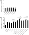
Genetic interactions between drag-1 mutants and sma mutants. Percentages of HSNs having no branches are shown. (a) sma-2(e502), sma-3(wk28), sma-4(e729), sma-6(wk7), and sma-9(wk55) mutants were compared with wild type. (b) drag-1(tk81) and drag-1(tm3773) mutants were compared with sma-1(e30), sma-5(n678), and sma-8(e2111) mutants and with double mutants consisting of drag-1(tk81) or drag-1(tm3773) in combination with individual sma mutations. Significant differences were determined by Fisher’s exact test. ***P < 0.001, **P < 0.01, *P < 0.05. NS not significant. n = 180 for each strain.
Figure 4
Figure 4
Rescue experiments of drag-1 mutants with modified DRAG-1 proteins. (a) Schematic representation of the GFP fusion constructs. The GFP or Venus coding sequence was inserted between amino acid (aa) 395 and 396 of the drag-1 coding region, just prior to the cleavage site of the C-terminal pro-peptide for drag-1p::drag-1::GFP::GPI. The C-terminal GPI-anchor signal (aa 387–408) was deleted from drag-1p::drag-1::GFP::GPI for drag-1p::drag-1::GFP::GPI. The lin-12 transmembrane domain (aa 907–934) (shown in purple) was connected with drag-1p::drag-1::GFP::GPI for drag-1p::drag-1::GFP::lin-12TM. (b) Percentages of HSNs having no branches are shown for the drag-1(tk81) mutants expressing the different rescuing constructs shown in (a). Data for drag-1p::drag-1::Venus::GPI is also shown. Significant differences were determined by Fisher’s exact test. *P < 0.05. NS not significant. n = 180, 107, 105, 91, and 109 for drag-1p::drag-1::Venus::GPI, drag-1p::drag-1::GFP::GPI, drag-1p::drag-1::GFP::∆GPI #1, drag-1p::drag-1::GFP::∆GPI #2, and drag-1p::drag-1::GFP::lin-12TM, respectively. The #1 and #2 refer to two independently isolated transgenic lines.
Figure 5
Figure 5
drag-1 expression. (a) drag-1p::Venus expression. Expression was detected from late embryos to the adult stage in the pharynx, intestine, and hypodermis. White arrows, dotted arrows and white arrowheads correspond to the pharynx, intestine and hypodermis, respectively. Yellow arrowheads and asterisk point to seam cells and developing vulval epithelium which do not express Venus, respectively. Scale bar: 100 μm. (b) Expression of drag-1p::drag-1::Venus::GPI. The drag-1p::drag-1::Venus::GPI plasmid was injected into unc-119(e2498) animals at 150 ng/μl with 30 ng/μl of pBSII KS(–) and 20 ng/μl pDP#MM016B. DIC (upper) and fluorescence (lower) images of an L4 stage animal are shown. Venus expression was detected only in the pharynx. Anterior is to the left. Scale bar: 100 μm. (c) Immunostaining using anti-DRAG-1. L4 to young-adult animals expressing drag-1p::drag-1::Venus::GPI were fixed and stained with anti-DRAG-1. DRAG-1 expression was detected in the pharynx, intestine, hypodermal seam cells, and ventral hypodermal cells (arrows) except the vulval hypodermis (asterisk). Scale bar: 50 μm.
Figure 6
Figure 6
Tissue-specific rescue experiments of drag-1 mutants. Percentages of HSNs having no branches are shown. drag-1(tk81) and drag-1(tm3773) mutants were compared with those transgenic for rol-6p::drag-1, myo-2p::drag-1, elt-2p::drag-1, SCMp::drag-1 and dab-1p::drag-1. Two independently isolated transgenic lines (#1 and #2) were examined for each transgene. Significant differences were determined by Fisher’s exact test relative to the corresponding drag-1 mutants. **P < 0.01, *P < 0.05. NS not significant. n = 180 for each strain.
Figure 7
Figure 7
drag-1 does not enhance unc-40/+with respect to HSN branching defects. Percentages of HSNs having no branches are shown. drag-1(tk81) and drag-1(tm3773) mutants were compared with unc-40(e271)/+heterozygotes and with drag-1(tk81) unc-40(e271)/+and drag-1(tm3773) unc-40(e271)/+double mutants. Significant differences were determined by Fisher’s exact test. **P < 0.01, *P < 0.05. NS not significant. n = 90 for each strain.

Similar articles

Cited by

References

    1. Sainath R, Gallo G. Cytoskeletal and signaling mechanisms of neurite formation. Cell Tissue Res. 2015;359:267–278. doi: 10.1007/s00441-014-1955-0. - DOI - PMC - PubMed
    1. Dent EW, Barnes AM, Tang F, Kalil K. Netrin-1 and semaphorin 3A promote or inhibit cortical axon branching, respectively, by reorganization of the cytoskeleton. J. Neurosci. 2004;24:3002–3012. doi: 10.1523/JNEUROSCI.4963-03.2004. - DOI - PMC - PubMed
    1. Mann F, Zhukareva V, Pimenta A, Levitt P, Bolz J. Membrane-associated molecules guide limbic and nonlimbic thalamocortical projections. J. Neurosci. 1998;18:9409–9419. doi: 10.1523/JNEUROSCI.18-22-09409.1998. - DOI - PMC - PubMed
    1. Yoshida J, Kubo T, Yamashita T. Inhibition of branching and spine maturation by repulsive guidance molecule in cultured cortical neurons. Biochem. Biophys. Res. Commun. 2008;372:725–729. doi: 10.1016/j.bbrc.2008.05.124. - DOI - PubMed
    1. Shibata K, et al. Repulsive guidance molecule a regulates hippocampal mossy fiber branching in vitro. NeuroReport. 2013;24:609–615. doi: 10.1097/WNR.0b013e3283632c08. - DOI - PubMed

Publication types

MeSH terms

Substances