Loss of ZNF677 expression is a predictive biomarker for lymph node metastasis in Middle Eastern Colorectal Cancer
- PMID: 34785764
- PMCID: PMC8595636
- DOI: 10.1038/s41598-021-01869-0
Loss of ZNF677 expression is a predictive biomarker for lymph node metastasis in Middle Eastern Colorectal Cancer
Abstract
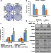
Zinc-finger proteins are transcription factors with a "finger-like" domain that are widely involved in many biological processes. The zinc-finger protein 677 (ZNF677) belongs to the zinc-finger protein family. Previous reports have highlighted the tumor suppressive role of ZNF677 in thyroid and lung cancer. However, its role in colorectal cancer (CRC) has not been explored. ZNF677 protein expression was analyzed by immunohistochemistry in a large cohort of 1158 CRC patients. ZNF677 loss of expression was more frequent in CRC tissues (45.3%, 525/1158), when compared to that of normal tissue (5.1%, 11/214) (p < 0.0001) and was associated with mucinous histology (p = 0.0311), advanced pathological stage (p < 0.0001) and lymph node (LN) metastasis (p = 0.0374). Further analysis showed ZNF677 loss to be significantly enriched in LN metastatic CRC compared to overall cohort (p = 0.0258). More importantly, multivariate logistic regression analysis showed that ZNF677 loss is an independent predictor of LN metastasis in CRC (Odds ratio = 1.41; 95% confidence interval 1.05-1.87; p = 0.0203).The gain- and loss-of-function studies in CRC cell lines demonstrated that loss of ZNF677 protein expression prominently increased cell proliferation, progression of epithelial-mesenchymal transition and conferred chemoresistance, whereas its overexpression reversed the effect. In conclusion, loss of ZNF677 protein expression is common in Middle Eastern CRC and contributes to the prediction of biological aggressiveness of CRC. Therefore, ZNF677 could not only serve as a marker in predicting clinical prognosis in patient with CRC but also as a potential biomarker for personalized targeted therapy.
© 2021. The Author(s).
Conflict of interest statement
The authors declare no competing interests.
Figures


Similar articles
-
Loss of ZNF677 Expression Is an Independent Predictor for Distant Metastasis in Middle Eastern Papillary Thyroid Carcinoma Patients.Int J Mol Sci. 2021 Jul 22;22(15):7833. doi: 10.3390/ijms22157833. Int J Mol Sci. 2021. PMID: 34360599 Free PMC article.
-
Proteomics analysis of differential protein expression identifies heat shock protein 47 as a predictive marker for lymph node metastasis in patients with colorectal cancer.Int J Cancer. 2017 Mar 15;140(6):1425-1435. doi: 10.1002/ijc.30557. Int J Cancer. 2017. PMID: 27925182
-
Serum miR-200c is a novel prognostic and metastasis-predictive biomarker in patients with colorectal cancer.Ann Surg. 2014 Apr;259(4):735-43. doi: 10.1097/SLA.0b013e3182a6909d. Ann Surg. 2014. PMID: 23982750 Free PMC article.
-
Clinicopathological and prognostic implications of polo-like kinase 1 expression in colorectal cancer: A systematic review and meta-analysis.Gene. 2019 Dec 30;721:144097. doi: 10.1016/j.gene.2019.144097. Epub 2019 Sep 4. Gene. 2019. PMID: 31493507
-
Distant Metastasis in Colorectal Cancer Patients-Do We Have New Predicting Clinicopathological and Molecular Biomarkers? A Comprehensive Review.Int J Mol Sci. 2020 Jul 24;21(15):5255. doi: 10.3390/ijms21155255. Int J Mol Sci. 2020. PMID: 32722130 Free PMC article.
Cited by
-
The Roles of Zinc Finger Proteins in Colorectal Cancer.Int J Mol Sci. 2023 Jun 16;24(12):10249. doi: 10.3390/ijms241210249. Int J Mol Sci. 2023. PMID: 37373394 Free PMC article. Review.
-
ZNF677 inhibits oral squamous cell carcinoma growth and tumor stemness by regulating FOXO3a.Hum Cell. 2023 Jul;36(4):1464-1476. doi: 10.1007/s13577-023-00910-w. Epub 2023 May 2. Hum Cell. 2023. PMID: 37129799
-
Overexpression of the pro-protein convertase furin predicts prognosis and promotes papillary thyroid carcinoma progression and metastasis through RAF/MEK signaling.Mol Oncol. 2023 Jul;17(7):1324-1342. doi: 10.1002/1878-0261.13396. Epub 2023 Feb 27. Mol Oncol. 2023. PMID: 36799665 Free PMC article.
References
-
- Alhurry AMAH, et al. A review of the incidence of colorectal cancer in the Middle East. Ann. Colorectal Res. 2017;5:e46292.
MeSH terms
Substances
LinkOut - more resources
Full Text Sources
Medical
Molecular Biology Databases

