. 2021 Nov 16;4(1):1292.
doi: 10.1038/s42003-021-02797-5.

Nipah virus W protein harnesses nuclear 14-3-3 to inhibit NF-κB-induced proinflammatory response

Affiliations

Nipah virus W protein harnesses nuclear 14-3-3 to inhibit NF-κB-induced proinflammatory response

François Enchéry et al. Commun Biol. .

Abstract

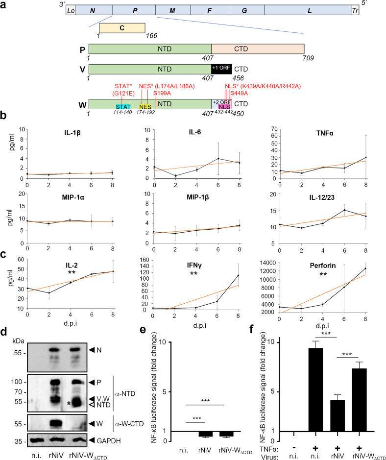
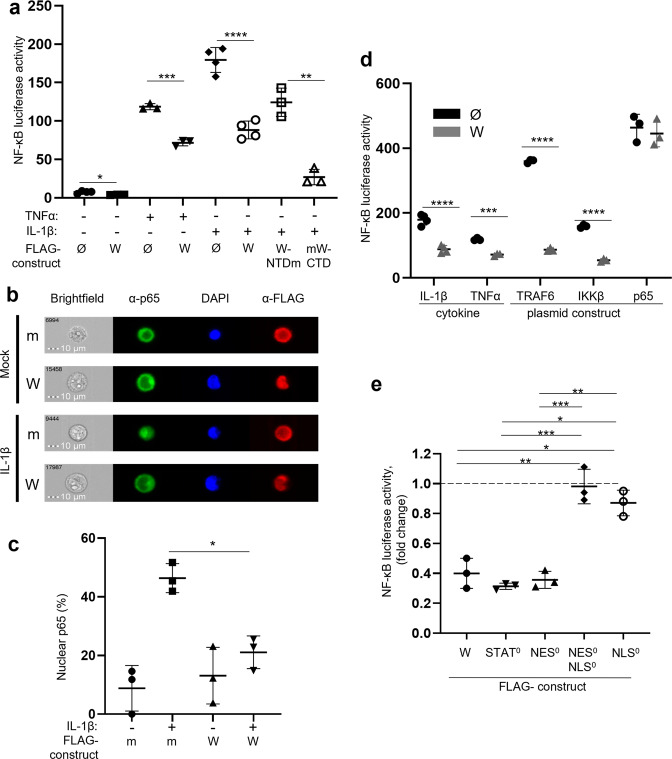
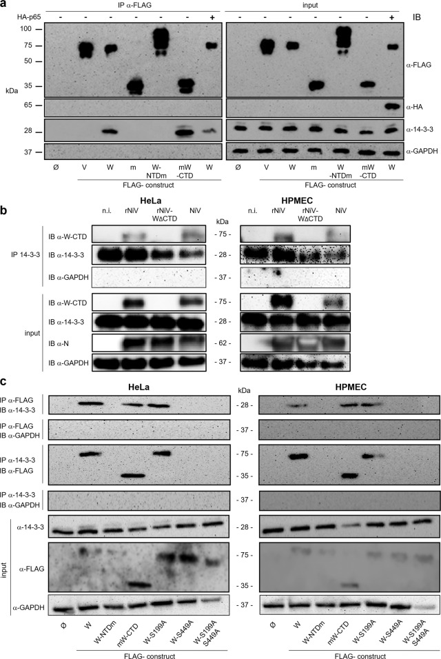
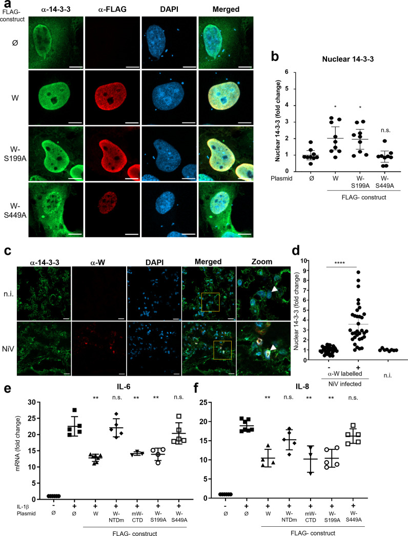
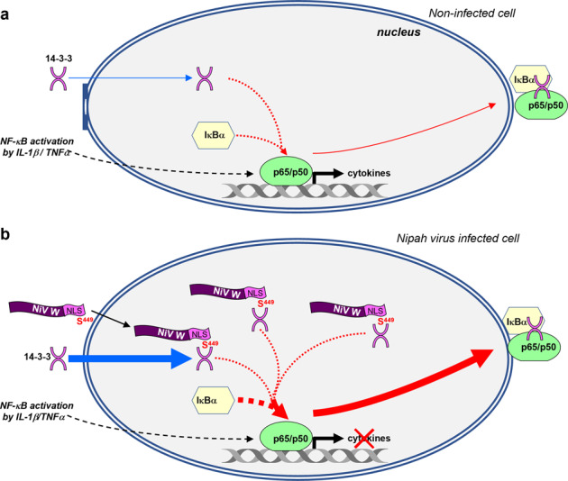
Nipah virus (NiV) is a highly pathogenic emerging bat-borne Henipavirus that has caused numerous outbreaks with public health concerns. It is able to inhibit the host innate immune response. Since the NF-κB pathway plays a crucial role in the innate antiviral response as a major transcriptional regulator of inflammation, we postulated its implication in the still poorly understood NiV immunopathogenesis. We report here that NiV inhibits the canonical NF-κB pathway via its nonstructural W protein. Translocation of the W protein into the nucleus causes nuclear accumulation of the cellular scaffold protein 14-3-3 in both African green monkey and human cells infected by NiV. Excess of 14-3-3 in the nucleus was associated with a reduction of NF-κB p65 subunit phosphorylation and of its nuclear accumulation. Importantly, W-S449A substitution impairs the binding of the W protein to 14-3-3 and the subsequent suppression of NF-κB signaling, thus restoring the production of proinflammatory cytokines. Our data suggest that the W protein increases the steady-state level of 14-3-3 in the nucleus and consequently enhances 14-3-3-mediated negative feedback on the NF-κB pathway. These findings provide a mechanistic model of W-mediated disruption of the host inflammatory response, which could contribute to the high severity of NiV infection.


Conflict of interest statement

The authors declare no competing interests.

Figures

Fig. 1
Fig. 1. W-CTD is necessary for the inhibition of the canonical NF-κB pathway following NiV infection.
a Schematic representation of the NiV genome and the proteins encoded by the P gene: P, C, V, and W proteins. Critical residues of NES, NLS, and STAT-binding sites within the W protein and residue substitutions made in this work are indicated in red. Le, Leader; Tr, Trailer. b, c Plasma from NiV-infected African green monkeys (n = 3) was collected at indicated days post infection (d.p.i.) and analyzed for the presence of NF-κB-controlled cytokines and perforin produced either by a large set of cell types (b) or principally by T and NK cells (c), using the Milliplex NHP Cytokine assay, with measurements for individual animals done in duplicate. Results are expressed as the mean of concentrations, with error bars representing the SD. Linear regressions were performed (red straight lines) to evaluate the statistical significance of the slopes (**p < 0.01). Details of statistical analyses are given in Supplementary Table 1. d HEK293T NF-κB-luc cells were infected with rNiV or rNiV-W∆CTD at an MOI = 3. At 24 h post infection, cells were lysed and the expression of N protein, P gene-encoded proteins (P, V, W, NTD fragment (common to P, V, and W proteins)), was assessed by western blotting using indicated anti-N, anti-NTD, or anti-W-CTD antibodies. V and W proteins migrate with similar apparent molecular mass and thus cannot be distinguished. The NTD truncated protein (open arrow, *) encoded by rNiV-W∆CTD migrates with a lower apparent molecular mass. e HEK293T NF-κB-luc cells were infected or not (n.i.) with rNiV or rNiV-W∆CTD (MOI = 3) and tested at 24 h.p.i. for NF-κB activity by luminescence quantification. Transfection with Renilla luciferase was used for the normalizing of the obtained results. f Same conditions as in e, with or without additional stimulation with 10 ng/mL of TNFα for 4 h prior to luminescence quantification. The data represent the mean values of at least three independent experiments with each point done in triplicate, presented as mean ± SD. ***p ≤ 0.001 using one-way ANOVA completed by a Tukey’s multiple-comparisons test.
Fig. 2
Fig. 2. W inhibits the canonical NF-κB pathway downstream of IKKβ by a mechanism depending on its nuclear import and inhibits nuclear accumulation of p65.
a HeLa cells were transfected with a NF-κB_luc plasmid together with either an empty vector (Ø) or plasmids encoding FLAG-tagged W protein variants. Transfection with Renilla luciferase was used for normalizing the results. Cells were stimulated 20 h after transfection with 10 ng/mL of TNFα or IL-1β for 4 h prior to measurement of NF-κB activity by luminescence quantification. b, c HeLa cells were transfected with FLAG-mCherry (m) or FLAG-tagged W (W) protein encoding plasmid and stimulated or not with 10 ng/mL IL-1β for 20 min. Cells were immunostained for NF-κB p65, FLAG, and DAPI, and the percentage of cells displaying nuclear localization of p65 was determined using ImageStreamX. d Cells were transfected with an empty vector (Ø) or a plasmid encoding the FLAG-tagged W protein and stimulated with either IL-1β or TNFα as detailed for a, or stimulated by transfection with a plasmid encoding either TRAF6, IKKβ, or NF-κB p65. e Cells were transfected with the NF-κB_luc vector together with the indicated plasmids encoding FLAG-tagged W protein or variants of it harboring disabled STAT1/4-binding site, NES, and/or NLS indicated as STAT0, NES0, NES0NLS0, and NLS0, respectively, and stimulated 20 h after transfection with 10 ng/mL of IL-1β. The data were expressed as the fold change of the signal observed with the empty vector (dotted line). NF-κB activity was assessed by luminescence quantification. The data represent mean values of three independent experiments showed as mean ± SD. Pairwise comparisons were carried out using a Student’s t-test and multiple comparisons were performed using one-way ANOVA completed by a Tukey’s multiple comparisons test (*p < 0.05, **p < 0.01 ***p < 0.001, ****p < 0.0001).
Fig. 3
Fig. 3. W protein requires CTD-S499 to interact with 14-3-3 in transfected and infected cells.
HeLa cells (ac) or HPMECs (b, c) were transfected with a plasmid encoding FLAG-tagged V or truncated/variant W proteins, fused or not with mCherry (shortened as m), or HA-p65; an empty vector (Ø) and a plasmid encoding FLAG-mCherry (m) were used as controls. a Coimmunoprecipitation of endogenous 14-3-3 protein or exogenous HA-p65 with FLAG-tag NiV proteins (IP) using anti-FLAG antibodies bound to magnetic beads and detected by western blotting (IB) using rabbit anti-pan 14-3-3 or mouse anti-HA antibodies. b HeLa and HPMECs were mock-infected (n.i.) or infected with rNiV, rNiV-W∆CTD, or wt NiV at MOI = 3. Cells were lysed 24 h later and immunoprecipitation was performed using anti-pan 14-3-3 antibodies coupled to magnetic beads. Input cell extracts and the eluate from beads were analyzed by western blotting using anti-NiV N, anti-W-CTD, anti-14-3-3, and anti-GAPDH antibodies. c Coimmunoprecipitation of endogenous 14-3-3 and FLAG-tagged W and W variant proteins in transfected HeLa and HPMECs was performed using either anti-pan 14-3-3 or anti-FLAG antibodies, and analyzed by western blotting as in a.
Fig. 4
Fig. 4. W-S449 is required for the inhibition of the canonical NF-κB pathway, and the phosphorylation and nuclear accumulation of NF-κB p65.
a HPMECs were cotransfected with a NF-κB_luc plasmid and a plasmid expressing the indicated FLAG-tagged W protein. Transfection with Renilla luciferase was used for the normalization of obtained results. Twenty hours after transfection, cells were stimulated with 10 ng/mL of IL-1β for 20 min and NF-κB activity was assessed by luminescence quantification. The data represent mean values of three independent experiments showed as mean ± SD”. b, HPMECs were transfected with the indicated plasmids. Forty-eight hours after transfection, cells were stimulated with 10 ng/mL of IL-1β for 20 min, and p65 and phosphorylated p65 at Ser536 (p65_Ser536P) were evaluated by western blotting. Data columns represent the mean ± SD of densitometry measurements done on samples from three independent experiments using ImageJ software. c HPMECs were transfected with the indicated plasmids and stimulated with 10 ng/mL of IL-1β for 20 min. After fixation and staining with NF-κB p65 and anti-FLAG antibodies, cells were analyzed by confocal microscopy (scale bar = 10 µm). d Mean fluorescence intensity of nuclear p65 protein. The nuclear signal obtained from anti-FLAG labeled cells was normalized to the nuclear signal from unlabeled cells and expressed as fold change. Error bars represent the confidence interval of the mean (CI 95%) for nine cells combined from three independent experiments. Statistical significance was assessed by one-way unpaired ANOVA completed with a multiple-comparisons Tukey’s test; only significant comparisons are shown (*p < 0.05, **p < 0.01, ***p < 0.001, ****p < 0.0001).
Fig. 5
Fig. 5. Expression of the 14-3-3 inhibitory peptide R18 disables W protein induced inhibition of NF-κB p65 nuclear localization.
a Representative images of HPMEC cells transfected or not (Ø) with plasmids encoding HA-R18 and indicated FLAG-tagged W proteins, and stimulated with 10 ng/mL of IL-1β for 20 min (scale bar = 10 µm). Cells were fixed and stained for NF-κB p65, anti-HA, and anti-FLAG, and analyzed by confocal microscopy. b The mean fluorescence intensity for p65 protein was measured in the cell nucleus. Signal obtained from W expressing cells (as labeled by anti-FLAG) was normalized to the signal from cells not labeled by anti-FLAG and expressed in fold change. Error bars represent the mean’s confidence interval (CI 95%) for nine cells, combined from three independent experiments. Each combination has been done in three different wells performed in two independent experiments. Statistical significance was assessed by a one-way unpaired ANOVA, multiple comparisons Tukey’s test; **p < 0.01 and ****p < 0.0001.
Fig. 6
Fig. 6. W protein induces 14-3-3 nuclear accumulation in transfected and in NiV-infected cells, and suppresses the production of proinflammatory cytokines in vitro.
a HPMECs were transfected with plasmids encoding FLAG-tagged W protein variants. Twenty hours later, cells were stimulated with 10 ng/mL of IL-1β for 20 min, fixed, permeabilized, stained with DAPI and anti-FLAG and anti-14-3-3 antibodies before analysis by confocal microscopy (scale bar = 10 µm). b Mean fluorescence intensity for 14-3-3 protein was measured in a IL-1β stimulated cell nucleus. Signal obtained from anti-W-labeled cells was normalized to the signal from unlabeled cells and expressed in terms of fold change. Error bars represent the confidence interval of the mean (CI 95%) for nine cells, combined from two independent experiments. Statistical significance was assessed by a one-way unpaired ANOVA completed with a multiple-comparisons Tukey’s test; *p < 0.05. c Nuclear accumulation of 14-3-3 in NiV antigen-positive cells from lungs of in vivo-infected AGM with a representative image including an enlarged portion of the merged images. A NiV-infected AGM was killed 10 days post infection and lung sections were labeled using DAPI, anti-W-CTD (red), and pan 14-3-3-specific rabbit polyclonal antibodies (scale bars = 20 µm). d Mean nuclear signal of 14-3-3 in infected cells as labeled with anti-W-CTD antibodies, expressed as the fold change of nuclear signal in non-infected cells as lacking W-CTD labeling within the same field. Error bars represent the confidence interval of the mean (CI 95%) from five different lung sections. Statistical significance was assessed by paired t-test between unlabeled and anti-W-CTD-labeled cells from NiV-infected slices (****p < 0.0001). Mean nuclear 14-3-3 signal in the nucleus of lung cells from a non-infected AGM (n.i.) was comparable to that observed in non-infected cells (i.e., lacking anti-W-CTD labeling) from NiV-infected AGM. e, f HPMECs were transfected with plasmids encoding FLAG-tagged W protein variants and stimulated 20 h later with 10 ng/mL of IL-1β for 4 h. Cells were lysed 24 h later and quantified for their contents in IL-6 and IL-8 mRNA by RT-qPCR. Data columns represent mean values ± SD of at least three independent experiments with each point done in triplicate (**p < 0.01 using ordinary one-way ANOVA followed by a Turkey’s multiple comparisons test).
Fig. 7
Fig. 7. Model of the W-mediated inhibition of the NF-κB pathway.
a IL-1β/TNFα stimulation (black arrows) leads to the entry of NF-κB p65/p50 complex into the cell nucleus and the activation of cytokine expression. The activation is controlled by regulatory proteins 14-3-3 and IκB-α (red arrows), resulting in the nuclear export of 14-3-3/p65/p50-IκBα complex, , . 14-3-3 remains in equilibrium between the cytoplasm and the nucleus. b NiV W protein is imported into the nucleus of the infected cell, mediated by its NLS within its CTD. Via its CTD-S449, the W protein binds to 14-3-3, resulting in an increased influx of 14-3-3 into the nucleus (thicker blue arrow). When the W protein is artificially debilitated by either NLS mutation or S449A substitution, it loses, respectively, its ability to enter into the nucleus, or to interact with nuclear 14-3-3 and, consequently, to inhibit NF-κB activity (not illustrated). Accumulated nuclear 14-3-3 enhances the export of the p65/p50/IκB-α complex from the nucleus (thicker red arrow). The increased translocation rate of NF-κB p65/p50 out of the nucleus prevents its efficient binding onto the promoters of proinflammatory cytokines and results in the inhibition of the host inflammatory response. Only details of the intranuclear part of the NF-κB pathway, which are critical for the comprehension of the action of the W protein revealed in this study, are presented in the figure. Of note, as the molecular mechanism used by 14-3-3 molecules to enter the nucleus (passively or actively) is still unknown, the 14-3-3 nuclear entry trafficking is not indicated by the classical double inverted arrows used to mean (dis)equilibrium. Additional details are described in the Discussion.

References

    1. Hayden MS, Ghosh S. Shared principles in NF-kappaB signaling. Cell. 2008;132:344–362. - PubMed
    1. Zhang Q, Lenardo MJ, Baltimore D. 30 Years of NF-κB: a blossoming of relevance to human pathobiology. Cell. 2017;168:37–57. - PMC - PubMed
    1. Rahman MM, McFadden G. Modulation of NF-κB signalling by microbial pathogens. Nat. Rev. Microbiol. 2011;9:291–306. - PMC - PubMed
    1. Christian, F., Smith, E. L. & Carmody, R. J. The regulation of NF-κB subunits by phosphorylation. Cells5, 12 (2016). - PMC - PubMed
    1. Milanovic M, Kracht M, Schmitz ML. The cytokine-induced conformational switch of nuclear factor κB p65 is mediated by p65 phosphorylation. Biochem. J. 2014;457:401–413. - PubMed

Publication types