

Emodin-Conjugated PEGylation of Fe3O4 Nanoparticles for FI/MRI Dual-Modal Imaging and Therapy in Pancreatic Cancer
- PMID: 34785894
- PMCID: PMC8579871
- DOI: 10.2147/IJN.S335588
Emodin-Conjugated PEGylation of Fe3O4 Nanoparticles for FI/MRI Dual-Modal Imaging and Therapy in Pancreatic Cancer
Erratum in
-
Erratum: Emodin-Conjugated PEGylation of Fe3O4 Nanoparticles for FI/MRI Dual-Modal Imaging and Therapy in Pancreatic Cancer [Corrigendum].Int J Nanomedicine. 2022 Feb 16;17:711-712. doi: 10.2147/IJN.S361728. eCollection 2022. Int J Nanomedicine. 2022. PMID: 35210770 Free PMC article.
Abstract
Background: Pancreatic cancer (PC) remains a difficult tumor to diagnose and treat. It is often diagnosed as advanced by reason of the anatomical structure of the deep retroperitoneal layer of the pancreas, lack of typical symptoms and effective screening methods to detect this malignancy, resulting in a low survival rate. Emodin (EMO) is an economical natural product with effective treatment and few side effects of cancer treatment. Magnetic nanoparticles (MNPs) can achieve multiplexed imaging and targeted therapy by loading a wide range of functional materials such as fluorescent dyes and therapeutic agents.
Purpose: The purpose of this study was to design and evaluate a multifunctional theranostic nanoplatform for PC diagnosis and treatment.
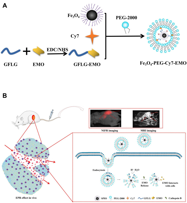
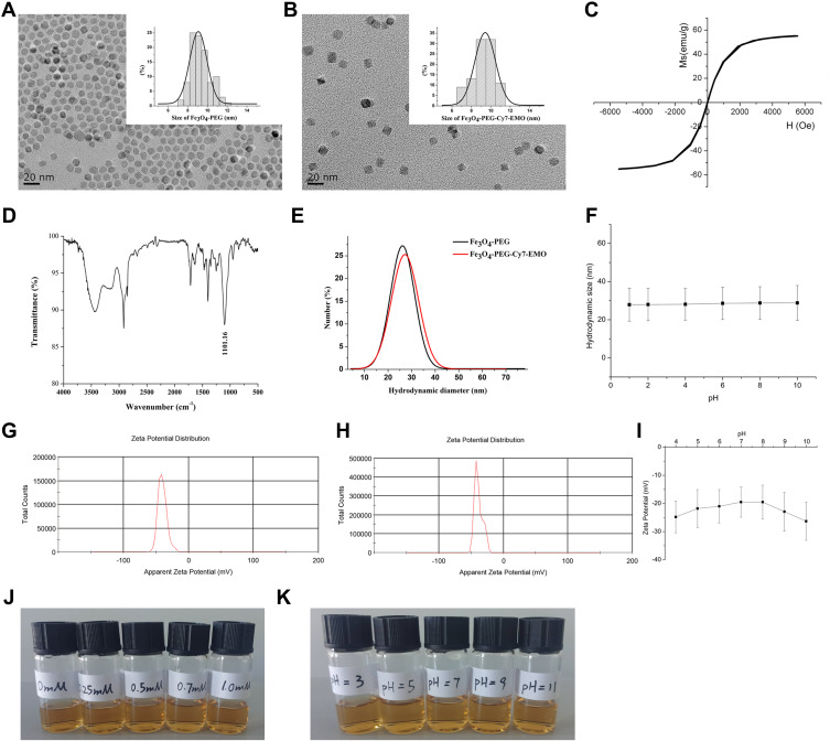
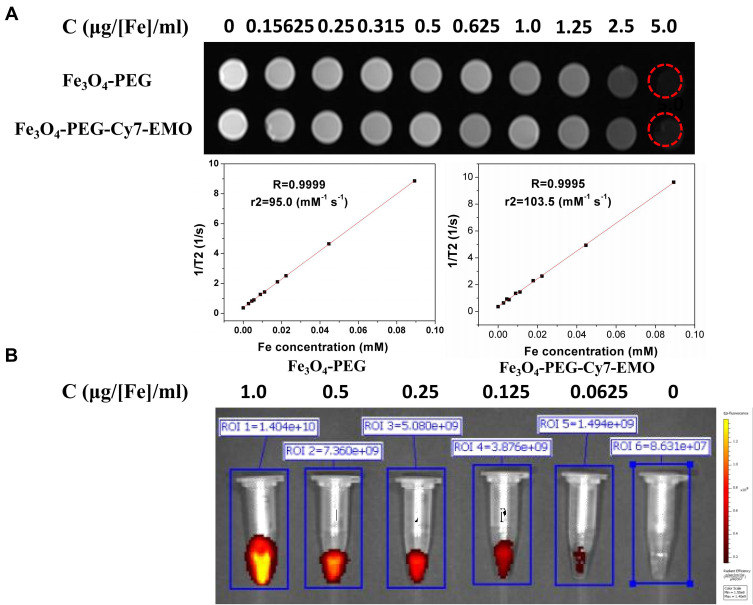
Methods: In this study, we successfully developed EMO-loaded, Cy7-functionalized, PEG-coated Fe3O4 (Fe3O4-PEG-Cy7-EMO). Characteristics including morphology, hydrodynamic size, zeta potentials, stability, and magnetic properties of Fe3O4-PEG-Cy7-EMO were evaluated. Fluorescence imaging (FI)/magnetic resonance imaging (MRI) and therapeutic treatment were examined in vitro and in vivo.
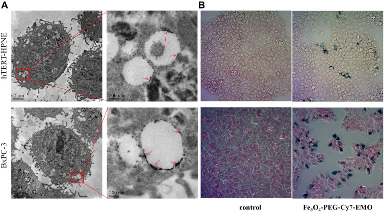
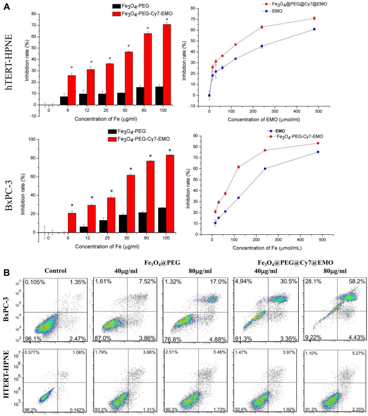
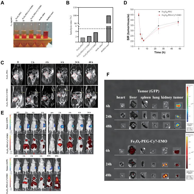
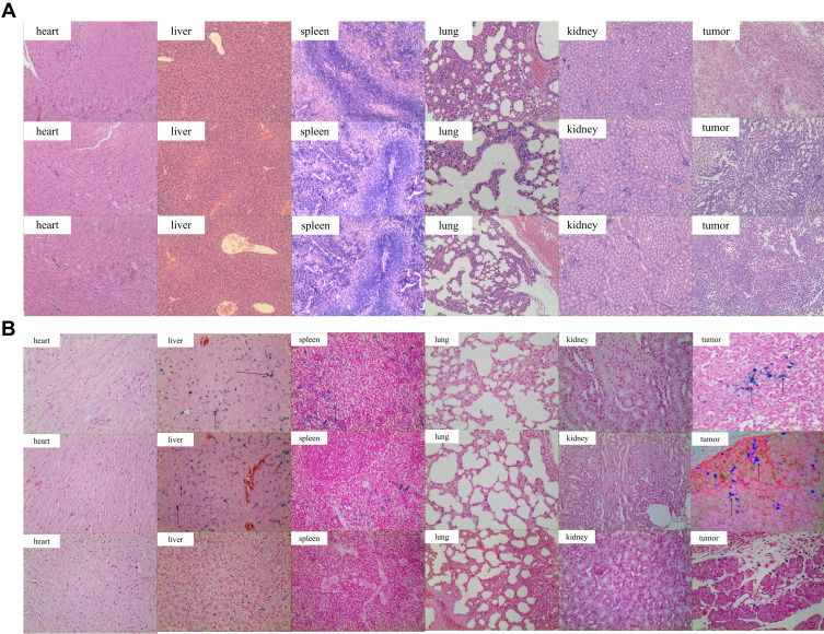
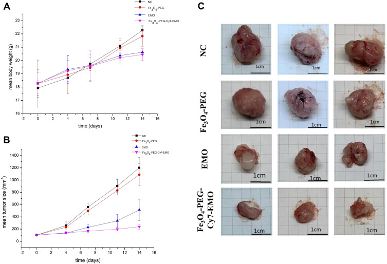
Results: Fe3O4-PEG-Cy7-EMO nanoparticles had a core size of 9.9 ± 1.2 nm, which showed long-time stability and FI/MRI properties. Bio-transmission electron microscopy (bio-TEM) results showed that Fe3O4-PEG-Cy7-EMO nanoparticles were endocytosed into BxPC-3 cells, while few were observed in hTERT-HPNE cells. Prussian blue staining also confirmed that BxPC-3 cells have a stronger phagocytic ability as compared to hTERT-HPNE cells. Additionally, Fe3O4-PEG-Cy7-EMO had a stronger inhibition effect on BxPC-3 cells than Fe3O4-PEG and EMO. The hemolysis experiment proved that Fe3O4-PEG-Cy7-EMO can be used in vivo experiments. In vivo analysis demonstrated that Fe3O4-PEG-Cy7-EMO enabled FI/MRI dual-modal imaging and targeted therapy in pancreatic tumor xenografted mice.
Conclusion: Fe3O4-PEG-Cy7-EMO may serve as a potential theranostic nanoplatform for PC.
Keywords: emodin; magnetic nanoparticles; pancreatic cancer; passive targeting.
© 2021 Ren et al.
Conflict of interest statement
The authors report no conflicts of interest concerning this article.
Figures
Comment in
-
Comments on "Emodin-Conjugated PEGylation of Fe3O4 Nanoparticles for FI/MRI Dual-Modal Imaging and Therapy in Pancreatic Cancer" [Letter].Int J Nanomedicine. 2022 Apr 6;17:1633-1634. doi: 10.2147/IJN.S357154. eCollection 2022. Int J Nanomedicine. 2022. PMID: 35418753 Free PMC article. No abstract available.
Similar articles
-
Oridonin-loaded and GPC1-targeted gold nanoparticles for multimodal imaging and therapy in pancreatic cancer.Int J Nanomedicine. 2018 Oct 24;13:6809-6827. doi: 10.2147/IJN.S177993. eCollection 2018. Int J Nanomedicine. 2018. PMID: 30425490 Free PMC article.
-
Comments on "Emodin-Conjugated PEGylation of Fe3O4 Nanoparticles for FI/MRI Dual-Modal Imaging and Therapy in Pancreatic Cancer" [Letter].Int J Nanomedicine. 2022 Apr 6;17:1633-1634. doi: 10.2147/IJN.S357154. eCollection 2022. Int J Nanomedicine. 2022. PMID: 35418753 Free PMC article. No abstract available.
-
Plectin-1 Targeted Dual-modality Nanoparticles for Pancreatic Cancer Imaging.EBioMedicine. 2018 Apr;30:129-137. doi: 10.1016/j.ebiom.2018.03.008. Epub 2018 Mar 15. EBioMedicine. 2018. PMID: 29574092 Free PMC article.
-
A Gold Nanocage Probe Targeting Survivin for the Diagnosis of Pancreatic Cancer.Pharmaceutics. 2023 May 19;15(5):1547. doi: 10.3390/pharmaceutics15051547. Pharmaceutics. 2023. PMID: 37242788 Free PMC article.
-
Advances in nano drug delivery systems for enhanced efficacy of emodin in cancer therapy.Int J Pharm X. 2024 Dec 25;9:100314. doi: 10.1016/j.ijpx.2024.100314. eCollection 2025 Jun. Int J Pharm X. 2024. PMID: 39834843 Free PMC article. Review.
Cited by
-
Pancreatic Cancer in High-Income Asia-Pacific: A Population-Based Study.Cancer Control. 2025 Jan-Dec;32:10732748251330713. doi: 10.1177/10732748251330713. Epub 2025 Apr 2. Cancer Control. 2025. PMID: 40176285 Free PMC article.
-
An overview of the feasibility of nanomedicine in pancreatic cancer theranostics.Explor Target Antitumor Ther. 2025 Jun 18;6:1002326. doi: 10.37349/etat.2025.1002326. eCollection 2025. Explor Target Antitumor Ther. 2025. PMID: 40547806 Free PMC article. Review.
-
The LAMB3-EGFR signaling pathway mediates synergistic Anti-Cancer effects of berberine and emodin in Pancreatic cancer.Biochem Pharmacol. 2024 Oct;228:116509. doi: 10.1016/j.bcp.2024.116509. Epub 2024 Aug 28. Biochem Pharmacol. 2024. PMID: 39214450
-
Role of emodin to prevent gastrointestinal cancers: recent trends and future prospective.Discov Oncol. 2025 Apr 5;16(1):468. doi: 10.1007/s12672-025-02240-9. Discov Oncol. 2025. PMID: 40186678 Free PMC article. Review.
-
Nanoparticle-mediated delivery of herbal-derived natural products to modulate immunosenescence-induced drug resistance in cancer therapy: a comprehensive review.Front Oncol. 2025 Apr 28;15:1567896. doi: 10.3389/fonc.2025.1567896. eCollection 2025. Front Oncol. 2025. PMID: 40356750 Free PMC article. Review.
References
-
- Kulkarni NM, Mannelli L, Zins M, et al. White paper on pancreatic ductal adenocarcinoma from society of abdominal radiology’s disease-focused panel for pancreatic ductal adenocarcinoma: part II, update on imaging techniques and screening of pancreatic cancer in high-risk individuals. Abdom Radiol. 2020;45(3):729–742. doi:10.1007/s00261-019-02290-y - DOI - PubMed
-
- Kulkarni NM, Soloff EV, Tolat PP, et al. White paper on pancreatic ductal adenocarcinoma from society of abdominal radiology’s disease-focused panel for pancreatic ductal adenocarcinoma: part I, AJCC staging system, NCCN guidelines, and borderline resectable disease. Abdom Radiol. 2020;45(3):716–728. doi:10.1007/s00261-019-02289-5 - DOI - PubMed
MeSH terms
Substances
LinkOut - more resources
Full Text Sources
Medical
