. 2021 Oct 15;13(10):11026-11047.
eCollection 2021.

Decellularized extracellular matrix loaded with IPFP-SC for repairing rabbit osteochondral defects

Affiliations

Decellularized extracellular matrix loaded with IPFP-SC for repairing rabbit osteochondral defects

Lexiang Li et al. Am J Transl Res. .

Abstract

Background: Tissue engineering is widely applied to treat osteochondral damage in osteoarthritis (OA). However, the superposition of seed cells, material scaffolds, inducing factors, and microenvironmental factors limit their practical application. We intended to develop a novel tissue engineering method for improving the repairment of osteochondral damage and to discuss its effect on repairing osteochondral defects.

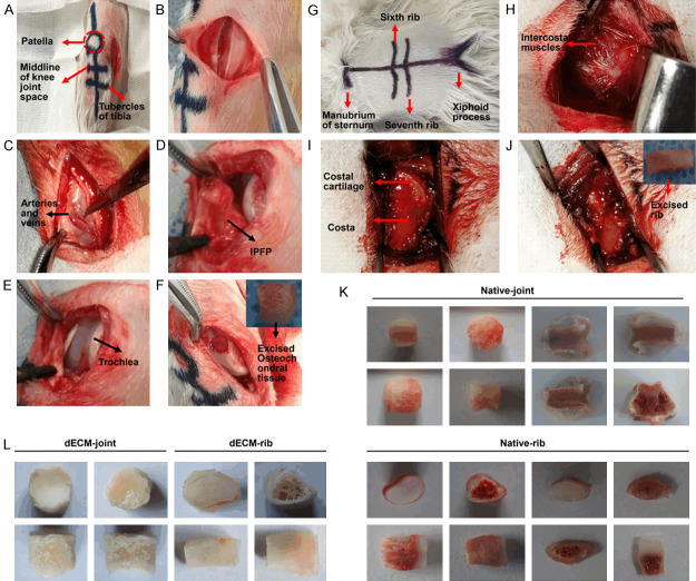
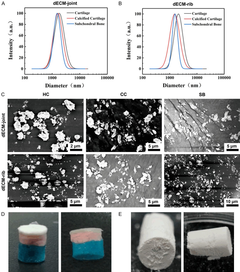
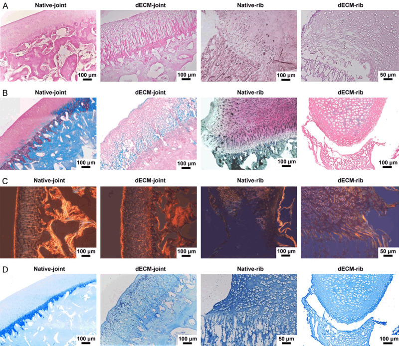
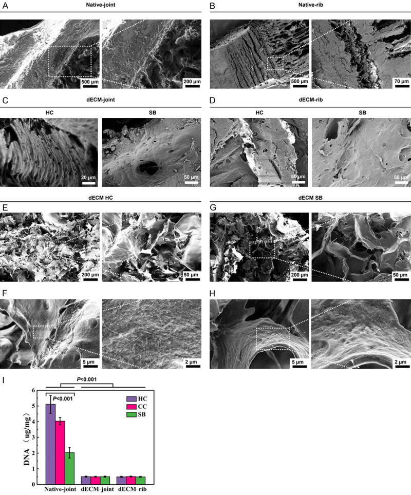
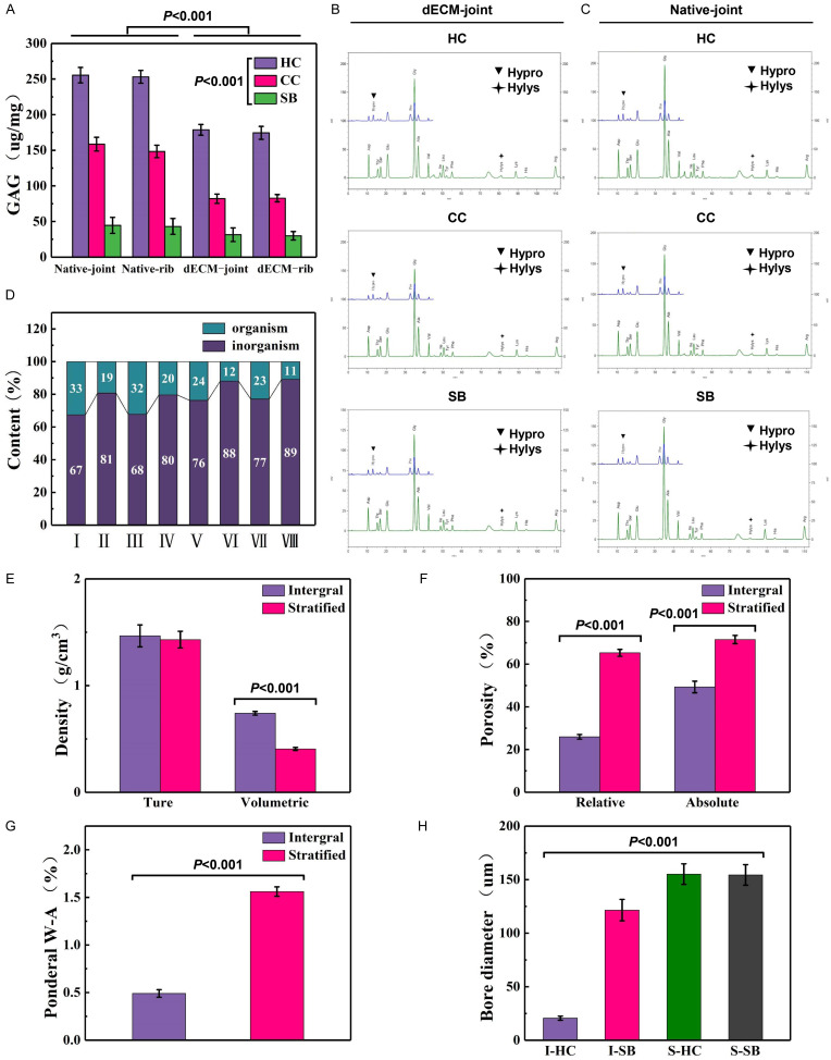
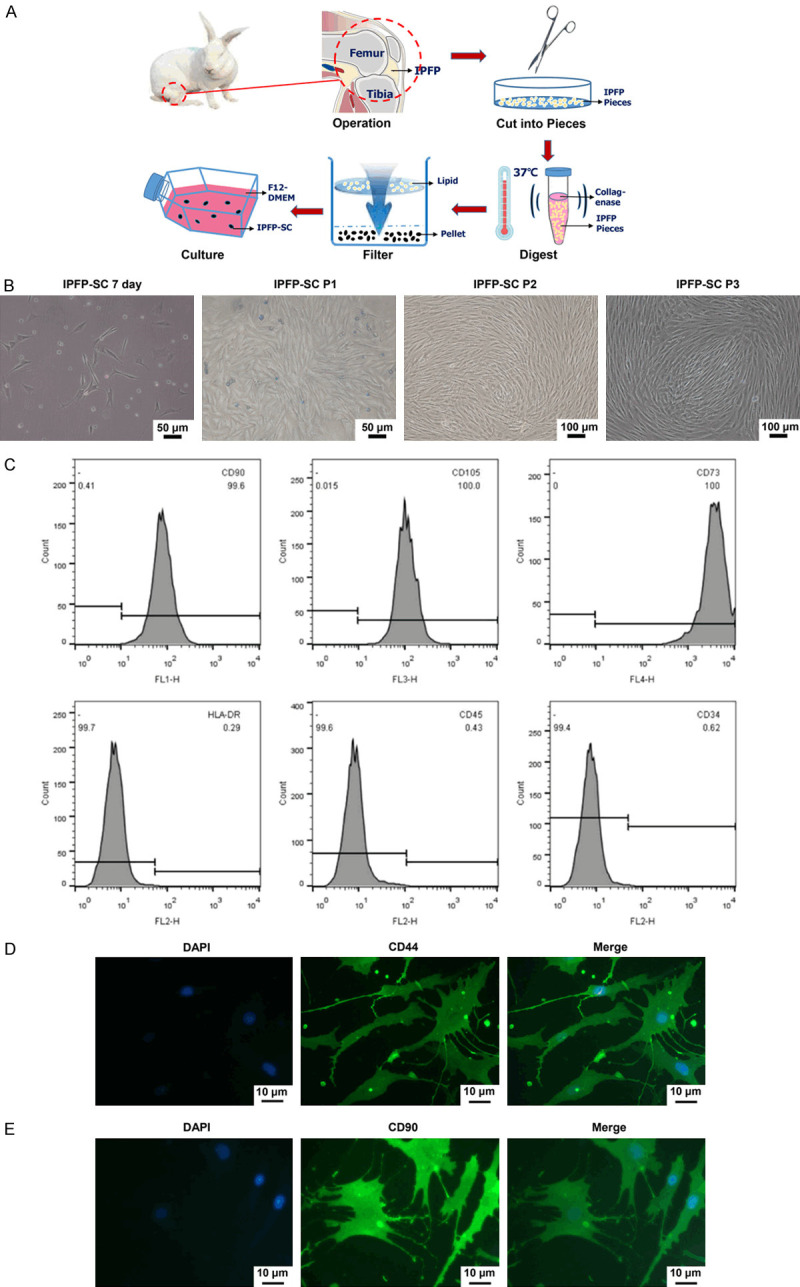
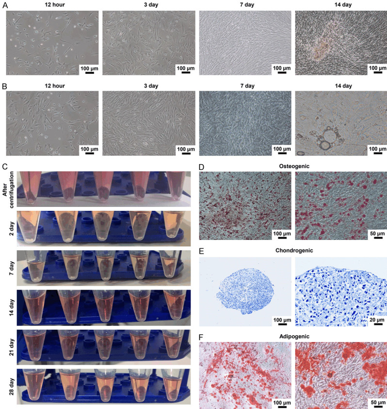
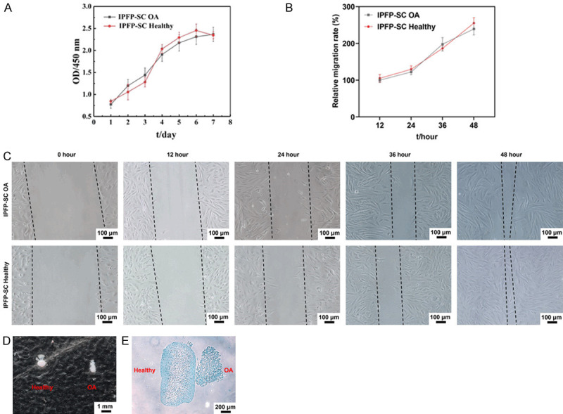
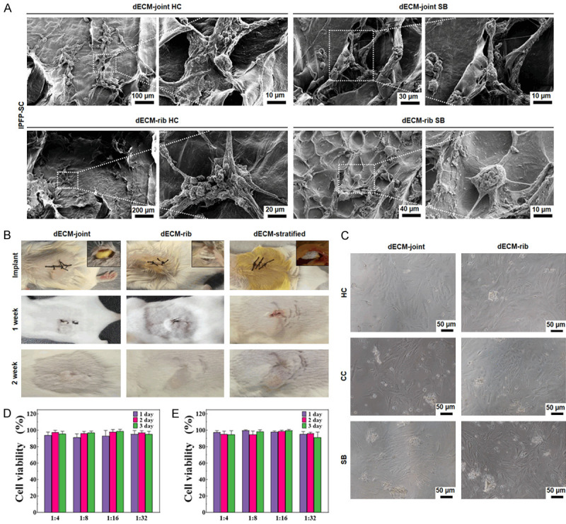
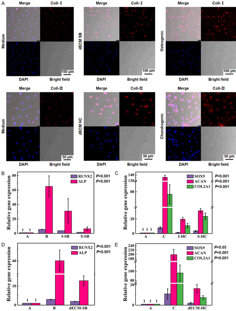
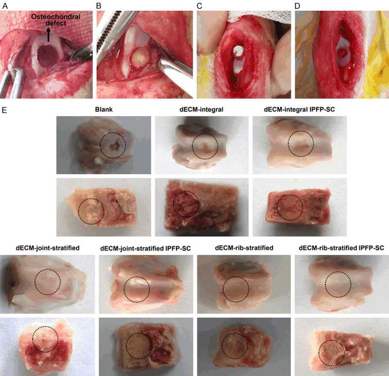
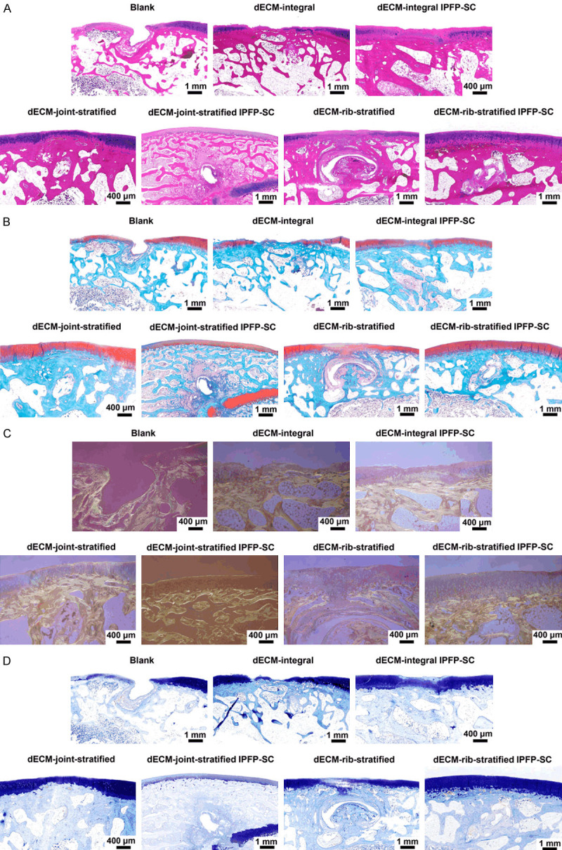
Methods: The combined decellularization methods of physics, chemistry and enzymes were used to decellularize rabbit rib cartilage and articular cartilage, and rabbit decellularizated osteochondral composite scaffolds were prepared. The structure and organization of the scaffolds were analyzed. We extracted and identified infrapatellar fat pad stem cells (IPFP-SCs) from healthy rabbits and OA rabbit, which were different in viability, migration, osteogenic and chondrogenic differentiation. Finally, a variety of decellularizated bone cartilage composite scaffolds were loaded with rabbit IPFP-SC for in vitro and in vivo studies.

Results: The decellularization effect was strong, and the organic ingredients were lost. The layered scaffold showed lower density, greater porosity, larger pore size and water absorption than the whole scaffold, but the mechanical properties of the two scaffolds were low. IPFP-SCs were successfully extracted, and the migration and cartilage ability of IPFP-SCs in OA group were weak. The decellularized scaffold showed a high biocompatibility. The structure and composition of osteochondral promoted osteogenic differentiation and chondrogenic differentiation of IPFP-SCs. Moreover, the decellularized extracellular matrix loaded with IPFP-SC had the strongest repairing effect.

Conclusion: The decellularized extracellular matrix loaded with IPFP-SC showed a better repair effect on rabbit osteochondral defects.

Keywords: Tissue engineering; decellularized extracellular matrix; infrapatellar fat pad stem cells; osteoarthritis; osteochondral defects.


Conflict of interest statement

None.

Figures

Figure 1
Figure 1
The overall removal process and structure diagram of rabbit knee joint osteochondral and rib cartilage. A. Schematic diagram of the internal incision and preparation of the rabbit knee joint. B. The skin and subcutaneous layers were incised. C. Attention was paid to avoid opening and closing the blood vessels and their branches inside the nodular sac. D. Picture of entering the joint capsule. E. The femoral pulley part was exposed. F. The bone cartilage in the middle of the pulley was drilled with a ring drill. G. Schematic diagram of arc-shaped incision and scoring of rabbit ribs. H. The skin and subcutaneous layers were incised to expose the intercostal muscles. I. The intercostal muscles were stripped to expose the rib cartilage. J. The rib cartilage was removed. K. The general view and three-layer structure of natural joints and rib cartilage. L. General view of decellularized joints and rib cartilage.
Figure 2
Figure 2
Fabrication of layered scaffolds for decellularized joints and rib cartilage. A. Size distribution of decellularized particles in rabbit joints. HC: Hyaline cartilage; CC: Calcified cartilage; SB: Subchondral bone. B. The size distribution of rabbit rib decellularized particles. C. SEM scan of decellularized hyaline cartilage, calcified cartilage and subchondral bone particles in rabbit joints and ribs. SEM: scanning electron microscope. D. Schematic model of layered osteochondral scaffold. E. Dry layered osteochondral scaffold.
Figure 3
Figure 3
Histological staining of the whole joint and rib cartilage after decellularization. A. Hematoxylin-eosin (HE) staining image of natural articular cartilage, decellularized articular osteochondral, normal rib cartilage, and decellularized rib cartilage. B. Safranin-fast green staining images of four groups of cartilage. C. Sirius scarlet staining image of four groups of cartilage. D. Toluidine blue staining of cartilage in four groups.
Figure 4
Figure 4
Validation of the decellularizing effect of the whole bone cartilage of joints and ribs and three matrix particles. A, B. SEM images of rabbit natural joints and rib cartilage. C, D. SEM images of rabbit decellularized osteochondral whole scaffold. E, F. The microstructure and ultrastructure of the HC layer of the rabbit decellularized osteochondral layered scaffold. G, H. Microstructure and ultrastructure of SB layer of rabbit decellularized osteochondral layered scaffold. I. The residual DNA content of the three matrix particles. P < 0.001 vs. Native-joint; P < 0.001 vs. HC.
Figure 5
Figure 5
Detection of the components of the decellularized osteochondral matrix of joints and ribs. A. Glycosaminoglycan (GAG) content of decellularized joints and rib cartilage matrix. B, C. The amino acid content of the three-layer structure of HC, CC and SB in decellularized joints and rib cartilage. D. The content of organic and inorganic substances in the two-layer structure of natural and decellularized joints and rib cartilage (I: natural joint CC, II: natural joint SB, III: natural rib CC, IV: natural rib SB, V: decellularized joint CC, VI: decellularized joint SB, VII: decellularized rib CC, VIII: decellularized rib SB). E-H. Physical parameters of rabbit decellularized osteochondral integral scaffolds and layered scaffolds (true density and volume density, effective and absolute porosity, mass water absorption, cartilage and subchondral bone layer pore size).
Figure 6
Figure 6
Isolation and identification of rabbit infrapatellar fat pad stem cells (IPFP-SCs). A. Schematic diagram of rabbit IPFP-SC extracted by collagenase digestion method. B. Morphology of rabbit IPFP-SC. C-E. Flow cytometry and immunofluorescence were used to identify molecular markers on the surface of IPFP-SC.
Figure 7
Figure 7
Osteogenesis, cartilage and adipogenesis of IPFP-SC. A. The morphology of rabbit IPFP-SC osteogenic differentiation. B. Morphology of adipogenic differentiation of rabbit IPFP-SCs. C. Rabbit IPFP-SC induced differentiation into cartilage. D. Alizarin red staining after osteogenic differentiation. E. Toluidine blue staining after chondrogenesis induced differentiation. F. Oil Red O staining image after adipogenesis induced differentiation.
Figure 8
Figure 8
Comparison of cell viability, migration and cartilage ability between OA rabbit and healthy rabbit IPFP-SCs. A. Cell counting kit-8 (CCK-8) was used to detect the viability of IPFP-SCs. B, C. Wound healing experiment to observe cell migration ability. D, E. Toluidine blue staining of healthy rabbits and OA rabbit IPFP-SC after induction into cartilage globules.
Figure 9
Figure 9
Detection of biocompatibility and biotoxicity of rabbit decellularized osteochondral matrix and scaffold. A. SEM scan of rabbit decellularized bone cartilage whole scaffold loaded with IPFP-SC. B. Decellularized joints and rib cartilage scaffolds were implanted subcutaneously in mice. C. Decellularized osteochondral granules were co-cultured with IPFP-SC. D, E. The IPFP-SC cell viability (%) in the extract of the joint and rib decellularized osteochondral scaffold was tested by CCK-8.
Figure 10
Figure 10
In vitro induction of rabbit decellularized osteochondral matrix and scaffold loaded with IPFP-SC. A. Immunofluorescence was used to determine the expressions of Coll-I and Coll-II. B, C. Real-time quantitative PCR (RT-qPCR) was used to determine the relative expressions of osteogenic cartilage genes after IPFP-SC stereo culture induction. A. Normal culture without inducing factors; B. Osteogenic induction liquid induction; C. Chondrogenic induction liquid induction; I-SB: Whole subchondral bone scaffold; S-SB: Layered subchondral bone scaffold; I-HC: Whole hyaline cartilage scaffold; S-HC: Layered hyaline cartilage scaffold. D, E. RT-qPCR was used to determine the relative expressions of osteogenic cartilage genes after co-culture of IPFP-SC decellularized particles. dECM-SB: co-cultured decellularized subchondral bone particles with IPFP-SC; dECM-HC: co-cultured decellularized hyaline cartilage particles with IPFP-SC.
Figure 11
Figure 11
A variety of decellularized osteochondral scaffolds were implanted into the osteochondral defect model and postoperative treatment. A. Fabrication of osteochondral defect in the middle of the pulley. B. Decellularized whole osteochondral scaffold implantation. C. Decellularized layered osteochondral scaffold implantation. D. Decellularized layered osteochondral scaffold loaded with IPFP-SC implantation. E. Observation of gross specimen after repairing osteochondral defect 6 months. dECM: decellularized extracellular matrix.
Figure 12
Figure 12
Histological observation of cartilage defect tissue repair specimens. A. HE staining image. B. Safranin solid green staining image. C. Stained image of Sirius. D. Toluidine blue staining image.

Similar articles

Cited by

References

    1. Nelson AE. Osteoarthritis year in review 2017: clinical. Osteoarthritis Cartilage. 2018;26:319–325. - PMC - PubMed
    1. Lieberthal J, Sambamurthy N, Scanzello CR. Inflammation in joint injury and post-traumatic osteoarthritis. Osteoarthritis Cartilage. 2015;23:1825–1834. - PMC - PubMed
    1. Goldring SR, Goldring MB. Changes in the osteochondral unit during osteoarthritis: structure, function and cartilage-bone crosstalk. Nat Rev Rheumatol. 2016;12:632–644. - PubMed
    1. Mapp PI, Walsh DA. Mechanisms and targets of angiogenesis and nerve growth in osteoarthritis. Nat Rev Rheumatol. 2012;8:390–398. - PubMed
    1. Makris EA, Gomoll AH, Malizos KN, Hu JC, Athanasiou KA. Repair and tissue engineering techniques for articular cartilage. Nat Rev Rheumatol. 2015;11:21–34. - PMC - PubMed

LinkOut - more resources