

The value of contrast-enhanced ultrasound versus shear wave elastography in differentiating benign and malignant superficial lymph node lesions
- PMID: 34786088
- PMCID: PMC8581941
The value of contrast-enhanced ultrasound versus shear wave elastography in differentiating benign and malignant superficial lymph node lesions
Abstract
Objective: To analyze the value of contrast-enhanced ultrasound (CEUS) versus shear wave elastography (SWE) in differentiating benign and malignant superficial lymph node lesions.
Methods: In this retrospective study, a total of 140 superficial lymph nodes from 140 patients pathologically confirmed to have an enlargement of their superficial lymph nodes were examined using CEUS and SWE. The results and diagnostic efficacy were analyzed.
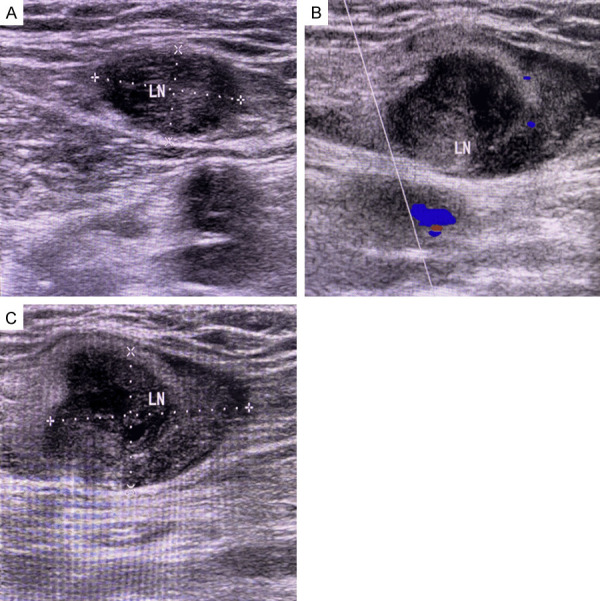
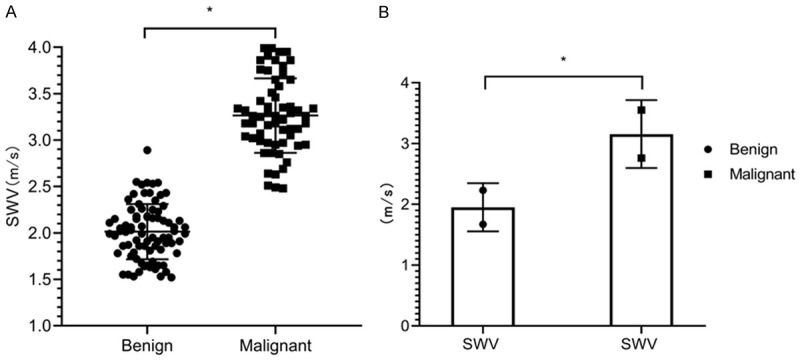
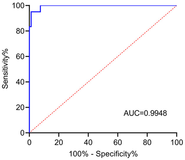
Results: Among the 67 benign lymph nodes, there were 38 cases of type I, 17 of type II, and 12 of types III and IV. Among the 73 malignant lymph nodes, there were 53 cases of type III, 11 of type IV, and 9 of types I and II. Among the patients with lymph nodes <1 cm, there were 20, 4, 8, and 5 cases of types I, II, III, and IV, respectively. Among the patients with 1-2 cm lymph nodes, there were 15, 10, 26 and 7 cases of types I, II, III, and IV, respectively. There were 6, 10, 27, and 2 cases of types I, II, III, and IV in the >2 cm lymph nodes, respectively. The accuracy, sensitivity, and specificity of CEUS in the diagnosis of malignant lymph nodes were 85.00%, 87.67%, and 82.09%, respectively, and those of SWE were 89.29%, 80.82%, and 98.51%, respectively. SWE showed higher specificity than CEUS (P<0.05). SWE showed mean shear wave velocity (SWV) values of (2.11±0.41) m/s for the benign lymph nodes and (3.22±0.79) m/s for the malignant lymph nodes (P<0.05). The receiver operating characteristic (ROC) curves of the SWV values for the benign and malignant lymph nodes showed AUC=0.9948.
Conclusion: Both CEUS and SWE are valuable in the differentiation of benign and malignant lymph node lesions, but SWE has a higher specificity. The SWV value of SWE is superior in the differentiation of benign and malignant lymph nodes. The combination of the two methods can achieve a higher accuracy.
Keywords: Superficial lymph nodes; benign; differentiation; malignant; shear wave elastography; ultrasonography.
AJTR Copyright © 2021.
Conflict of interest statement
None.
Figures
Similar articles
-
Value of Shear Wave Elastography Versus Contrast-Enhanced Sonography for Differentiating Benign and Malignant Superficial Lymphadenopathy Unexplained by Conventional Sonography.J Ultrasound Med. 2017 Jan;36(1):189-199. doi: 10.7863/ultra.16.01014. Epub 2016 Nov 30. J Ultrasound Med. 2017. PMID: 27925679
-
Determining whether the diagnostic value of B-ultrasound combined with contrast-enhanced ultrasound and shear wave elastography in breast mass-like and non-mass-like lesions differs: a diagnostic test.Gland Surg. 2023 Feb 28;12(2):282-296. doi: 10.21037/gs-23-51. Epub 2023 Feb 24. Gland Surg. 2023. PMID: 36915819 Free PMC article.
-
Diagnostic accuracy of contrast-enhanced ultrasound synchronized with shear wave elastography in the differential diagnosis of benign and malignant breast lesions: a diagnostic test.Gland Surg. 2023 Jan 1;12(1):54-66. doi: 10.21037/gs-22-684. Epub 2023 Jan 13. Gland Surg. 2023. PMID: 36761482 Free PMC article.
-
Comparative Diagnostic Accuracy of Contrast-Enhanced Ultrasound and Shear Wave Elastography in Differentiating Benign and Malignant Lesions: A Network Meta-Analysis.Front Oncol. 2019 Mar 5;9:102. doi: 10.3389/fonc.2019.00102. eCollection 2019. Front Oncol. 2019. PMID: 30891425 Free PMC article.
-
Diagnostic Accuracy Evaluation of Two-Dimensional Shear Wave Elastography in the Differentiation Between Benign and Malignant Thyroid Nodules: Systematic Review and Meta-analysis.J Ultrasound Med. 2020 Sep;39(9):1729-1741. doi: 10.1002/jum.15271. Epub 2020 Mar 30. J Ultrasound Med. 2020. PMID: 32227500
Cited by
-
Diagnostic value of high-frame-rate contrast-enhanced ultrasound and contrast vector imaging for superficial lymph node lesions.BMC Cancer. 2025 Apr 27;25(1):785. doi: 10.1186/s12885-025-14190-0. BMC Cancer. 2025. PMID: 40289072 Free PMC article.
-
High-frequency contrast-enhanced ultrasound in discriminating benign and malignant superficial lymph nodes: a diagnostic comparison.BMC Cancer. 2025 May 28;25(1):961. doi: 10.1186/s12885-025-14238-1. BMC Cancer. 2025. PMID: 40437406 Free PMC article.
-
Differential Diagnosis Value of Shear-Wave Elastography for Superficial Enlarged Lymph Nodes.Front Oncol. 2022 Jun 30;12:908085. doi: 10.3389/fonc.2022.908085. eCollection 2022. Front Oncol. 2022. PMID: 35847906 Free PMC article.
References
-
- Null M, Agarwal M. Treasure Island (FL): StatPearls Publishing; 2021. Anatomy, lymphatic system. - PubMed
-
- Bogoslowski A, Kubes P. Lymph nodes: the unrecognized barrier against pathogens. ACS Infect Dis. 2018;4:1158–1161. - PubMed
-
- Yin SS, Cui QL, Fan ZH, Yang W, Yan K. Diagnostic value of arrival time parametric imaging using contrast-enhanced ultrasonography in superficial enlarged lymph nodes. J Ultrasound Med. 2019;38:1287–1298. - PubMed
LinkOut - more resources
Full Text Sources