. 2021 Nov 17:10:e71252.
doi: 10.7554/eLife.71252.

Modulation of pulsatile GnRH dynamics across the ovarian cycle via changes in the network excitability and basal activity of the arcuate kisspeptin network

Affiliations

Modulation of pulsatile GnRH dynamics across the ovarian cycle via changes in the network excitability and basal activity of the arcuate kisspeptin network

Margaritis Voliotis et al. Elife. .

Abstract

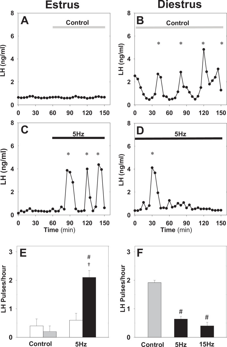
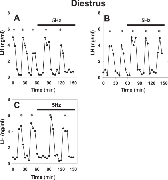
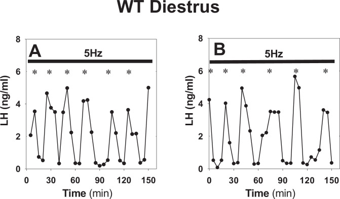
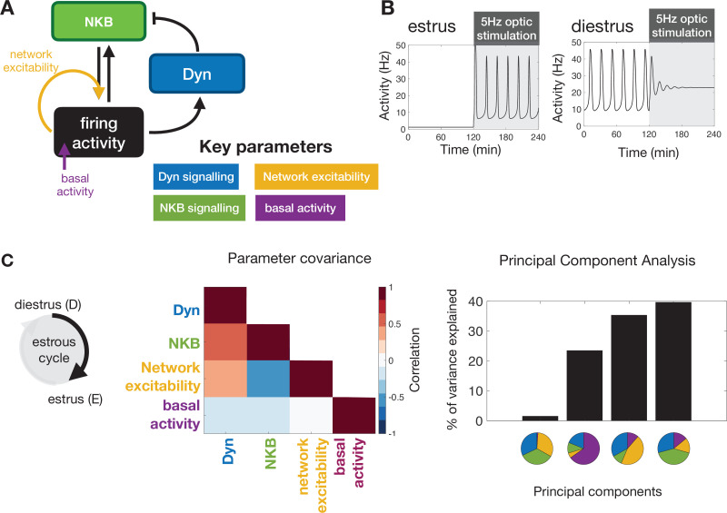
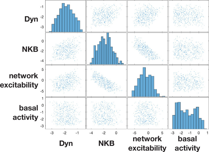
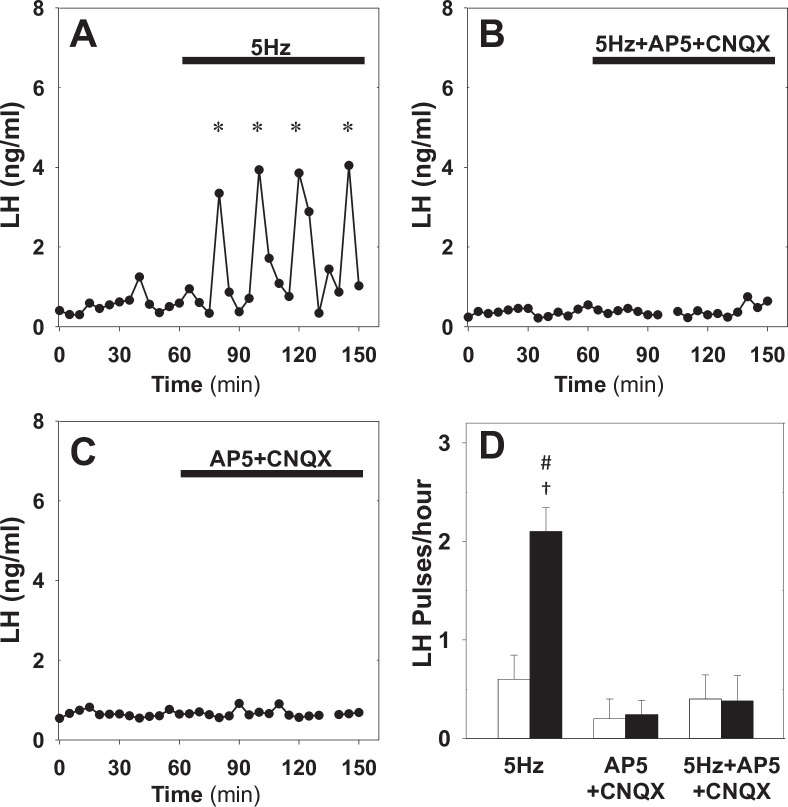
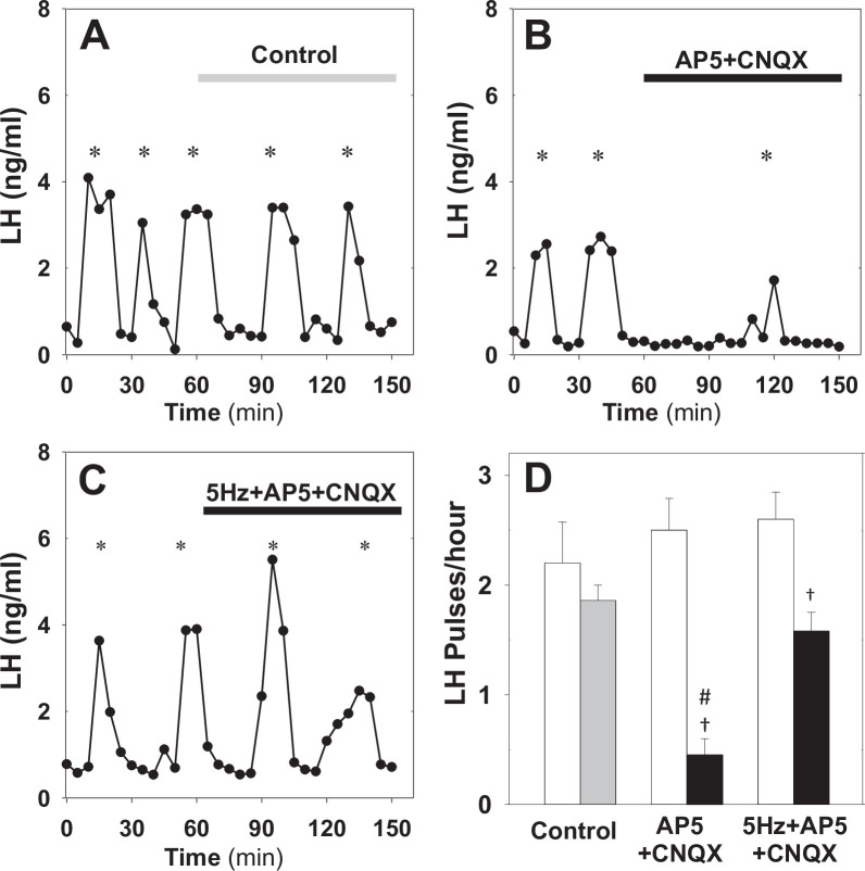
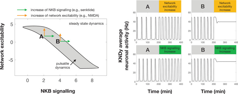
Pulsatile GnRH release is essential for normal reproductive function. Kisspeptin secreting neurons found in the arcuate nucleus, known as KNDy neurons for co-expressing neurokinin B, and dynorphin, drive pulsatile GnRH release. Furthermore, gonadal steroids regulate GnRH pulsatile dynamics across the ovarian cycle by altering KNDy neurons' signalling properties. However, the precise mechanism of regulation remains mostly unknown. To better understand these mechanisms, we start by perturbing the KNDy system at different stages of the estrous cycle using optogenetics. We find that optogenetic stimulation of KNDy neurons stimulates pulsatile GnRH/LH secretion in estrous mice but inhibits it in diestrous mice. These in vivo results in combination with mathematical modelling suggest that the transition between estrus and diestrus is underpinned by well-orchestrated changes in neuropeptide signalling and in the excitability of the KNDy population controlled via glutamate signalling. Guided by model predictions, we show that blocking glutamate signalling in diestrous animals inhibits LH pulses, and that optic stimulation of the KNDy population mitigates this inhibition. In estrous mice, disruption of glutamate signalling inhibits pulses generated via sustained low-frequency optic stimulation of the KNDy population, supporting the idea that the level of network excitability is critical for pulse generation. Our results reconcile previous puzzling findings regarding the estradiol-dependent effect that several neuromodulators have on the GnRH pulse generator dynamics. Therefore, we anticipate our model to be a cornerstone for a more quantitative understanding of the pathways via which gonadal steroids regulate GnRH pulse generator dynamics. Finally, our results could inform useful repurposing of drugs targeting the glutamate system in reproductive therapy.

Keywords: GnRH pulse generator; KNDy; mathematical model; mouse; neuroscience; optogenetics.


Conflict of interest statement

MV, XL, RD, GL, DI, CM, KO, KT No competing interests declared

Figures

Figure 1.
Figure 1.. Expression of arcuate nucleus (ARC) kisspeptin neurones with ChR2-mCherry in Kiss-Cre mouse.
Coronal section showing red mCherry fluorescence positive neurons in the ARC which indicates ChR2 receptor expressing kisspeptin neurones, following unilateral injection of AAV9.EF1.dflox.hChR2(H134R)-mCherry.WPRE.hGH into the ARC of Kiss-Cre mouse. Note the absence of mCherry fluorescence in the other side of ARC. 3 V, Third ventricle.
Figure 2.
Figure 2.. Differential effect of optic stimulation of ARC kisspeptin neurons in estrous and diestrous Kiss-Cre mice.
(A–B) Representative examples showing LH secretion in response to no stimulation (grey bar) or sustained blue light (473 nm, 5 ms pulse width, black bar) activation of kisspeptin neurons at 5 Hz in estrous (C) and diestrous (D) mice. (E) Summary showing mean ± SEM LH pulse frequency over the 60 min control period (white bars) and over the subsequent stimulation period (black bar) in estrous mice. (F) Summary showing mean ± SEM LH pulse frequency in the control (grey bar) and stimulated (black bars) diestrous mice. *Denote LH pulses. #p < 0.05 vs control; p < 0.05 vs pre-stimulation; n = 5–6 per group.
Figure 2—figure supplement 1.
Figure 2—figure supplement 1.. Optogenetic stimulation of ARC kisspeptin neurons in diestrous Kiss-Cre mice using the original protocol.
(A–C) Representative examples showing LH secretion in diestrous Kiss-Cre mice over a 60 min control period followed by a 90 min period of sustained blue light (473 nm, 5 ms pulse width, black bar) activation of kisspeptin neurons at 5 Hz. There is a tendency for LH pulses to slow down during the activation period: (A) 0.05 vs 0.222 pulses/h (slow down); (B), 0.03 vs 0.03 pulses/h (no change); and (C) 0.03 vs 0.222 pulse/h (slow down). To investigate the possibility of an effect in more depth, we revised the protocol by removing the control period and extending stimulating to 150 min (see Figure 1 in main text). *Denote LH pulses.
Figure 2—figure supplement 2.
Figure 2—figure supplement 2.. Sustained optogenetic stimulation in control WT animals.
(A–B) Representative examples showing normal LH secretion in diestrous WT animals under sustained blue light (473 nm, 5 ms pulse width, black bar) activation of kisspeptin neurons at 5 Hz. *Denote LH pulses.
Figure 3.
Figure 3.. Model predictions on the key mechanisms modulating the behaviour of the KNDy pulse generator across the ovarian cycle.
(A) Schematic illustration of the coarse-grained model of the ARC KNDy population. The model comprises three dynamical variables representing the average levels of Dyn and NKB secreted by the population, and its average firing activity. We hypothesise that four key parameters modulate the behaviour of the system across the ovarian cycle: (i) Dyn signalling strength; (ii) NKB signalling strength; (iii) network excitability; and (iv) basal neuronal activity. Estimates for the four parameters in estrus and diestrus are inferred from LH pulse frequency data in estrus and diestrus animals; with or without 5 Hz optic stimulation (Figure 2E&F) (B) System response to low frequency stimulation during estrus and diestrus, using the maximum a-posteriori estimate of the parameter values inferred from the frequency data. (C) Analysis of parameter changes across the cycle. For each of the four parameter (θi;i=1,2,3,4), the diestrus-to-estrus change is defined as the log-ratio between the corresponding parameter values, that is, log10(θestrusi/θdiestrusi). Normalised covariance (correlation) matrix of parameter changes reveals negative correlation between changes in NKB signalling strength and network excitability, and positive correlation between Dyn signalling strength and both NKB signalling. Eigen-parameters are visualised as pie charts. The eigen-parameter explaining the least of the variance in the posterior distribution corresponds to the stiffest parameter combination to which the system is most sensitive.
Figure 3—figure supplement 1.
Figure 3—figure supplement 1.. Posterior distributions of diestrus-to-estrus parameter changes inferred from data.
The behaviour of the model depends upon four parameters: (i) Dyn signalling strength; (ii) NKB signalling strength; (iii) network excitability and (iv) basal neuronal activity. For each of the four parameter (θi;i=1,2,3,4), we quantify the diestrus-to-estrus change as the log-ratio between the corresponding parameters, that is, log10θestrusiθdiestrusi, and we infer them from frequency of LH pulses using Approximate Bayesian Computation based on sequential Monte Carlo (see main text).
Figure 4.
Figure 4.. Effect of NMDA + AMPA receptor antagonists on pulsatile LH secretion in estrus.
Representative examples showing LH secretion in estrous mice in response to optic stimulation (5 Hz blue light, 473 nm, 5 ms pulse width) (A) and optic stimulation combined with the NMDA + AMPA receptor antagonist (bolus ICV injection [12 nmol AP5 +5 nmol CNQX] over 5 min, followed by a continuous infusion [20 nmol AP5 and 10 nmol CNQX] for the remaining 90 min) treatment (B). NMDA + AMPA receptor antagonist alone had no effect (C). (D) Summary showing mean ± SEM LH pulse frequency over the 60 min non-stimulatory period (white bars) and over the subsequent 90 min stimulation period or appropriate non-stimulatory period in presence of. NMDA + AMPA receptor antagonist alone (black bar) in diestrous mice. *Denote LH pulses. p < 0.05 vs pre-stimulation. #p < 0.05 compared to antagonist treatment groups; n = 5–6 per group.
Figure 5.
Figure 5.. Effect of NMDA + AMPA receptor antagonists on pulsatile LH secretion in diestrus.
Representative examples showing pulsatile LH secretion in response to ICV administration of aCSF as control (A), treatment with NMDA + AMPA receptor antagonists (AP5+ CNQX: bolus ICV injection [12 nmol AP5 +5 nmol CNQX] over 5 min, followed by a continuous infusion [20 nmol AP5 and 10 nmol CNQX] for the remaining 90 min) (B) and combined NMDA/AMPA receptor antagonist treatment and sustained optic stimulation (blue light 473 nm, 5 ms pulse width) at 5 Hz (C). (D) Summary showing mean ± SEM LH pulse frequency over the 60 min non-stimulatory period (white bars) and over the subsequent 90 min stimulation period in control mice (grey bar) and mice receiving treatment (black bar). *Denote LH pulses. p < 0.05 vs pre-stimulation. #p < 0.05 compared to 5 Hz stimulation plus antagonist treatment and aCSF control groups; n = 5–6 per group.
Figure 6.
Figure 6.. Differential effect of perturbations on the dynamics of the pulse generator.
Two-parameter (NKB signalling and network excitability) bifurcation diagram showing the region in the parameter space for which the system exhibits pulsatile dynamics (grey area). Two points (denoted by A and B) illustrate how an increase in NKB signalling or network excitability could have a differential effect on the dynamics of the system. For point A an increase in network excitability or NKB signalling could lead to an increase in the frequency and width of pulses. However, for point B a similar increase leads to pulse inhibition and steady state system dynamics. Furthermore, a negative correlation in how NKB signalling and network excitability co-vary (i.e. aligned with the direction of the pulsatile regime) make the system dynamics less sensitive to small perturbations and enable more robust control over the cyle.
Figure 6—figure supplement 1.
Figure 6—figure supplement 1.. Dynamic behaviour of the KNDY system as a function of dynorphin and NKB singalling.
Positive correlation in the regulation of these two parameters (see positive co-regulation arrows) allows robust control over the system’s dynamics, that is, large enough changes (regardless their actual magnitude) will move the system from the quiescent into the pulsatile regime. Negative correlation in the regulation of the two parameters (see negative co-regulation arrows) makes system dynamics more sensitive to the magnitude of the change (arrow length), for example, large changes can fail to trigger LH pulses.

Similar articles

Cited by

References

    1. Arias P, Jarry H, Leonhardt S, Moguilevsky JA, Wuttke W. Estradiol modulates the LH release response to N-methyl-D-aspartate in adult female rats: studies on hypothalamic luteinizing hormone-releasing hormone and neurotransmitter release. Neuroendocrinology. 1993;57:710–715. doi: 10.1159/000126429. - DOI - PubMed
    1. Bonavera JJ, Sahu A, Kalra SP, Kalra PS. The hypothalamic peptides, beta-endorphin, neuropeptide K and interleukin-1 beta, and the opiate morphine, enhance the excitatory amino acid-induced LH release under the influence of gonadal steroids. Journal of Neuroendocrinology. 1994;6:557–564. doi: 10.1111/j.1365-2826.1994.tb00619.x. - DOI - PubMed
    1. Brann DW, Mahesh VB. Excitatory amino acid regulation of gonadotropin secretion: modulation by steroid hormones. The Journal of Steroid Biochemistry and Molecular Biology. 1992;41:847–850. doi: 10.1016/0960-0760(92)90436-m. - DOI - PubMed
    1. Christian CA, Moenter SM. The neurobiology of preovulatory and estradiol-induced gonadotropin-releasing hormone surges. Endocrine Reviews. 2010;31:544–577. doi: 10.1210/er.2009-0023. - DOI - PMC - PubMed
    1. Cravo RM, Margatho LO, Osborne-Lawrence S, Donato J, Jr, Atkin S, Bookout AL, Rovinsky S, Frazão R, Lee CE, Gautron L, Zigman JM, Elias CF. Characterization of Kiss1 neurons using transgenic mouse models. Neuroscience. 2011;173:37–56. doi: 10.1016/j.neuroscience.2010.11.022. - DOI - PMC - PubMed

Publication types