. 2021 Dec 22;6(6):e0074521.
doi: 10.1128/mSphere.00745-21. Epub 2021 Nov 17.

Adaptive Role of Cell Death in Yeast Communities Stressed with Macrolide Antifungals

Affiliations

Adaptive Role of Cell Death in Yeast Communities Stressed with Macrolide Antifungals

N A Kireeva et al. mSphere. .

Abstract

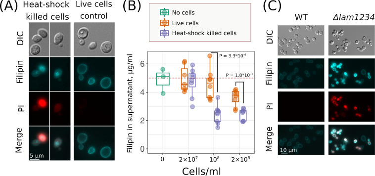
Microorganisms cooperate with each other to protect themselves from environmental stressors. An extreme case of such cooperation is regulated cell death for the benefit of other cells. Dying cells can provide surviving cells with nutrients or induce their stress response by transmitting an alarm signal; however, the role of dead cells in microbial communities is unclear. Here, we searched for types of stressors the protection from which can be achieved by death of a subpopulation of cells. Thus, we compared the survival of Saccharomyces cerevisiae cells upon exposure to various stressors in the presence of additionally supplemented living versus dead cells. We found that dead cells contribute to yeast community resistance against macrolide antifungals (e.g., amphotericin B [AmB] and filipin) to a greater extent than living cells. Dead yeast cells absorbed more macrolide filipin than control cells because they exposed intracellular sterol-rich membranes. We also showed that, upon the addition of lethal concentrations of AmB, supplementation with AmB-sensitive cells but not with AmB-resistant cells enabled the survival of wild-type cells. Together, our data suggest that cell-to-cell heterogeneity in sensitivity to AmB can be an adaptive mechanism helping yeast communities to resist macrolides, which are naturally occurring antifungal agents. IMPORTANCE Eukaryotic microorganisms harbor elements of programmed cell death (PCD) mechanisms that are homologous to the PCD of multicellular metazoa. However, it is still debated whether microbial PCD has an adaptive role or whether the processes of cell death are an aimless operation in self-regulating molecular mechanisms. Here, we demonstrated that dying yeast cells provide an instant benefit for their community by absorbing macrolides, which are bacterium-derived antifungals. Our results illustrate the principle that the death of a microorganism can contribute to the survival of its kin and suggest that early plasma membrane permeabilization improves community-level protection. The latter makes a striking contrast to the manifestations of apoptosis in higher eukaryotes, the process by which plasma membranes maintain integrity.

Keywords: antifungals; bioflocculation; environmental stress; macrolides; programmed cell death; stress response; yeast.


Figures

FIG 1
FIG 1
Scheme of experiment to test how an excess of viable and inviable auxotrophic cells alter the survival of prototrophic cells under various stress conditions. (A) Mixtures of histidine auxotrophic (aux) (his) heat shock-killed cells and histidine prototrophic (HIS+) living cells (i), histidine auxotrophic (his) control cells and histidine prototrophic (HIS+) living cells (ii), and histidine prototrophic living cells without histidine auxotrophic cells (iii) were subjected to various stressors and transferred to an SD-his agar plate. Unstressed control histidine prototrophic (HIS+) cells (iv) were plated before supplementation of stress factors. (B) Representative experiments in which AmB (7 μg/ml) was used as the stressor. In a typical experiment, we assessed 3.6 × 107 cells/ml auxotrophic cells (his) and 4 × 106 cells/ml prototroph cells (HIS+).
FIG 2
FIG 2
Excess of live and dead yeast cells in the suspension alleviates the lethality of some environmental stressors and antifungals. (A) Heatmap indicates relative survival of prototrophic yeast cells supplemented with different amounts of living or heat shock-killed auxotrophic cells. The values were normalized to the survival of prototrophic yeast cells treated with the same stress but not supplemented with auxotrophic (aux) cells. (B) Stressors are ranked according to whether dead or living prototroph cells contribute more or less to the survival of the auxotroph’s suspension. Zero value indicates equal contribution of dead and living cells to the community stress resistance. (C) The panel shows the survival of prototroph yeast cells under the indicated stress conditions without the addition of auxotrophic cells (no aux cells).
FIG 3
FIG 3
Supplementation of dead yeast cells protects yeast suspensions against macrolide antifungals better than supplementation of additional living cells. (A) Protection of prototrophic HIS+ yeast cells against AmB by auxotrophic his cells killed by heat shock of different intensities. The protection provided by heat-shocked his yeast cells to prototrophic yeast cells is proportional to the percentage of inviable auxotrophic yeast cells in the suspension. With 2.2 μg/ml AmB, Kendall’s tau was 0.54 and the P value was 1.77 × 10−13; with 4.4 μg/ml AmB, tau was 0.443 and the P value was 1.76 × 10−7. To perform this experiment, we treated auxotrophic (his or trp) yeast cells with different temperatures (t) (30°C to 70°C), added them to the corresponding prototrophic (HIS+ or TRP+) strain, and then subjected them to AmB for 3 h. (B) Supplementation of heat shock-killed his cells (heat shock-killed aux cells [violet]) but not his living cells (live aux cells [orange]) protected HIS+ prototrophic cells from macrolides AmB, filipin, natamycin, and nystatin.
FIG 4
FIG 4
AmB-killed auxotrophic his yeast cells protect prototroph HIS+ yeast from AmB. Wild-type HIS+ cells (4 × 106 cells/ml) were supplemented into AmB-killed his cells (3.6 × 107 cells/ml) or untreated his cells (3.6 × 107 cells/ml) and treated with AmB. Thereafter, we plated yeast suspensions on SD-his agar and calculated the percentage of HIS+ cells that survived AmB exposure. We calculated P values by the unpaired Mann-Whitney test.
FIG 5
FIG 5
AmB-sensitive Δlam1 Δlam2 Δlam3 Δlam4 cells protect wild-type yeast cells from AmB better than the same amount of control cells. (А) Growth of Δpmp3, Δlam1 Δlam2 Δlam3 Δlam4lam1234), and control cells in the presence of AmB (0.8 μg/ml). (В) Scheme of the experiment. (C) Survival of wild-type HIS+ cells treated with AmB. Wild-type (W303 or BY4741) HIS+ cells (4 × 107 cells/ml) were supplemented with an auxotrophic strain, either Δpmp3 his or control BY4741 his cells (top panel) or either Δlam1 Δlam2 Δlam3 Δlam4lam1234) or W303 control (bottom panel). Concentrations of auxotrophic cells are indicated in the top row of the panel.
FIG 6
FIG 6
Cell mixture of WT and AmB-hypersensitive cells survive AmB better than homogenic WT cells. (A) Scheme of the experiment and figure legend. In all cases, we equalized the final concentration of cells in the testing tubes. Numbers designate the final concentration of cells per milliliter. (B) Average yeast cell survival in the wild-type (W303), Δlam1 Δlam2 Δlam3 Δlam4lam1234), and the wild-type/Δlam1234 mixed suspensions treated with 10 or 20 μg/ml of AmB. In these experiments, we assessed cell survival by calculating the number of CFU in YPD plates after 3 h of AmB treatment (e.g., we did not distinguish the strain of surviving cells from the mixed suspensions). Shaded gray lines connect data points from separate day experiments. P = 0.027 according to paired Wilcoxon signed rank test for a comparison of the wild-type/Δlam1234 mixed suspension with the wild-type suspension.
FIG 7
FIG 7
Overexpression of CTT1 in trp cells provides no increase in AmB resistance for TRP+ cells in the same suspension. (A) Overexpression of CTT1 provides resistance to hydrogen peroxide (3 h of incubation times). To increase CTT1 expression, we incubated the PGAL-CTT1 strain in galactose-containing rich medium (YPGal) overnight. (B) Control trp or PGAL-CTT1 trp cells were supplemented to TRP+ cells. The trp cells were killed in advance with heat shock (dead aux cells) or remained untreated (viable aux cells). Cell mixtures were treated with 7 μg/ml AmB; the incubation time with AmB was 3 h. Otherwise, the experimental design was as described in the legend to Fig. 1 and in Materials and Methods.
FIG 8
FIG 8
Dead yeast cells absorb macrolide filipin with intracellular compartments. (A) Different localization of the filipin signal in heat shock-killed and live control cells. DIC, differential interference contrast; PI, propidium iodide. (B) Heat shock-killed cells absorb more filipin compared to viable control cells. Suspension of yeast cells was supplemented with filipin (5 μg/ml) and then centrifuged. Integral fluorescence spectra in the supernatant were measured. P values were calculated according to the unpaired Mann-Whitney test. (C) Filipin staining induced permeabilization of Δlam1 Δlam2 Δlam3 Δlam4 (lam1234) strain but not the wild-type strain. Yeast cells were treated with filipin (5 μg/ml; incubation time, 5 min).

References

    1. Denyer SP, Maillard JY. 2002. Cellular impermeability and uptake of biocides and antibiotics in Gram-negative bacteria. Symp Ser Soc Appl Microbiol 92:35S–45S. doi:10.1046/j.1365-2672.92.5s1.19.x. - DOI - PubMed
    1. Cannon RD, Lamping E, Holmes AR, Niimi K, Baret PV, Keniya MV, Tanabe K, Niimi M, Goffeau A, Monk BC. 2009. Efflux-mediated antifungal drug resistance. Clin Microbiol Rev 22:291–321. doi:10.1128/CMR.00051-08. - DOI - PMC - PubMed
    1. Bush K, Bradford PA. 2019. Interplay between β-lactamases and new β-lactamase inhibitors. Nat Rev Microbiol 17:295–306. doi:10.1038/s41579-019-0159-8. - DOI - PubMed
    1. Wloch-Salamon DM. 2014. Sociobiology of the budding yeast. J Biosci 39:225–236. doi:10.1007/s12038-013-9344-5. - DOI - PubMed
    1. Rodríguez-Rojas A, Baeder DY, Johnston P, Regoes RR, Rolff J. 2021. Bacteria primed by antimicrobial peptides develop tolerance and persist. PLoS Pathog 17:e1009443. doi:10.1371/journal.ppat.1009443. - DOI - PMC - PubMed

Publication types

MeSH terms

LinkOut - more resources