

Basic ultrasound head-to-toe skills for intensivists in the general and neuro intensive care unit population: consensus and expert recommendations of the European Society of Intensive Care Medicine
- PMID: 34787687
- PMCID: PMC8596353
- DOI: 10.1007/s00134-021-06486-z
Basic ultrasound head-to-toe skills for intensivists in the general and neuro intensive care unit population: consensus and expert recommendations of the European Society of Intensive Care Medicine
Abstract
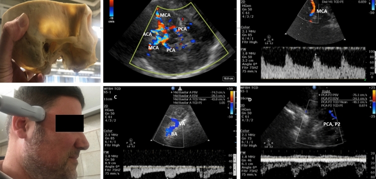
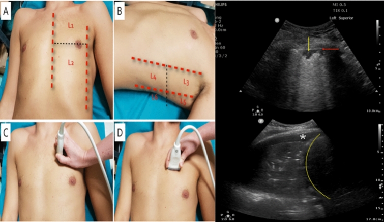
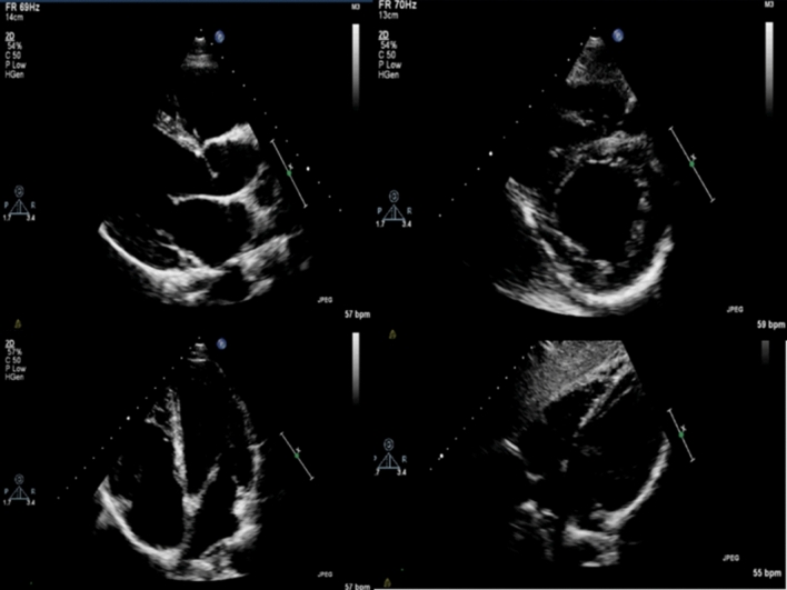
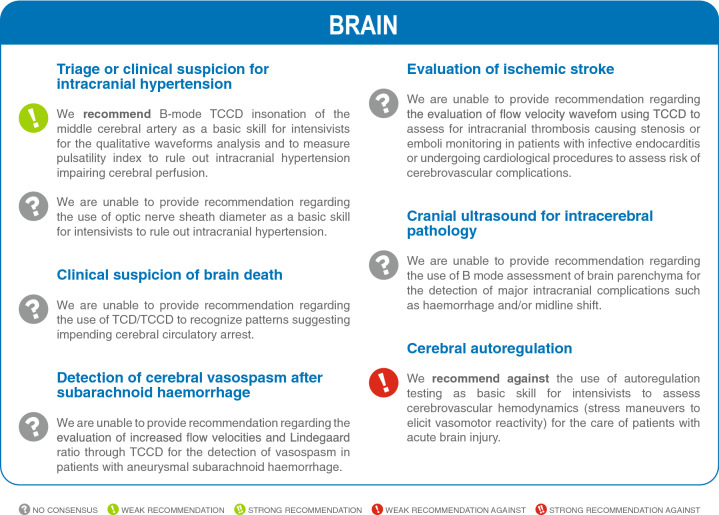
Purpose: To provide consensus, and a list of experts' recommendations regarding the basic skills for head-to-toe ultrasonography in the intensive care setting.
Methods: The Executive Committee of the European Society of Intensive Care (ESICM) commissioned the project and supervised the methodology and structure of the consensus. We selected an international panel of 19 expert clinicians-researchers in intensive care unit (ICU) with expertise in critical care ultrasonography (US), plus a non-voting methodologist. The panel was divided into five subgroups (brain, lung, heart, abdomen and vascular ultrasound) which identified the domains and generated a list of questions to be addressed by the panel. A Delphi process based on an iterative approach was used to obtain the final consensus statements. Statements were classified as a strong recommendation (84% of agreement), weak recommendation (74% of agreement), and no recommendation (less than 74%), in favor or against.
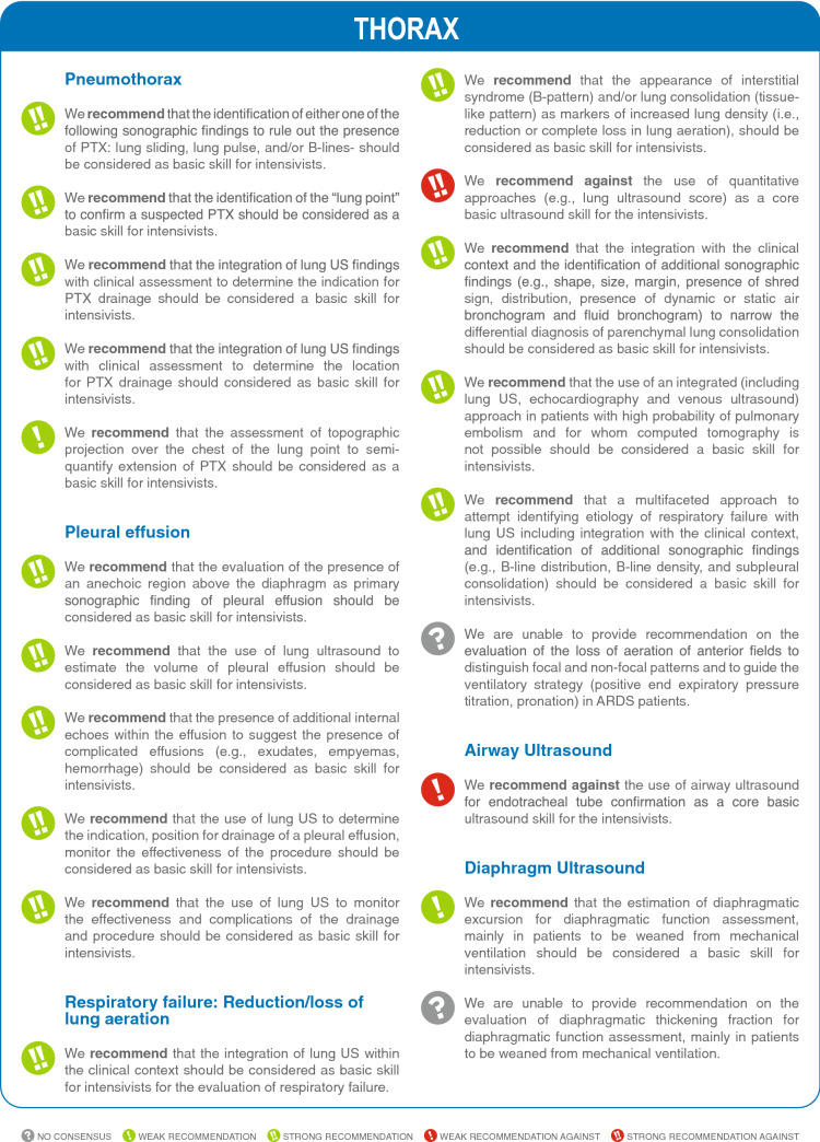
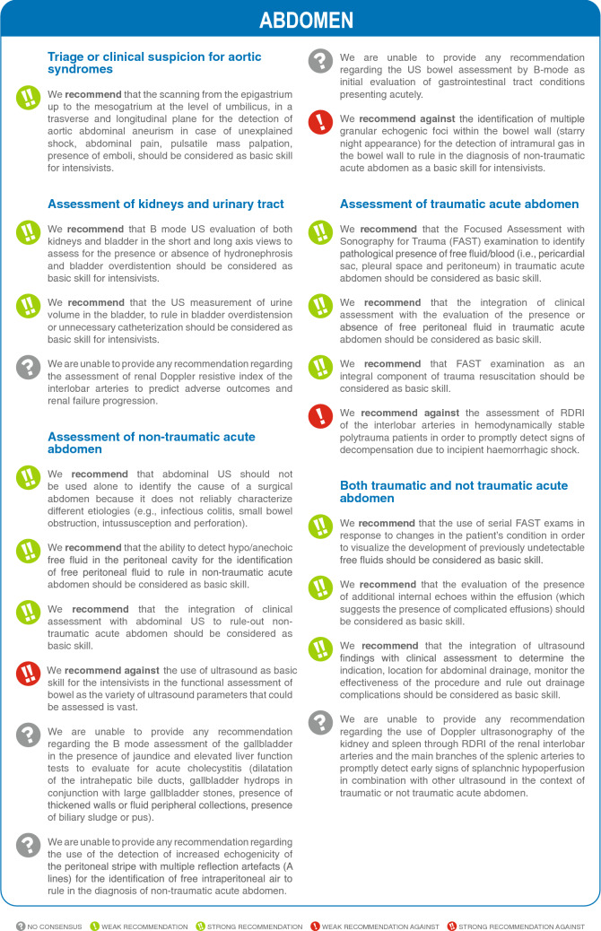
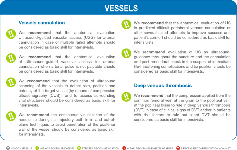
Results: This consensus produced a total of 74 statements (7 for brain, 20 for lung, 20 for heart, 20 for abdomen, 7 for vascular Ultrasound). We obtained strong agreement in favor for 49 statements (66.2%), 8 weak in favor (10.8%), 3 weak against (4.1%), and no consensus in 14 cases (19.9%). In most cases when consensus was not obtained, it was felt that the skills were considered as too advanced. A research agenda and discussion on training programs were implemented from the results of the consensus.
Conclusions: This consensus provides guidance for the basic use of critical care US and paves the way for the development of training and research projects.
Keywords: Abdominal ultrasound; Brain ultrasound; Consensus; Echocardiography; Intensive care unit; Lung ultrasound; Ultrasonography; Vascular ultrasound.
© 2021. The Author(s).
Conflict of interest statement
AW: received honorarium for delivery of educational content from GE, Medasense and Vygon. SM: received fees for lectures from General Electric, outside the present work. ML: received honoraria for lecture from MSD, Edwards, Medtronic and Teleflex. Advisory board for Masimo and Edwards. AM: received travel expenses and registration for meetings, congresses, and courses and lecture fees from Vygon, Edwards and Philips. AS: honorarium and travel/lodging reimbursement for continuing medical education courses and workshops by Society of Critical Care Medicine, Neurocritical care society and American Academy of Neurology. FST: scientific advisor for Nihon Khoden, Eurosets and Neuropics. Received lecture fees for BD and Zoll. AVB: research grant from GSK company. All other authors have no conflicts of interest to declare.
Figures


Comment in
-
Defining basic (lung) ultrasound skills: not so basic after all?Intensive Care Med. 2022 May;48(5):628-629. doi: 10.1007/s00134-022-06666-5. Epub 2022 Mar 30. Intensive Care Med. 2022. PMID: 35355097 No abstract available.
-
Defining basic (lung) ultrasound skills; not so basic after all? Author's reply.Intensive Care Med. 2022 May;48(5):630-631. doi: 10.1007/s00134-022-06682-5. Epub 2022 Mar 31. Intensive Care Med. 2022. PMID: 35357545 No abstract available.
-
Basic ultrasound skill for intensivists: future scope for expansion of the recommendations of the European Society of Intensive Care Medicine.Intensive Care Med. 2022 Jul;48(7):971-972. doi: 10.1007/s00134-022-06717-x. Epub 2022 May 16. Intensive Care Med. 2022. PMID: 35577993 No abstract available.
-
Basic ultrasound skill for intensivists: future scope for expansion of the recommendations of the European Society of Intensive Care Medicine. Author's reply.Intensive Care Med. 2022 Jul;48(7):973-974. doi: 10.1007/s00134-022-06673-6. Epub 2022 May 17. Intensive Care Med. 2022. PMID: 35579688 No abstract available.
