. 2022 Feb;18(2):642-659.
doi: 10.1007/s12015-021-10301-z. Epub 2021 Nov 17.

Application of Human Epineural Conduit Supported with Human Mesenchymal Stem Cells as a Novel Therapy for Enhancement of Nerve Gap Regeneration

Affiliations

Application of Human Epineural Conduit Supported with Human Mesenchymal Stem Cells as a Novel Therapy for Enhancement of Nerve Gap Regeneration

Maria Siemionow et al. Stem Cell Rev Rep. 2022 Feb.

Abstract

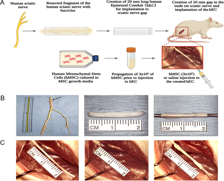
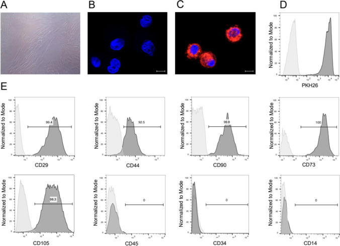
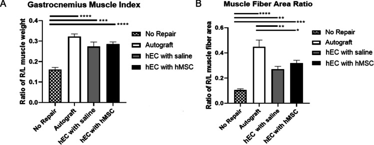
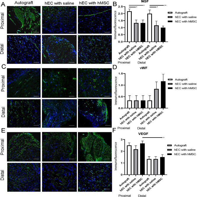
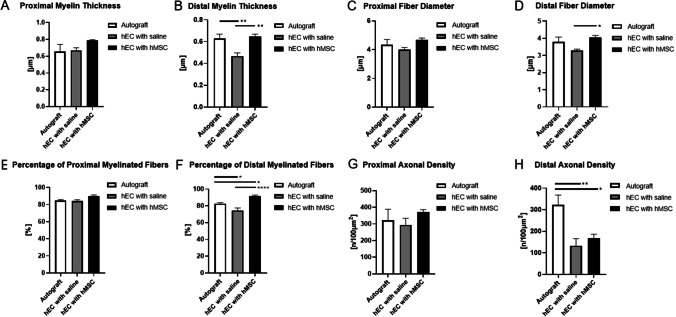
Various therapeutic methods have been suggested to enhance nerve regeneration. In this study, we propose a novel approach for enhancement of nerve gap regeneration by applying human epineural conduit (hEC) supported with human mesenchymal stem cells (hMSC), as an alternative to autograft repair. Restoration of 20 mm sciatic nerve defect with hEC created from human sciatic nerve supported with hMSC was tested in 4 experimental groups (n = 6 each) in the athymic nude rat model (Crl:NIH-Foxn1rnu): 1 - No repair control, 2 - Autograft control, 3 - Matched diameter hEC filled with 1 mL saline, 4 - Matched diameter hEC supported with 3 × 10⁶ hMSC. Assessments included: functional tests: toe-spread and pinprick, regeneration assessment by immunofluorescence staining: HLA-1, HLA-DR, NGF, GFAP, Laminin B, S-100, VEGF, vWF and PKH26 labeling; histomorphometric analysis of myelin thickness, axonal density, fiber diameter and myelinated nerve fibers percentage; Gastrocnemius Muscle Index (GMI) and muscle fiber area ratio. Best sensory and motor function recovery, as well as GMI and muscle fiber area ratio, were observed in the autograft group, and were comparable to the hEC with hMSC group (p = 0.038). Significant improvements of myelin thickness (p = 0.003), fiber diameter (p = 0.0296), and percentage of myelinated fibers (p < 0.0001) were detected in hEC group supported with hMSC compared to hEC with saline controls. At 12-weeks after nerve gap repair, hEC combined with hMSC revealed increased expression of neurotrophic and proangiogenic factors, which corresponded with improvement of function comparable with the autograft control. Application of our novel hEC supported with hMSC provides a potential alternative to the autograft nerve repair.

Keywords: Autograft; Human Epineural conduit; Mesenchymal stem cells; Nerve regeneration; Peripheral nerve repair; Regenerative medicine.


Conflict of interest statement

M.S. is the inventor of the patent application related to Methods of Engineering of Neural Tissue (US/2012/171172A1) and holds a patent on the use of epineural sheath grafts for neural regeneration and protection (WO/2009/124170A1). The authors M.S., M.M.S., K.K., S.B., W.G.K., and J.C. do not have any non-financial conflict of interest.

Figures

Fig. 1
Fig. 1
The outline of experimental study design for creation and application of the human epineural conduit (hEC) as a novel therapy for enhancement of nerve gap regeneration. A Schematic representation of the creation and application of the human epineural conduit (hEC) supported with human mesenchymal stem cells (hMSC). B Creation of the human epineural conduit (hEC) from the sciatic nerve. Human sciatic nerve with branches purchased from the Musculoskeletal Transplant Foundation. The arrow marks the human sciatic nerve (left picture). Epineural sheath after removal of the fascicles during harvesting (middle picture). Empty human epineural conduit, ready for implantation to fill the sciatic nerve gap (right picture). C Implantation of human epineural conduit into the sciatic nerve gap. 20 mm long segment of the rat sciatic nerve before resection (left picture). Creation of a 20 mm gap in the rat sciatic nerve (middle picture). Implantation of hEC into the 20 mm gap followed by injection of either hMSC or saline into the conduit (right picture)
Fig. 2
Fig. 2
Phenotype characterization of the human mesenchymal stem cells (hMSC) for the application as a supportive therapy for human epineural conduit (hEC). A “Fibroblast- like” morphology of hMSC in cell culture after 8 passages. B, C Representative images confirming the efficacy of PKH26 hMSC labelling via confocal microscopy: (B) Unlabeled hMSC, (C) PKH26 labelled hMSC; for merging: Blue-DAPI, Red-PKH26, scale 10 μm; (D) Flow cytometry histogram confirming the efficacy of PKH26 hMSC labelling: unstained hMSC (light grey histogram on the left) superimposed on the PKH26 labeled hMSC (dark grey histogram on the right). E Flow cytometry evaluation of hMSC phenotype. The representative histograms confirm the presence of CD29, CD44, CD90, CD105, CD73 positive cells and lack of expression of hematopoietic markers: CD45, CD34 and CD14
Fig. 3
Fig. 3
Assessment of muscle denervation atrophy by Gastrocnemius Muscle Index (GMI) and muscle fiber area ratio at 12-weeks follow-up after sciatic nerve repair with the hEC. A GMI was significantly lower in the no repair group when compared to the autograft group, the hEC with hMSC group and the hEC with saline group. B Significant differences in the muscle fiber area ratio were revealed between the no repair group when compared to the autograft, the hEC with MSC and the hEC with the saline group. Differences were also statistically significant between the autograft when compared to the hEC with hMSC and the hEC with saline group. The graphs represent mean values with SEM, statistical significance is marked with asterisks: *p < 0.05, **p < 0.01, ***p < 0.001, ****p < 0.0001
Fig. 4
Fig. 4
Functional assessment of nerve regeneration by toe-spread and pinprick tests up to 12-weeks after sciatic nerve repair with hEC. A Improvement of motor function assessed by toe-spread test began at week 6th in both conduit groups and the autograft. Significant difference was observed between the no repair and the autograft as well as the no repair group and the hEC with hMSC groups at 9-weeks. Similar trend was observed at 12-weeks after nerve repair between the no repair group and the autograft group as well as the no repair and the hEC with hMSC group. B Sensory function assessed by a pinprick test revealed significant difference between the no repair group when compared to the autograft and both, the hEC with saline and the hEC with hMSC at 6-weeks. At 9-weeks and 12-weeks follow-up the same trend was observed. Moreover, at 12-weeks after nerve repair, the statistical significance was observed between the autograft and the hEC with saline group. The graphs represent mean values with SEM, statistical significance is marked with asterisks: *p < 0.05, **p < 0.01, ***p < 0.001, ****p < 0.0001 and with crosses ++ p < 0.01
Fig. 5
Fig. 5
Presence of PKH26 labeled cells and expression of HLA-1 and HLA-DR within the proximal and distal end of the conduit assessed by fluorescence and immunofluorescence staining at 12-weeks after nerve repair with the hEC. A, B PKH26 labeled cells were detectable at the proximal and distal ends of the hEC supported with hMSC group. C, D HLA-1 expression was observed only in the hEC supported with hMSC group, confirming stem cell presence in the conduits. E, F The presence of HLA-DR was observed only in the hEC supported with hMSC group. Magnification 200x, scale bar 20 μm. The graphs represent mean values with SEM, statistical significance is marked with asterisks: *p < 0.05, **p < 0.01, ***p < 0.001, ****p < 0.0001
Fig. 6
Fig. 6
Expression of GFAP, S-100 and Laminin B within proximal and distal ends of the conduit in autograft, hEC with saline and hEC with hMSC groups assessed by immunofluorescent staining at 12-weeks after nerve repair with the hEC. A, B GFAP expression was the highest in the autograft group and moderate in the hEC with hMSC group at the proximal end, while in the hEC with saline group expression of GFAP was weak. C, D S-100 expression in the hEC group supported with hMSC was comparable to the autograft control group at both - proximal and distal ends of the conduit. In the hEC group filled with saline, S-100 expression was weak at both – proximal and distal ends of the conduit. (E, F) The highest level of Laminin B expression was detected at the proximal and distal end of the conduit in the hEC with hMSC when compared with the hEC with saline and the autograft group. Magnification 200x, scale bar 20 μm. The graphs represent mean values with SEM, statistical significance is marked with asterisks: *p < 0.05, **p < 0.01, ***p < 0.001, ****p < 0.0001
Fig. 7
Fig. 7
Expression of NGF, vWF and VEGF within proximal and distal ends of the conduit in the autograft, hEC with saline and hEC with hMSC groups assessed by immunofluorescent staining at 12-weeks after nerve repair with hEC. A, B Strong expression of NGF was noted in the autograft group at proximal and distal conduit end. Moderate expression was observed at both ends of the conduit in the hEC supported with hMSC and the hEC with the saline group. C, D Expression of vWF was weak in all groups at the proximal end. At the distal conduit end vWF expression was the highest in the hEC group supported with hMSC. Moderate level of vWF expression was detected in the hEC with saline group, whereas in the autograft group expression of vWF was the weakest. E, F The highest VEGF expression level was revealed in the hEC with hMSC group within proximal and distal conduit end. Magnification 200x, scale bar 20 μm. The graphs represent mean values with SEM, statistical significance is marked with asterisks: *p < 0.05, **p < 0.01, ***p < 0.001, ****p < 0.0001
Fig. 8
Fig. 8
Histological assessment of the proximal and distal conduit ends at 12-weeks after nerve defect repair. A Proximal myelin thickness was increased in the hEC with hMSC group when compared to the hEC with saline and autograft groups. B Distal myelin thickness was significantly increased in the hEC with hMSC group when compared to the hEC with saline group and between the autograft and the hEC with saline group. C The largest proximal fiber diameter was observed in the hEC with hMSC treated group, followed by the autograft and the hEC with saline group. No significant results were observed. D Distal fiber diameter size was significantly greater in the hEC with hMSC when compared to the hEC with saline group. E The highest percentage of myelinated fibers at the proximal end was found in the hEC with hMSC group, followed by the autograft and the hEC with saline group. F The highest percentage of distal myelinated fibers was found in the hEC with hMSC group, followed by the autograft and the hEC with saline group. G The highest proximal axonal density was observed in the hEC with hMSC group, with no significant differences between the hEC with saline and the autograft groups. H The largest distal axonal density was observed in the autograft group, followed by the hEC with hMSC and the hEC with the saline group. The graphs represent mean values with SEM, statistical significance is marked with asterisks: *p < 0.05, **p < 0.01, ***p < 0.001, ****p < 0.0001

Similar articles

Cited by

References

    1. Noble J, Munro C, Prasad V, Midha R. Analysis of upper and lower extremity peripheral nerve injuries in a population of patients with multiple injuries. The Journal of Trauma. 1998;45(1):116–122. - PubMed
    1. Kouyoumdjian JA, Graça CR, FMF V. Peripheral nerve injuries: A retrospective survey of 1124 cases. Neurology India. 2017;65(3):551–555. - PubMed
    1. Daly W, Yao L, Zeugolis D, Windebank A, Pandit A. A biomaterials approach to peripheral nerve regeneration: Bridging the peripheral nerve gap and enhancing functional recovery. Journal of the Royal Society, Interface. 2012;9(67):202–221. - PMC - PubMed
    1. Kelsey, J.L., Praemer, A., Nelson, L.M., Felberg, A., Rice, D.P. (1997). Upper extremity disorders: Frequency, impact and cost. New York, Churchill Livingstone, p.26–42.
    1. Campbell WW. Evaluation and management of peripheral nerve injury. Clinical Neurophysiology. 2008;119(9):1951–1965. - PubMed

Publication types