. 2022 Apr;45(2):753-767.
doi: 10.1007/s10753-021-01581-1. Epub 2021 Nov 17.

Emodin Attenuates LPS-Induced Acute Lung Injury by Inhibiting NLRP3 Inflammasome-Dependent Pyroptosis Signaling Pathway In vitro and In vivo

Affiliations

Emodin Attenuates LPS-Induced Acute Lung Injury by Inhibiting NLRP3 Inflammasome-Dependent Pyroptosis Signaling Pathway In vitro and In vivo

Yuhan Liu et al. Inflammation. 2022 Apr.

Abstract

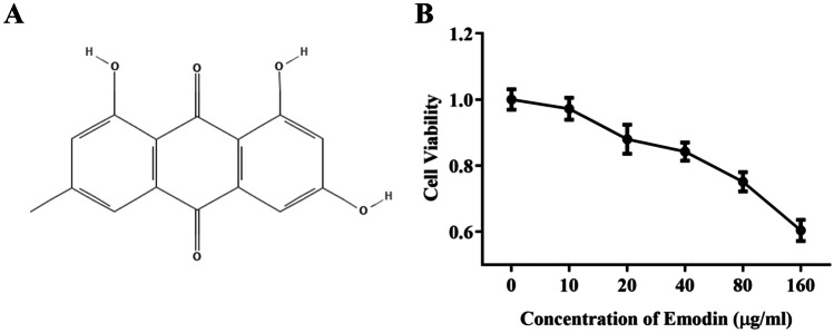
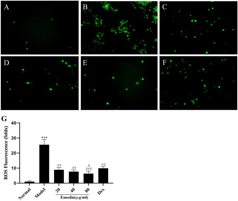
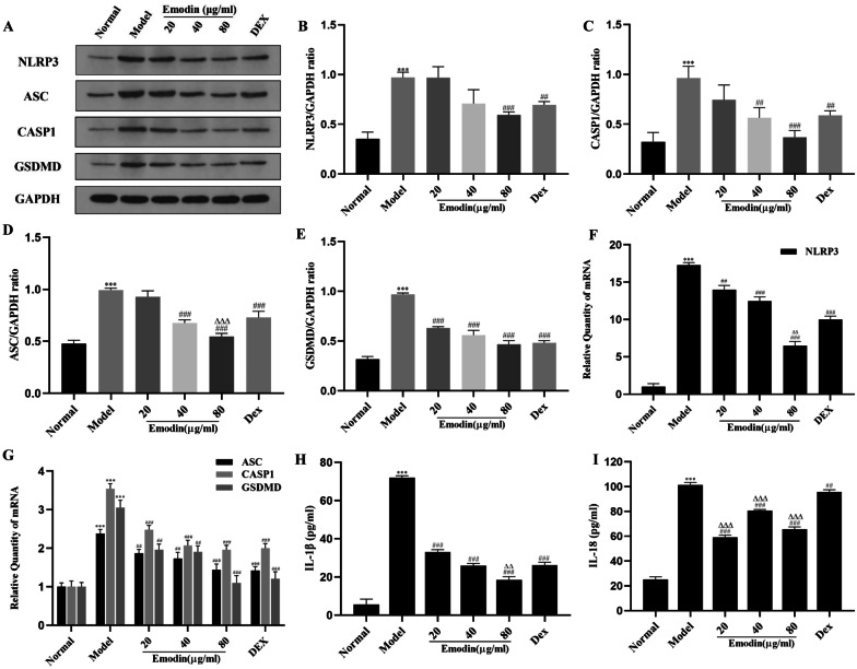
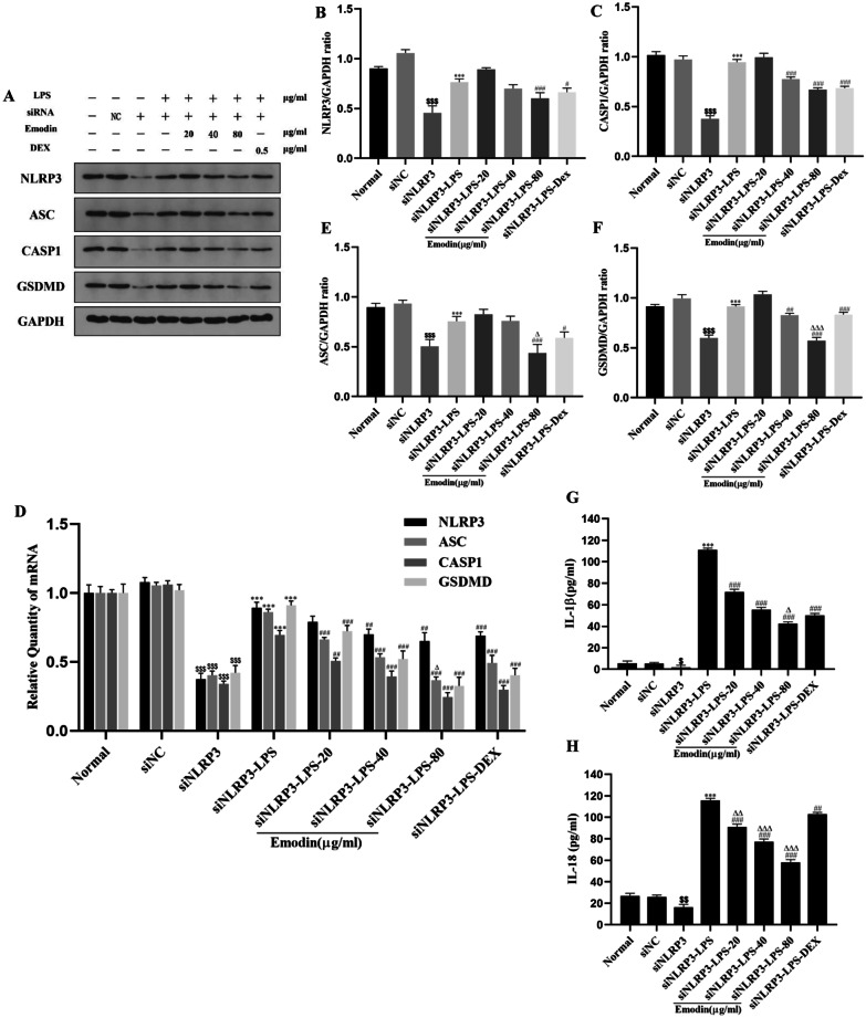
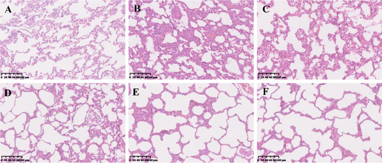
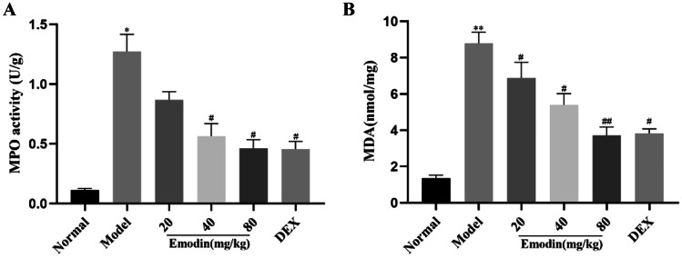
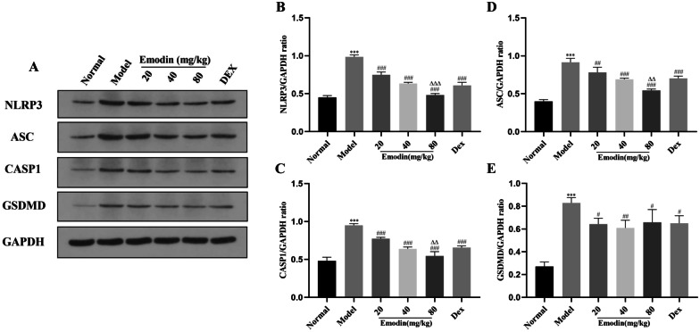
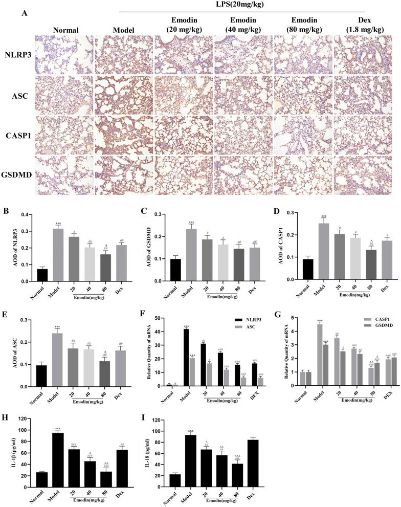
Emodin, the effective component of the traditional Chinese medicine Dahuang, has anti-inflammatory effects. However, the protective effects and potential mechanisms of emodin are not clear. This study investigated the protective effects and potential mechanisms of emodin on lipopolysaccharide (LPS)-induced acute lung injury (ALI) in vitro and in vivo. In vivo, we designed an LPS-induced ALI rat model. In vitro, we chose the J774A.1 cell line to establish an inflammatory cellular model, and knocked down NOD-like receptor family pyrin domain containing 3 (NLRP3) using small interfering RNA. The mRNA and protein expression of NLRP3, a C-terminal caspase recruitment domain (ASC), caspase 1 (CASP1), and gasdermin D (GSDMD) in cells and lung tissues were detected by western blot and real-time quantitative polymerase chain reaction (PCR). The expression levels of interleukin 1 beta (IL-1β) and IL-18 in the serum and supernatant were determined by the enzyme-linked immunosorbent assay. The degree of pathological injury in lung tissue was evaluated by hematoxylin and eosin (H&E) staining. In vitro, we demonstrated that emodin could inhibit NLRP3 and then inhibit the expression of ASC, CASP1, GSDMD, IL-1β, and IL-18. In vivo, we confirmed that emodin had protective effects on LPS-induced ALI and inhibitory effects on NLRP3 inflammasome -dependent pyroptosis. Emodin showed excellent protective effects against LPS-induced ALI by regulating the NLRP3 inflammasome-dependent pyroptosis signaling pathway.

Keywords: LPS; NLRP3 inflammasome; acute lung injury; emodin; pyroptosis.


Conflict of interest statement

The authors declare no competing interests.

Figures

Fig. 1
Fig. 1
A The chemical structure of emodin. B A CCK-8 assay of J774A.1 cell viability after emodin treatment. Values are expressed as the mean ± SD of three individual experiments.
Fig. 2
Fig. 2
Effect of emodin on ROS in J774A.1 cells after LPS stimulation. A Normal group. B Model group. C Emodin (20 μg/mL) group. D Emodin (40 μg/mL) group. E Emodin (80 μg/mL) group. F DEX group. The images were captured using fluorescence microscopy at 400 × magnification. G ROS fluorescence intensities were analyzed using ImageJ and expressed in fold change. Values are expressed as the mean ± SD of three individual experiments. ***P < 0.001 vs. normal group. ##P < 0.01, ###P < 0.001 vs. model group; P < 0.05 vs. DEX group.
Fig. 3
Fig. 3
Effects of emodin on NLRP3 and downstream molecules in LPS-stimulated J774A.1 cells. AE The protein levels of NLRP3, ASC, CASP1, and GSDMD were detected by western blotting, and the grayscale values of the bands were analyzed using ImageJ software. F, G The mRNA levels of NLRP3, CASP1, ASC, and GSDMD were detected by RT-PCR. H, I The expression of IL-1β and IL-18 in the cell supernatant was detected by ELISA. Values are expressed as the mean ± SD of three individual experiments. ***P < 0.001 vs. normal group. #P < 0.05, ##P < 0.01, ###P < 0.001 vs. model group; ∆∆P < 0.01, ∆∆∆P < 0.001 vs. DEX group.
Fig. 4
Fig. 4
Effects of emodin on NLRP3 and downstream molecules in LPS-stimulated J774A.1 cells after NLRP3 knockdown. A, D The mRNA and protein levels of NLRP3, ASC, CASP1, and GSDMD in J774A.1 cells after transfection were detected by RT-PCR and western blotting. B, C, E, F The grayscale values of the bands were analyzed using ImageJ software. G, H The expression of IL-1β and IL-18 in the cell supernatant after transfection was detected by ELISA. Values are expressed as the mean ± SD of three individual experiments. $P < 0.05, $$P < 0.01, $$$P < 0.01 vs. siNC group; ***P < 0.001 vs. siNLRP3 group; #P < 0.05, ##P < 0.01, ###P < 0.001 vs. siNLRP3-LPS group; P < 0.05, ∆∆P < 0.01, ∆∆∆P < 0.001 vs. siNLRP3-DEX group. siNLRP3: NLRP3 was knocked down in J774A.1 cells by siRNA.
Fig. 5
Fig. 5
Pathological changes in the lung tissues were observed by H&E staining (original magnification 200 ×). A Normal group. B Model group. C Emodin (20 mg/kg) group. D Emodin (40 mg/kg) group. E Emodin (80 mg/kg) group. F DEX group. The lung tissues of rats treated with different concentrations of emodin and DEX looked healthier than those of the model rats.
Fig. 6
Fig. 6
Effects of emodin on LPS-induced activity of MPO and MDA in the lung tissues. A MPO activity. B MDA activity in lung tissues. All data are presented as the mean ± SD (n = 8). *P < 0.05, **P < 0.01 vs. normal group. #P < 0.05, ##P < 0.01 vs. model group.
Fig. 7
Fig. 7
Effects of emodin on NLRP3 inflammasome-dependent signaling pathway-associated proteins in a rat model. AE The protein levels of NLRP3, ASC, CASP1, and GSDMD were detected by western blotting, and the grayscale values of the bands were analyzed using ImageJ software. All data are presented as the mean ± SD (n = 8). ***P < 0.001 vs. normal group. #P < 0.05, ##P < 0.01, ###P < 0.001 vs. model group; ∆∆P < 0.01, ∆∆∆P < 0.001 vs. DEX group.
Fig. 8
Fig. 8
Effects of emodin on NLRP3 inflammasome-dependent signaling pathway in rat lung tissues. A Semi-quantitative evaluation of NLRP3, CASP1, ASC, and GSDMD immunohistochemical staining (original magnification 200 ×). BE The average OD was determined by Image-Pro Plus 6.0 software. FG The mRNA levels of NLRP3, CASP1, ASC, and GSDMD in rat lung tissues were determined by RT-PCR. HI Effects of emodin on the expression of IL-1β and IL-18 in rat serum was determined by ELISA. Data are shown as the mean ± SD (n = 8). ***P < 0.001 vs. normal group; #P < 0.05, ##P < 0.01, ###P < 0.001 vs. model group; ΔP < 0.05, ΔΔP < 0.01, ΔΔΔP < 0.001 vs. DEX group.

Similar articles

Cited by

References

    1. Butt Y, Kurdowska A, Allen TC. Acute lung injury: A clinical and molecular review. Archives of pathology & laboratory medicine. 2016;140:345–350. doi: 10.5858/arpa.2015-0519-RA. - DOI - PubMed
    1. Ware LB, Matthay MA. The acute respiratory distress syndrome. The New England journal of medicine. 2000;342:1334–1349. doi: 10.1056/NEJM200005043421806. - DOI - PubMed
    1. Ding J, Wang K, Liu W, She Y, Sun Q, Shi J, et al. Pore-forming activity and structural autoinhibition of the gasdermin family. Nature. 2016;535:111–116. doi: 10.1038/nature18590. - DOI - PubMed
    1. He Y, Hara H, Nunez G. Mechanism and regulation of NLRP3 inflammasome activation. Trends in biochemical sciences. 2016;41:1012–1021. doi: 10.1016/j.tibs.2016.09.002. - DOI - PMC - PubMed
    1. Wang S, Zhao J, Wang H, Liang Y, Yang N, Huang Y. Blockage of P2X7 attenuates acute lung injury in mice by inhibiting NLRP3 inflammasome. International immunopharmacology. 2015;27:38–45. doi: 10.1016/j.intimp.2015.04.035. - DOI - PMC - PubMed

LinkOut - more resources