. 2022 Jan 4;145(1):31-44.
doi: 10.1161/CIRCULATIONAHA.121.056019. Epub 2021 Nov 17.

Retention of the NLRP3 Inflammasome-Primed Neutrophils in the Bone Marrow Is Essential for Myocardial Infarction-Induced Granulopoiesis

Affiliations

Retention of the NLRP3 Inflammasome-Primed Neutrophils in the Bone Marrow Is Essential for Myocardial Infarction-Induced Granulopoiesis

Gopalkrishna Sreejit et al. Circulation. .

Abstract

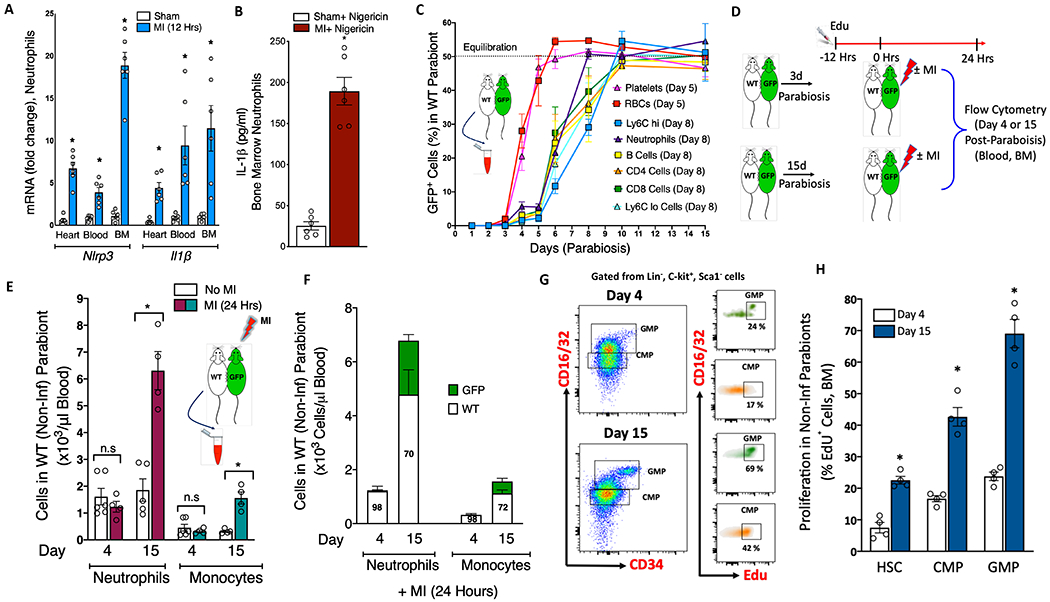
Background: Acute myocardial infarction (MI) results in overzealous production and infiltration of neutrophils to the ischemic heart. This is mediated in part by granulopoiesis induced by the S100A8/A9-NLRP3-IL-1β signaling axis in injury-exposed neutrophils. Despite the transcriptional upregulation of the NLRP3 (Nod Like Receptor Family Pyrin Domain-Containing 3) inflammasome and associated signaling components in neutrophils, the serum levels of IL-1β (interleukin-1β), the effector molecule in granulopoiesis, were not affected by MI, suggesting that IL-1β is not released systemically. We hypothesize that IL-1β is released locally within the bone marrow (BM) by inflammasome-primed and reverse-migrating neutrophils.

Methods: Using a combination of time-dependent parabiosis and flow cytometry techniques, we first characterized the migration patterns of different blood cell types across the parabiotic barrier. We next induced MI in parabiotic mice by permanent ligation of the left anterior descending artery and examined the ability of injury-exposed neutrophils to permeate the parabiotic barrier and induce granulopoiesis in noninfarcted parabionts. Last, using multiple neutrophil adoptive and BM transplant studies, we studied the molecular mechanisms that govern reverse migration and retention of the primed neutrophils, IL-1β secretion, and granulopoiesis. Cardiac function was assessed by echocardiography.

Results: MI promoted greater accumulation of the inflammasome-primed neutrophils in the BM. Introducing a time-dependent parabiotic barrier to the free movement of neutrophils inhibited their ability to stimulate granulopoiesis in the noninfarcted parabionts. Previous priming of the NLRP3 inflammasome is not a prerequisite, but the presence of a functional CXCR4 (C-X-C-motif chemokine receptor 4) on the primed-neutrophils and elevated serum S100A8/A9 levels are necessary for homing and retention of the reverse-migrating neutrophils. In the BM, the primed-neutrophils secrete IL-1β through formation of gasdermin D pores and promote granulopoiesis. Pharmacological and genetic strategies aimed at the inhibition of neutrophil homing or release of IL-1β in the BM markedly suppressed MI-induced granulopoiesis and improved cardiac function.

Conclusions: Our data reveal a new paradigm of how circulatory cells establish a direct communication between organs by delivering signaling molecules (eg, IL-1β) directly at the sites of action rather through systemic release. We suggest that this pathway may exist to limit the off-target effects of systemic IL-1β release.

Keywords: NLRP3 inflammasome; S100A8/A9; granulopoiesis; interleukin-1beta; leukocytosis; myocardial infarction; neutrophils.


Figures

Figure 1.
Figure 1.. Reverse migration of injury exposed-neutrophils is essential to sustain MI-induced granulopoiesis in the BM.
A, Nlrp3 and Il1β mRNA levels in neutrophils sorted from different tissues of sham and MI mice (12 Hrs. post-MI). *P<0.05 compared to the corresponding sham group for each gene (Unpaired t test). n=6/group. MI, Myocardial infarction. B, Quantification of IL-1β released from BM neutrophils following stimulation with nigericin (10 mM, 30 minutes). *P<0.05 compared to the sham group (Unpaired t test). C, Percentage of GFP+ cells in the blood of WT parabionts at different time points following parabiosis. n =6-9/group. Data were collected using multiple pairs of parabionts. The day in the parentheses indicate the earliest day at which the GFP+ cells appearing in WT parabionts were significantly different (P<0.05, Kruskal-Wallis test and Dunn’s multiple comparison) from prior days for each cell type. D, Experimental design depicting the strategy for inducing MI in GFP parabionts at day 3 or day 14 post-parabiosis. EdU was injected 12 hours prior to MI and mice were killed ~ 24 hours post-MI. E, Number of GFP+ cells in WT parabionts at day 4 and day 15 post-parabiosis. MI was induced in GFP mice on day 3 or day 14 post-parabiosis and blood was collected from WT parabionts. F, Number of GFP+ and WT cells in WT parabionts at day 4 and day 15 post-parabiosis and MI. The number in the bars indicate the percentage of WT and GFP+ cells in WT parabionts (non-infarcted). Statistical tests for (E) and (F): * P<0.05 compared to the corresponding no-MI group for each cell type (Mann-Whitney test), n.s, not significant. n =4-6/group. Flow cytometric gating (G) and quantification (H) of proliferation (EdU+) of hematopoietic stem and progenitor cells in the BM of non-infarcted parabionts at day 4 and day 15 post-parabiosis and MI. HSC, Hematopoietic stem cells; CMP, Common myeloid progenitors and GMP, Granulocyte macrophage progenitors. All cells were gated after eliminating debris, dead and clustered cells. CMP and GMPs were gated from myeloid progenitor cells (Lin, ckit+, Sca1) while HSCs (CD48, CD150+) were gated from LSK (Lin, ckit, Sca1+). n =4/ group * P<0.05 compared to day 4 for each corresponding cell type (unpaired t test). All data are means ± SEM.
Figure 2.
Figure 2.. S100A8/A9 but not inflammasome priming is required for retention of neutrophils in the BM.
A, Experimental design depicting the strategy for adoptive transfer (AT) of neutrophils (donor) isolated from WT sham, WT+ MI and S100a9−/− + MI mice (24 hours post-MI) into WT mice. Purified neutrophils (~99.4 %) from the blood and hearts of donors were pooled, labeled with CMFDA and injected in equal numbers to WT recipients. Blood from the recipients was collected at 3 hours and at termination (~16 hours post-AT). B, A pie chart representing spatial distribution of AT neutrophils in different organs of the recipients and their (C) quantification. The numbers in the parentheses indicate the percent of total donor cells remaining in recipients at 16 hours after AT. D, Endogenous neutrophil (non-AT) number in the blood of recipients following AT. F, Quantification of IL-1β in the BM extracellular fluid (consolidated from 2 femurs and 2 tibias/ mouse) in recipients. Statistical tests for figures (C to E): *P<0.05 compared to all groups, # vs. WT+ MI > WT group (1-way ANOVA and Holm-Sidak’s post-hoc test). n=5 per group. F, Experimental plan for AT of neutrophils from WT+ MI and Nlrp3−/− + MI mice to WT mice. Purified neutrophils from the blood and hearts of donors (24 hours post-MI) were pooled, labeled with CMFDA and injected in equal numbers to WT recipients. The recipients were euthanized at ~ 16 hours after AT. G, Flow cytometry plots showing accumulation of donor neutrophils and their (H) quantification in the BM of recipients. The number in the parentheses represent the total number of neutrophils or CMFDA neutrophils as a percent of total neutrophils in the BM. n=5 per group, n.s, not-significant (unpaired t test). Flow cytometric gating (I) and quantification (J) of proliferation (EdU+) of hematopoietic stem and progenitor cells in the BM of recipients. CMP and GMPs were gated from myeloid progenitor cells (Lin, ckit+, Sca1) while HSCs (CD48, CD150+) were gated from LSK (Lin, ckit, Sca1+). n =5/group * P<0.05 compared to corresponding groups that received neutrophils from WT+MI mice (unpaired t test). K, mRNA expression of various adhesion molecules in BM sinusoidal endothelial cells (BMSECs) treated with recombinant S100A8/A9 or TNF-α for 6 hours. P<0.05 compared to vehicle treated group for each corresponding gene (1-way ANOVA and Holm-Sidak’s post-hoc test). n =4/group. L, Experimental design for adhesion assay. BMSECs were cultured to confluence and stimulated with either rS100A8/A9 or TNF-α for 6 hours. Purified BM neutrophils from GFP mice were primed with LPS for 3 hours, washed off excess LPS, counted and overlayed on BMCES in equal numbers under static conditions. After 2 hours, non-adherent neutrophils were removed and counted using a cellometer. The adherent cells were fixed, stained (CD31) and visualized (M) under a fluorescent microscope. N, Number of neutrophils adherent to BMSECs were determined by subtracting the number of non-adherent cells from the total number of neutrophils added. *P<0.05 compared to untreated and unstimulated group (1-way ANOVA and Holm-Sidak’s post-hoc test). n =3. The characters on bars represent the corresponding images in figure M. All data are means ± SEM.
Figure 3.
Figure 3.. Inflammasome-primed neutrophils require a functional CXCR4 to induce granulopoiesis after MI.
A, Experimental design depicting the strategy for inhibition of CXCR4 using AMD3100 followed by adoptive transfer (AT) of CD45.2 WT neutrophils to CD45.1 WT mice. Blood and heart neutrophils from MI mice (24 Hrs. post-MI) were incubated with AMD3100 (CXCR4 antagonist) or vehicle, labelled with CMFDA green and injected to WT mice. B, Representative flow cytometry plots (left panel) showing homing of AT neutrophils and their quantification (right panel) in the BM, spleen and lungs of recipient mice. *P<0.05 compared to vehicle-treated group corresponding to each tissue (unpaired t test). n=6 per group. C, Confocal fluorescent microscopic images of non-decalcified femur sections of recipients illustrating the AT neutrophils (white arrow). The images to the right are Z-stacks of the BM (60 mM thickness) presented at maximum projection. Red; Endomucin and Green; CD45.2 neutrophils. D, Quantification of IL-1β in the BMEF of recipient mice. E, The number of neutrophils in the BM of recipients. Flow cytometric gating (F) and quantification of the (G) number and (H) proliferation (EdU+) of hematopoietic stem and progenitor cells in the BM of recipients. CMP and GMPs were gated from myeloid progenitor cells (Lin, ckit+, Sca1) while HSCs (CD48, CD150+) were gated from LSK (Lin, ckit, Sca1+). Statistical tests for figures B, D, E, G and H: * P<0.05 compared to corresponding vehicle treated group for each cell type (Unpaired t test). All data are means ± SEM.
Figure 4.
Figure 4.. Granulopoiesis induced by the reverse-migrating neutrophils requires GSDMD-dependent IL-1β release in the BM.
A, Representative flowcytometric images (ImageStream) of the N-terminal gasdermin D (GSDMD-N) expression (top panel) in AT neutrophils in the BM of WT recipients (from Fig. 2A) and their quantification (bottom panel). *P<0.05 compared to all groups, # vs. WT+ MI > WT group (1-way ANOVA and Holm-Sidak’s post-hoc test). n=5 per group. B, IL-1β release in response to stimulation by S100A8/A9 (or LPS) and nigericin from WT and Gsdmd−/− blood neutrophils. *P<0.05 compared to WT S100A8/A9 + nigericin or WT LPS + nigericin groups (unpaired t test). n= 3 independent experiments. C, Experimental overview: MI was induced in CD45.2 WT and Gsdmd−/− mice following which (~20 Hrs. later), the blood and heart neutrophils were harvested and adoptively transferred (AT) to CD45.1 WT recipients. EdU (0.2 mg/mouse) was injected 30 minutes before AT and 12 hours later, the recipients were killed and their blood and BM analyzed for leukocytes and HSPCs. D, Representative flow cytometry plots showing the homing of CD 45.2 neutrophils (AT) in the BM of CD45.1 recipients. The number in the top panels indicate the percent of CD45.2 cells while the number in the bottom panels are CD45.1 leukocytes in recipient mice. E, The number of CD45.1 neutrophils and monocytes produced in recipient mice in response to AT of CD45.2 neutrophils from WT or Gsdmd−/− mice after MI. Representative flow cytometry plots (F) showing the effect of AT of MI-primed neutrophils on HSPC proliferation and their quantification (G) in the BM of recipient mice. The number in flow panels indicate the percent of EdU+ cells in the BM. Statistical tests for (E) and (G): *P<0.05 compared to the corresponding WT group for each cell type (unpaired t test), n= 6. H, Representative flow cytometry plots of leukocytes in the blood (top panels) and hearts (bottom panels) in WT and Gsdmd−/− mice 24 Hrs. post-MI. The numbers in flow panels represent the percentage of neutrophils. Quantification of the number of leukocytes is shown in (I) for blood and (J) for hearts. Statistical test for (I) *P<0.05 compared to the corresponding WT group for each cell type (Mann-Whitney test), n= 7. Statistical test for (J) *P<0.05 compared to the corresponding WT group for each cell type (unpaired t test), n= 7. All data are means ± SEM.
Figure 5.
Figure 5.. Hematopoietic stem cell deletion of caspase 1 or gasdermin D improves cardiac function.
A, Experimental overview: Bone marrow (BM) from C57BL6 WT, Caspase 1 −/− and Gsdmd −/− mice in BL6 background was transplanted to WT recipients and allowed to reconstitute for 6 weeks following which MI was induced by LAD ligation. B, H&E staining showing the infarcted area (boundary marked) and (C) quantification of the scar size in chimeric mice at termination (30 days post-MI). Statistical tests for (C): 1-way ANOVA and Tukey’s multiple comparison test, *P<0.05 compared to the sham group, # P<0.05 compared to WT> WT+ MI group. n= 6/ group. D, Vector diagrams showing the direction and magnitude of myocardial contraction. Three-dimensional regional wall displacement illustrations, demonstrating contraction (yellow–red) or relaxation (blue) of consecutive cardiac cycle results at 30 days post-MI. Measurement of left ventricular functional parameters including (E) Ejection fraction, (F) End systolic volume, (G) Stroke volume and, (H) Cardiac output. Statistical tests for (E) through (H): 1-way ANOVA and Holm-Sidak’s post-hoc test, *P<0.05 compared to all other groups, # P<0.05 compared to WT+ MI group for each parameter. All data are means ± SEM.
Figure 6.
Figure 6.. Graphical representation of the proposed signaling pathway.
(A) In response to myocardial infarction (MI), circulating neutrophils are attracted to the ischemic heart where some of them may undergo NETosis and release S100A8/A9 locally within the ischemic microenvironment. (B) S100A8/A9 binds to TLR4 on naïve incoming neutrophils and prime the NLRP3 inflammasome. (C) Additionally, the soluble S100A8/A9 released from neutrophils travel to the BM and upregulate various adhesion molecules on BM sinusoidal and arterial endothelial cells. (D) The inflammasome primed-neutrophils upregulate CXCR4, a receptor that is crucial for reverse migration and return to the BM. (E) In the BM, the reverse-migrating neutrophils are firmly held by adhesion molecules on endothelial cells. (F) An elusive signal 2 facilitate the release of IL-1β locally via gasdermin D pore formation in close proximity to IL-1β sensing stem and progenitor cells and (G) stimulate granulopoiesis that in turn help sustain leukocyte supply to the heart.

Comment in

References

    1. Nahrendorf M, Swirski FK, Aikawa E, Stangenberg L, Wurdinger T, Figueiredo JL, Libby P, Weissleder R, Pittet MJ. The healing myocardium sequentially mobilizes two monocyte subsets with divergent and complementary functions. J Exp Med. 2007;204:3037–3047. doi: 10.1084/jem.20070885. - DOI - PMC - PubMed
    1. Horckmans M, Ring L, Duchene J, Santovito D, Schloss MJ, Drechsler M, Weber C, Soehnlein O, Steffens S. Neutrophils orchestrate post-myocardial infarction healing by polarizing macrophages towards a reparative phenotype. European heart journal. 2017;38:187–197. doi: 10.1093/eurheartj/ehw002. - DOI - PubMed
    1. Kyne L, Hausdorff JM, Knight E, Dukas L, Azhar G, Wei JY. Neutrophilia and congestive heart failure after acute myocardial infarction. Am Heart J. 2000;139:94–100. doi: 10.1016/s0002-8703(00)90314-4. - DOI - PubMed
    1. Kirtane AJ, Bui A, Murphy SA, Barron HV, Gibson CM. Association of peripheral neutrophilia with adverse angiographic outcomes in ST-elevation myocardial infarction. The American journal of cardiology. 2004;93:532–536. doi: 10.1016/j.amjcard.2003.11.013. - DOI - PubMed
    1. O’Donoghue M, Morrow DA, Cannon CP, Guo W, Murphy SA, Gibson CM, Sabatine MS. Association between baseline neutrophil count, clopidogrel therapy, and clinical and angiographic outcomes in patients with ST-elevation myocardial infarction receiving fibrinolytic therapy. European heart journal. 2008;29:984–991. doi: 10.1093/eurheartj/ehn112. - DOI - PubMed

Publication types

Substances