. 2022 Feb 9;96(3):e0194921.
doi: 10.1128/JVI.01949-21. Epub 2021 Nov 17.

Short-Stalk Isoforms of CADM1 and CADM2 Trigger Neuropathogenic Measles Virus-Mediated Membrane Fusion by Interacting with the Viral Hemagglutinin

Affiliations

Short-Stalk Isoforms of CADM1 and CADM2 Trigger Neuropathogenic Measles Virus-Mediated Membrane Fusion by Interacting with the Viral Hemagglutinin

Ryuichi Takemoto et al. J Virol. .

Abstract

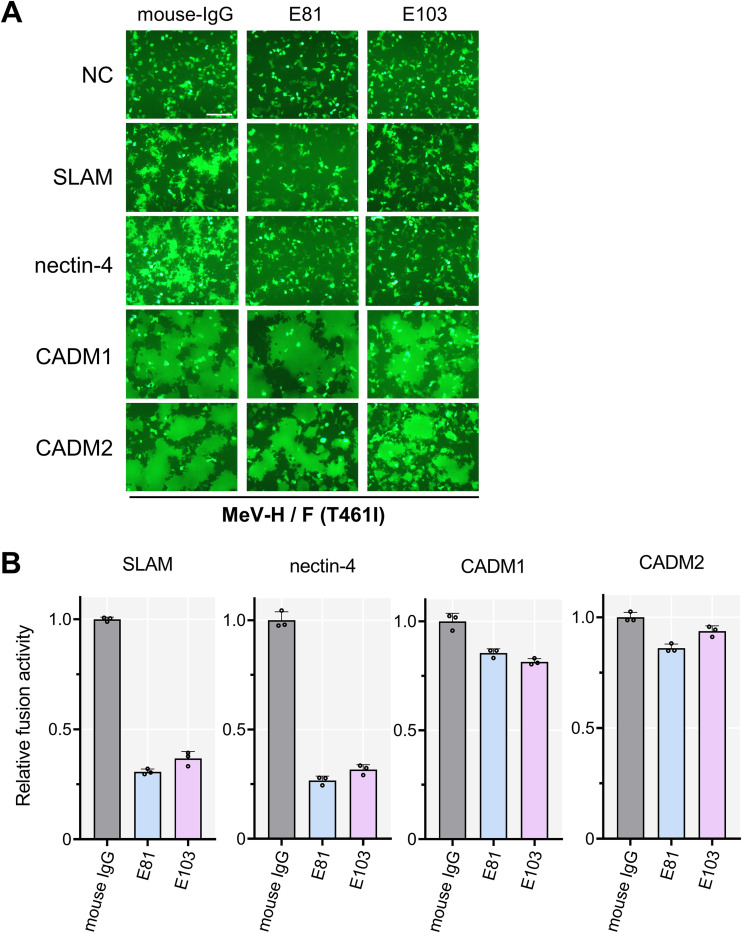
Measles virus (MeV), an enveloped RNA virus in the family Paramyxoviridae, usually causes acute febrile illness with skin rash but in rare cases persists in the brain, causing a progressive neurological disorder, subacute sclerosing panencephalitis (SSPE). MeV bears two envelope glycoproteins, the hemagglutinin (H) and fusion (F) proteins. The H protein possesses a head domain that initially mediates receptor binding and a stalk domain that subsequently transmits the fusion-triggering signal to the F protein. We recently showed that cell adhesion molecule 1 (CADM1; also known as IGSF4A, Necl-2, and SynCAM1) and CADM2 (also known as IGSF4D, Necl-3, and SynCAM2) are host factors enabling cell-cell membrane fusion mediated by hyperfusogenic F proteins of neuropathogenic MeVs as well as MeV spread between neurons lacking the known receptors. CADM1 and CADM2 interact in cis with the H protein on the same cell membrane, triggering hyperfusogenic F protein-mediated membrane fusion. Multiple isoforms of CADM1 and CADM2 containing various lengths of their stalk regions are generated by alternative splicing. Here, we show that only short-stalk isoforms of CADM1 and CADM2 predominantly expressed in the brain induce hyperfusogenic F protein-mediated membrane fusion. While the known receptors interact in trans with the H protein through its head domain, these isoforms can interact in cis even with the H protein lacking the head domain and trigger membrane fusion, presumably through its stalk domain. Thus, our results unveil a new mechanism of viral fusion triggering by host factors. IMPORTANCE Measles, an acute febrile illness with skin rash, is still an important cause of childhood morbidity and mortality worldwide. Measles virus (MeV), the causative agent of measles, may also cause a progressive neurological disorder, subacute sclerosing panencephalitis (SSPE), several years after acute infection. The disease is fatal, and no effective therapy is available. Recently, we reported that cell adhesion molecule 1 (CADM1) and CADM2 are host factors enabling MeV cell-to-cell spread in neurons. These molecules interact in cis with the MeV attachment protein on the same cell membrane, triggering the fusion protein and causing membrane fusion. CADM1 and CADM2 are known to exist in multiple splice isoforms. In this study, we report that their short-stalk isoforms can induce membrane fusion by interacting in cis with the viral attachment protein independently of its receptor-binding head domain. This finding may have important implications for cis-acting fusion triggering by host factors.

Keywords: CADM1; CADM2; SSPE; cis-acting fusion triggering; hemagglutinin; measles virus; membrane fusion; neuropathogenicity; splice isoform; subacute sclerosing panencephalitis.


Conflict of interest statement

The authors declare no conflict of interest.

Figures

FIG 1
FIG 1
Hamster CADM1 having exon 8 and CADM2 having exon 9 do not induce membrane fusion. (A) Fusion assay with human, mouse, and hamster CADM1 (left) and CADM2 (right). pCA7 expression plasmids encoding the H protein, the F(T461I) protein, and EGFP with pCA7 encoding human, mouse, or hamster CADM1/2 or pCA7 alone as a negative control (NC) were used to transfect 293FT cells. Cells were observed by fluorescence microscopy 30 h after transfection. Exon organizations of CADM1 and CADM2 used in this experiment are also shown. The numbers above rectangles are exon numbers. The numbering of the exons follows previous studies (39, 44–46). The exons present in all isoforms are indicated by gray rectangles, and the exons that are present in only some isoforms are indicated by red, yellow, or magenta rectangles. Bar = 250 μm. (B) Fusion assay with different isoforms of hamster CADM1 (left) and CADM2 (right). pCA7 expression plasmids encoding the H protein, the F(T461I) protein, and EGFP with pCA7 encoding one of the hamster CADM1/2 isoforms or pCA7 alone (NC) were used to transfect 293FT cells. Cells were observed by fluorescence microscopy 30 h after transfection. Bar = 250 μm. (C and D) DSP assay with human (h), mouse (m), and hamster (ham) CADM1 (C) and CADM2 (D) isoforms. pCA7 expression plasmids encoding the H protein and the F(T461I) protein with pCA7 encoding one of the CADM1/2 isoforms or pCA7 alone were used to transfect cocultured 293FT/DSP1 and 293FT/DSP2 cells. Renilla luciferase activity was measured 24 h after transfection (n = 3, means ± standard deviations [SD]). (E and F) Cell surface biotinylation assay with hamster CADM1 (E) and CADM2 (F) isoforms. pCA7 encoding E-cadherin (a cell surface protein control) with pCA7 encoding Flag-tagged EGFP (an intracellular protein control), one of the Flag-tagged hamster CADM1/2 isoforms, or pCA7 alone was used to transfect 293FT cells. Precipitates of biotinylated cell surface proteins and cell lysates were detected by Western blotting using anti-E-cadherin (top) or anti-Flag Ab (bottom).
FIG 2
FIG 2
Various isoforms of human CADM1 and CADM2. Exon organizations of human CADM1 (A) and CADM2 (B) are shown. Exons present in all isoforms are indicated by gray rectangles, and exons present in only some isoforms are indicated by red, blue, green, yellow, magenta, or black rectangles. The canonical isoform is designated CADM1 or CADM2, and the other isoforms are designated by appending numbers of exons not present in the canonical isoform (e.g., CADM1+ex8). One of the noncanonical CADM2 isoforms, CADM2+ex9(1), has the same exon 1 as the canonical CADM2, whereas another noncanonical isoform, CADM2+ex9(2), has a different exon 1. Schematic domain organizations of CADM1/2 are shown below the corresponding exons. Amino acids encoded by exon 8, exon 9, and exon 10 of CADM1 and exon 9 of CADM2 in the stalk regions are also shown. V, Ig-like V-type domain; C, Ig-like C2-type domain; TM, transmembrane domain.
FIG 3
FIG 3
Specific isoforms of CADM1 and CADM2 induce membrane fusion. (A) Fusion assay with various isoforms of human CADM1 (left) and CADM2 (right). pCA7 plasmids encoding the H protein, the F(T461I) protein, and EGFP with pCA7 encoding human CADM1/2 [CADM1, CADM1+ex8, CADM1+ex9(*), CADM1+ex10(*), CADM1+ex8/9, CADM1+ex9/10, CADM1+ex8/9/10, CADM2, CADM2+ex9(1), or CADM2+ex9(2)] or pCA7 alone as a negative control (NC) were used to transfect 293FT cells. Cells were observed by fluorescence microscopy 30 h after transfection. Exon organizations of respective isoforms are also shown. The isoforms indicated with asterisks are not found in the GTEx database. Bar = 250 μm. (B) DSP assay with various isoforms of human CADM1/2. pCA7 expression plasmids encoding the H protein and the F(T461I) protein with pCA7 encoding one CADM1/2 isoform or pCA7 alone were used to transfect cocultured 293FT/DSP1 and 293FT/DSP2 cells. Renilla luciferase activity was measured 24 h after transfection (n = 3; means ± SD). (C) Fusion assay using different hyperfusogenic F proteins with isoforms of human CADM1/2. Bar = 200 μm. (D) Cell surface biotinylation assay with isoforms of human CADM1/2. pCA7 encoding E-cadherin (a cell surface protein control) with pCA7 encoding one of the Flag-tagged human CADM1/2 isoforms was used to transfect 293FT cells. Precipitates of biotinylated cell surface proteins and cell lysates were detected by Western blotting using anti-E-cadherin (top) or anti-Flag Ab (bottom).
FIG 4
FIG 4
E-cadherin expression does not rescue the fusion-triggering function of long-stalk CADM1/2 isoforms. (A) Fusion assay with E-cadherin. pCA7 plasmids encoding the H protein, the F(T461I) protein, EGFP, and one of the isoforms of human CADM1/2 (CADM1, CADM1+ex8/9/10, CADM2, and CADM2+ex9) with pCA7 encoding E-cadherin or pCA7 alone as a negative control (NC) were used to transfect 293FT cells. Cells were observed by fluorescence microscopy 30 h after transfection. Bar = 200 μm. (B) DSP assay with E-cadherin. pCA7 plasmids encoding the H protein, the F(T461I) protein, and one of the isoforms of human CADM1/2 (CADM1, CADM1+ex8/9/10, CADM2, and CADM2+ex9) with pCA7 encoding E-cadherin or pCA7 alone were used to transfect cocultured 293FT/DSP1 and 293FT/DSP2 cells. Renilla luciferase activity was measured 24 h after transfection (n = 3; means ± SD).
FIG 5
FIG 5
Tissue expression patterns of alternatively spliced mRNA isoforms of CADM1 and CADM2. (A and B) Tissue expression patterns of human CADM1/2 isoforms were obtained from the GTEx consortium using the GTEx Portal (https://www.gtexportal.org/home/gene/CADM1 and https://www.gtexportal.org/home/gene/CADM2). The bars in the graphs indicate the total expression levels of all isoforms of CADM1 (A) or CADM2 (B) (left y axis). The lines in the graphs indicate the expression levels of respective isoforms of human CADM1 and CADM2 (right y axis). TPM, transcripts per million. (C and D) Expression of mouse (C) or hamster (D) CADM1/2 isoforms in the brain was examined by PCR using cDNA templates derived from mouse primary neurons (m-cDNA) and hamster brains (ham-cDNA), respectively. DNA fragments encompassing exons 6 to 11 for CADM1 or exons 7 to 10 for CADM2 were amplified. pCA7 plasmids encoding mouse CADM1, mouse CADM1+ex8, mouse CAMD2, mouse CADM2+ex9, hamster CADM1, hamster CADM1+ex8, hamster CADM2, and hamster CADM2+ex9 were used as control templates for PCR. Nucleotide sequences of amplified PCR fragments were confirmed by direct sequencing. Red and black asterisks indicate fusion-triggering (short-stalk) isoforms and other isoforms of CADM1/2, respectively.
FIG 6
FIG 6
Nontriggering isoforms of CADM1 and CADM2 do not affect membrane fusion triggered by the canonical isoforms. (A) Fusion assay. pCA7 plasmids encoding the H protein, the F(T461I) protein and EGFP with pCA7 encoding the canonical isoform, the nontriggering isoform, or both were used to transfect 293FT cells. Cells were observed by fluorescence microscopy 30 h after transfection. Bar = 200 μm. (B) DSP assay. pCA7 plasmids encoding the H protein and the F(T461I) protein with pCA7 encoding the canonical isoform, the nontriggering isoform, or both were used to transfect cocultured 293FT/DSP1 and 293FT/DSP2 cells. Renilla luciferase activity was measured 24 h after transfection (n = 3; means ± SD). The data were analyzed by the unpaired two-tailed Student's t test. *, P = 0.0098; ns, not significant (P = 0.4818).
FIG 7
FIG 7
The tetramerized headless H protein supports membrane fusion induced by fusion-triggering isoforms of CADM1 and CADM2. (A) Schematic diagrams of the HA-tagged full-length H protein [H(full)], the HA-tagged headless H protein (residues 1 to 180 of the H protein) [H(headless)] and the HA-tagged H(headless) protein with a tetramerization domain [H(headless)-TD]. CP, cytoplasmic domain; TM, transmembrane domain; TD, tetramerization domain. (B) Expression plasmids encoding H(headless) and H(headless)-TD were used to transfect 293FT cells. Cell lysates were detected by Western blotting using anti-beta-actin (top) or anti-HA Ab (bottom). (C) Fusion assay with various H constructs. pCA7 plasmids encoding the F(T461I) protein and one of the H constructs [H(full), H(headless), or H(headless)-TD] with pCA7 encoding one of the host proteins (CADM1, CADM1+ex8, CADM2, CADM2+ex9, SLAM, or nectin-4) or pCA7 alone (NC) were used to transfect 293FT cells. Cells were observed by fluorescence microscopy 48 h after transfection. Bar = 200 μm. (D) DSP assay with the H(headless)-TD protein. pCA7 plasmids encoding H(full) or H(headless)-TD and the F(T461I) protein with pCA7 encoding one of the host proteins (CADM1, CADM1+ex8, CADM2, CADM2+ex9, SLAM, or nectin-4) were used to transfect cocultured 293FT/DSP1 and 293FT/DSP2 cells. Renilla luciferase activity was measured 48 h after transfection (n = 3; means ± SD). The data were analyzed by the unpaired two-tailed Student's t test. *1, P < 0.0001; *2, P = 0.8059; *3, P < 0.0001; *4, P = 0.4118; *5, P = 0.001; *6, P = 0.2676.
FIG 8
FIG 8
The canonical isoforms of CADM1/2 interact with the headless H protein. Interactions of the HA-tagged H(full) protein (A) or the HA-tagged H(headless) protein (B) with the His-tagged CADM1, CADM1+ex8/9/10, CADM2, and CADM2+ex9 proteins were examined by coimmunoprecipitation assay. Immunoprecipitates (IP) and total lysates (INPUT) were detected by immunoblotting (IB) using anti-HA (top) or anti-His Ab (bottom).
FIG 9
FIG 9
Monoclonal Abs against the H protein head domain, E81 and E103, do not inhibit membrane fusion induced by CADM1/2. (A) Fusion assay in the presence of the monoclonal Abs against the H head domain. pCA7 plasmids encoding the H protein, the F(T461I) protein and EGFP with pCA7 encoding SLAM, nectin-4, CADM1, or CADM2 or pCA7 alone were used to transfect 293FT cells. At 2 h after transfection, one monoclonal Ab (E81 or E103) (50 μg per mL) or mouse IgG (negative control) was added to culture media. Cells were observed under fluorescence microscopy 30 h after transfection. Bar = 200 μm. (B) The DSP assay in the presence of the monoclonal Abs against the H protein head domain. pCA7 plasmids encoding the H protein and the F(T461I) protein with pCA7 encoding CADM1, CADM2, SLAM, nectin-4, or pCA7 alone, were used to transfect cocultured 293FT/DSP1 and 293FT/DSP2 cells. At 2 h after transfection, one monoclonal Ab (E81 or E103) (50 μg per mL) or mouse IgG (negative control) was added to culture media. Renilla luciferase activity was measured 24 h after transfection (n = 3; means ± SD).
FIG 10
FIG 10
The graphical summary of interactions between the MeV H protein and trans- or cis-acting host factors. (A) Interactions of the MeV H protein with trans-acting viral receptors, SLAM and nectin-4, through the H protein head domain. (B) Interactions of the full-length and headless MeV H proteins with isoforms of cis-acting host factors, CADM1 and CADM2. The isoform containing a short stalk region functionally interacts with the full-length and headless H proteins. “YES” indicates the functional interaction leading to fusion triggering; “NO” indicates no fusion triggering. TD, tetramerization domain.

Similar articles

Cited by

References

    1. Griffin DE. 2013. Measles virus, p 1042–1069. In Knipe DM, Howley PM, Cohen JI, Griffin DE, Lamb RA, Martin MA, Racaniello VR, Roizman B (ed), Fields virology, 6th ed. Lippincott Williams & Wilkins, Philadelphia, PA.
    1. Coughlin MM, Beck AS, Bankamp B, Rota PA. 2017. Perspective on global measles epidemiology and control and the role of novel vaccination strategies. Viruses 9:11. 10.3390/v9010011. - DOI - PMC - PubMed
    1. Bellini WJ, Rota JS, Lowe LE, Katz RS, Dyken PR, Zaki SR, Shieh WJ, Rota PA. 2005. Subacute sclerosing panencephalitis: more cases of this fatal disease are prevented by measles immunization than was previously recognized. J Infect Dis 192:1686–1693. 10.1086/497169. - DOI - PubMed
    1. Mekki M, Eley B, Hardie D, Wilmshurst JM. 2019. Subacute sclerosing panencephalitis: clinical phenotype, epidemiology, and preventive interventions. Dev Med Child Neurol 61:1139–1144. 10.1111/dmcn.14166. - DOI - PubMed
    1. Schönberger K, Ludwig MS, Wildner M, Weissbrich B. 2013. Epidemiology of subacute sclerosing panencephalitis (SSPE) in Germany from 2003 to 2009: a risk estimation. PLoS One 8:e68909. 10.1371/journal.pone.0068909. - DOI - PMC - PubMed

Publication types