

Automatic detection of anteriorly displaced temporomandibular joint discs on magnetic resonance images using a deep learning algorithm
- PMID: 34788124
- PMCID: PMC8925876
- DOI: 10.1259/dmfr.20210341
Automatic detection of anteriorly displaced temporomandibular joint discs on magnetic resonance images using a deep learning algorithm
Abstract
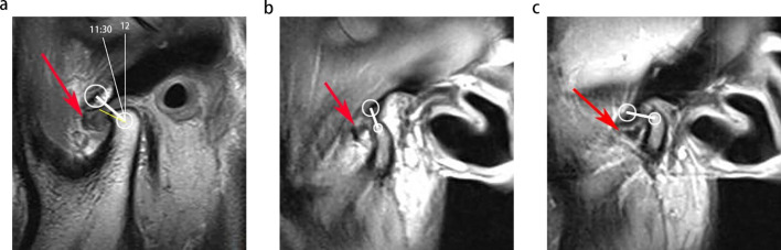
Objectives: This study aimed to develop models that can automatically detect anterior disc displacement (ADD) of the temporomandibular joint (TMJ) on MRIs before orthodontic treatment to reduce the risk of developing serious complications after treatment.
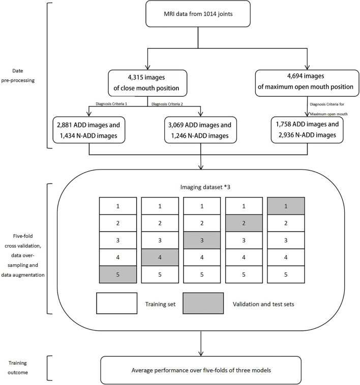
Methods: We used 9009 sagittal MRI of the TMJ as input and constructed three sets of deep learning models to detect ADD automatically. Deep learning models were developed using a convolutional neural network (CNN) based on the ResNet architecture and the "Imagenet" database. Five-fold cross-validation, oversampling, and data augmentation techniques were applied to reduce the risk of overfitting the model. The accuracy and area under the curve (AUC) of the three models were compared.
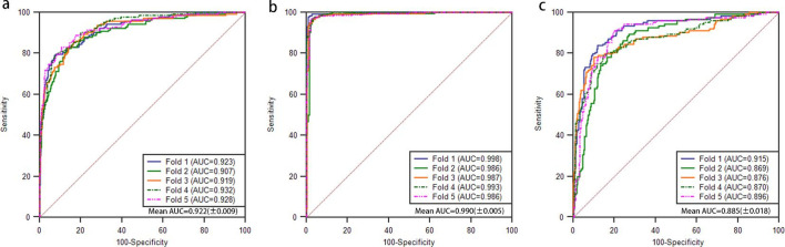
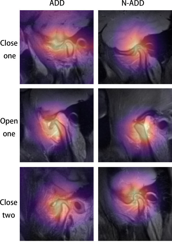
Results: The performance of the maximum open mouth position model was excellent with accuracy and AUC of 0.970 (±0.007) and 0.990 (±0.005), respectively. For closed mouth position models, the accuracy and AUC of diagnostic Criteria 1 were 0.863 (±0.008) and 0.922 (±0.009), respectively significantly higher than that of diagnostic Criteria 2 with 0.839 (±0.013) (p = 0.009) and AUC of 0.885 (±0.018) (p = 0.003). The classification activation heat map also improved our understanding of the models and visually displayed the areas that play a key role in the model recognition process.
Conclusion: Our CNN model resulted in high accuracy and AUC in detecting ADD and can therefore potentially be used by clinicians to assess ADD before orthodontic treatment, and hence improve treatment outcomes.
Keywords: Deep Learning; Magnetic Resonance Imaging; Temporomandibular Joint Disc.
Figures
References
-
- Manfredini D, Guarda-Nardini L, Winocur E, Piccotti F, Ahlberg J, Lobbezoo F. Research diagnostic criteria for temporomandibular disorders: a systematic review of axis I epidemiologic findings. Oral Surg Oral Med Oral Pathol Oral Radiol Endod 2011; 112: 453–62. doi: 10.1016/j.tripleo.2011.04.021 - DOI - PubMed
