Review
. 2022 Jan 19;35(1):e0009421.
doi: 10.1128/CMR.00094-21. Epub 2021 Nov 17.

Pathogenesis of Respiratory Viral and Fungal Coinfections

Affiliations
Review

Pathogenesis of Respiratory Viral and Fungal Coinfections

Fabián Salazar et al. Clin Microbiol Rev. .

Abstract

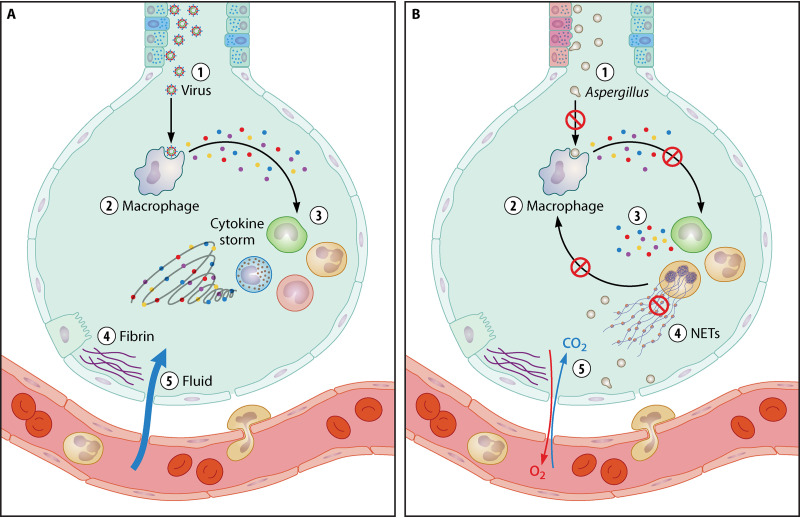
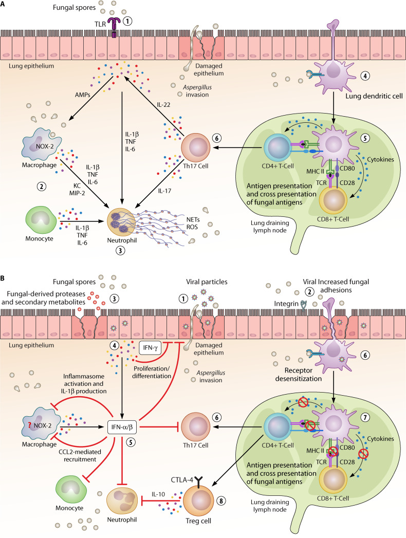
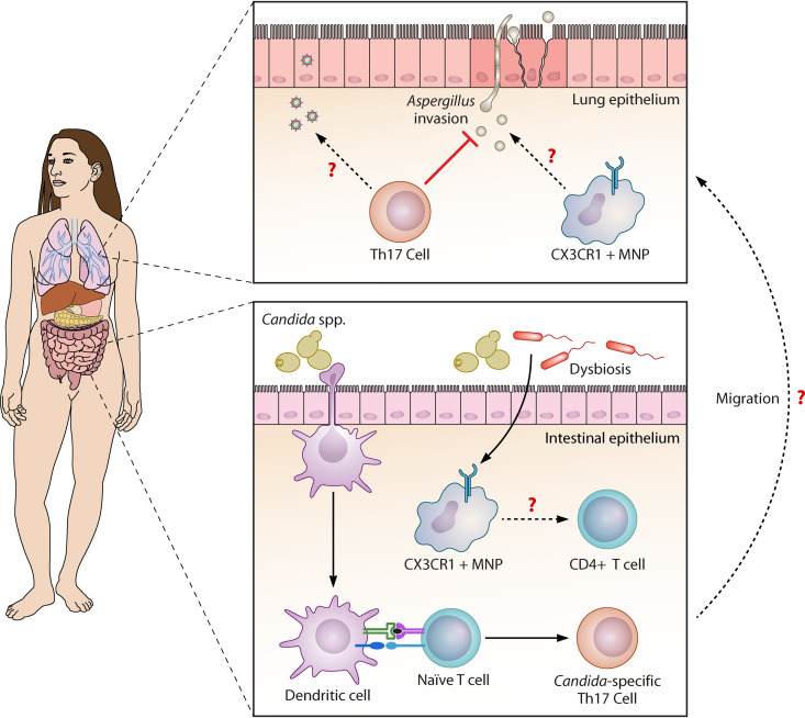
Individuals suffering from severe viral respiratory tract infections have recently emerged as "at risk" groups for developing invasive fungal infections. Influenza virus is one of the most common causes of acute lower respiratory tract infections worldwide. Fungal infections complicating influenza pneumonia are associated with increased disease severity and mortality, with invasive pulmonary aspergillosis being the most common manifestation. Strikingly, similar observations have been made during the current coronavirus disease 2019 (COVID-19) pandemic. The copathogenesis of respiratory viral and fungal coinfections is complex and involves a dynamic interplay between the host immune defenses and the virulence of the microbes involved that often results in failure to return to homeostasis. In this review, we discuss the main mechanisms underlying susceptibility to invasive fungal disease following respiratory viral infections. A comprehensive understanding of these interactions will aid the development of therapeutic modalities against newly identified targets to prevent and treat these emerging coinfections.

Keywords: SARS-CoV; antifungal immunity; aspergillosis; coinfection; copathogenesis; fungal pathogens; influenza; respiratory viruses.


Figures

FIG 1
FIG 1
Progression of respiratory viral-fungal coinfections into the alveolar space determines disease severity. (A) Progression of a viral infection into the alveolar space. (1) The virus infects airway epithelium. (2) Alveolar macrophages recognize the virus and in response produce cytokines. (3) Cytokines attract more immune cells, including neutrophils and monocytes, which in turn produce more cytokines, creating a cycle of inflammation that damages the lung tissue. (4) Damage can further occur through the formation of fibrin and scar tissue. (5) Weakened blood vessels allow fluid to seep in and fill the lung cavities, leading to respiratory failure. (B) Progression of a fungal infection into the alveolar space following severe viral pneumonia. (1) When Aspergillus enters the airways, damaged epithelium facilitates adhesion of fungal conidia and subsequent invasion. (2) Phagocytosis, fungal killing, and cytokine production by alveolar macrophages are impaired. (3) Recruitment of neutrophils and their cross talk with macrophages are also affected. (4) Loss of neutrophils compromises their cytokine production and neutrophil extracellular trap (NET)-mediated fungal killing. (5) The release of fibrinous material can cause the obstruction of the small airways, decreasing oxygen and carbon dioxide diffusion capacities and creating a hypoxic milieu that changes Aspergillus virulence properties and the outcome of host-Aspergillus interaction. (This figure was created with BioRender.)
FIG 2
FIG 2
Interplay between cellular mechanisms underlying respiratory viral-fungal coinfections. (A) Effector cellular mechanisms against fungal infections. (1) Fungal recognition by the airway epithelium leads to the production of proinflammatory cytokines that can activate other immune cells, including macrophages and neutrophils. (2) Monocytes and alveolar macrophages play pivotal roles during fungal infections, including phagocytosis and cytokine and chemokine production. (3) Neutrophils form neutrophil extracellular traps (NETs) and produce reactive oxygen species (ROS) that contribute to fungal killing. (4) Lung dendritic cells recognize, ingest, and kill Aspergillus conidia, acquire a fully mature state, and then migrate to draining lymph nodes. (5) Antigen presentation of fungus-derived peptides to naive CD4+ and CD8+ T cells occurs in the draining lymph nodes. (6) T cell activation leads to Th17 differentiation, which is pivotal for control of fungal infections at the airways. In particular, Th17 cells support neutrophil activation and the production of antimicrobial peptides (AMPs) by epithelial cells. (B) Mechanisms responsible for increased susceptibility to fungal infections in patients suffering severe viral pneumonia. (1 to 3) The lung epithelium undergoes different changes over the course of respiratory viral infections, including tissue disruption that facilitates secondary fungal invasion (1) and expression and/or exposure of receptors to which fungal pathogens can adhere (2). In addition, germinated fungal spores themselves release molecules with the potential to increase permeability and tissue damage, such as proteases and mycotoxins (3). (4) The airway epithelium produces type I and type III interferons (IFNs), which have a significant impact on antifungal immunity at different levels. (5) IFN-α/β are also produced by alveolar macrophages. IFNs reduce epithelial cell proliferation and differentiation, increasing susceptibility to coinfections. IFNs suppress monocyte, macrophage, and neutrophil recruitment and effector responses that are essential for fighting fungal infections. IFNs act as negative regulators of inflammasome activation in response to fungal pathogens, thus affecting fungal clearance. IFNs dampen Th17 responses, leading to attenuation of AMP production and neutrophil recruitment that are required for antifungal clearance. (6) Desensitization of pattern recognition receptors (PRRs), which are essential for fungal recognition and antifungal immunity, contributes to susceptibility to coinfections. (7) Viral infections also interfere with antigen-presenting cell functionalities, affecting the subsequent immune response to fungal antigens. For instance, viral infection affects antigen presentation through interference with any of the three signals required for T cell activation, namely, MHC presentation, expression of cosignaling molecules, and/or production of cytokines. (8) Regulatory T cells (Tregs) induced during the recovery and resolution phase of a viral infection persist for long enough to interfere with immunity (i.e., neutrophil functionalities) during subsequent fungal infections. Some questions remain, including the role of the NADPH oxidase 2 (NOX-2) complex in the context of viral-fungal coinfections. (This figure was created with BioRender.)
FIG 3
FIG 3
Gut-lung axis in the context of respiratory viral-fungal coinfections. The mycobiota plays a significant role in immunity and homeostasis in the intestine, which can influence immune responses at the airways. C. albicans-specific Th17 cells can confer protection against Aspergillus infection in the lungs. Importantly, intestinal microbial dysbiosis could affect functionalities of immune cells in the gut-lung axis, such as CX3CR1+ mononuclear phagocytes (MNPs), which in turn could influence susceptibility to fungal coinfections. Additional important questions remained unanswered. For instance, do these immune responses develop locally or traffic from the gut, or both? If they do migrate, what is their route of migration? What signals control it? (This figure was created with BioRender.)

References

    1. Brown GD, Denning DW, Gow NA, Levitz SM, Netea MG, White TC. 2012. Hidden killers: human fungal infections. Sci Transl Med 4:165rv13. 10.1126/scitranslmed.3004404. - DOI - PubMed
    1. White PL, Dhillon R, Cordey A, Hughes H, Faggian F, Soni S, Pandey M, Whitaker H, May A, Morgan M, Wise MP, Healy B, Blyth I, Price JS, Vale L, Posso R, Kronda J, Blackwood A, Rafferty H, Moffitt A, Tsitsopoulou A, Gaur S, Holmes T, Backx M. 2020. A national strategy to diagnose COVID-19 associated invasive fungal disease in the ICU. Clin Infect Dis 73:e1634–e1644. 10.1093/cid/ciaa1298. - DOI - PMC - PubMed
    1. Bongomin F, Gago S, Oladele RO, Denning DW. 2017. Global and multi-national prevalence of fungal diseases—estimate precision. J Fungi (Basel) 3:57. 10.3390/jof3040057. - DOI - PMC - PubMed
    1. Krammer F, Smith GJD, Fouchier RAM, Peiris M, Kedzierska K, Doherty PC, Palese P, Shaw ML, Treanor J, Webster RG, Garcia-Sastre A. 2018. Influenza. Nat Rev Dis Primers 4:3. 10.1038/s41572-018-0002-y. - DOI - PMC - PubMed
    1. McCullers JA. 2014. The co-pathogenesis of influenza viruses with bacteria in the lung. Nat Rev Microbiol 12:252–262. 10.1038/nrmicro3231. - DOI - PubMed

Publication types