. 2022 Jun 3;145(5):1684-1697.
doi: 10.1093/brain/awab409.

De novo FZR1 loss-of-function variants cause developmental and epileptic encephalopathies

Collaborators, Affiliations

De novo FZR1 loss-of-function variants cause developmental and epileptic encephalopathies

Sathiya N Manivannan et al. Brain. .

Abstract

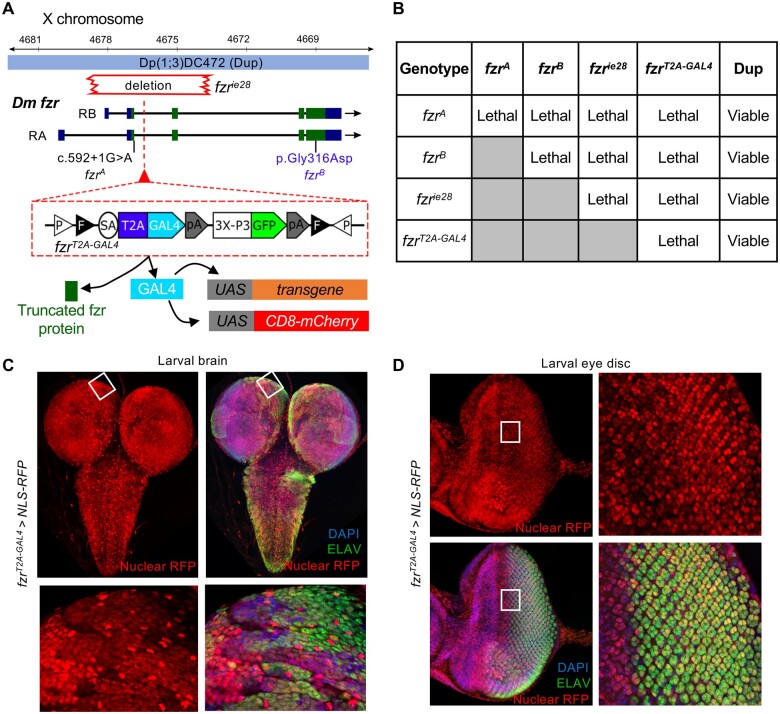
FZR1, which encodes the Cdh1 subunit of the anaphase-promoting complex, plays an important role in neurodevelopment by regulating the cell cycle and by its multiple post-mitotic functions in neurons. In this study, evaluation of 250 unrelated patients with developmental and epileptic encephalopathies and a connection on GeneMatcher led to the identification of three de novo missense variants in FZR1. Whole-exome sequencing in 39 patient-parent trios and subsequent targeted sequencing in an additional cohort of 211 patients was performed to identify novel genes involved in developmental and epileptic encephalopathy. Functional studies in Drosophila were performed using three different mutant alleles of the Drosophila homologue of FZR1 fzr. All three individuals carrying de novo variants in FZR1 had childhood-onset generalized epilepsy, intellectual disability, mild ataxia and normal head circumference. Two individuals were diagnosed with the developmental and epileptic encephalopathy subtype myoclonic atonic epilepsy. We provide genetic-association testing using two independent statistical tests to support FZR1 association with developmental and epileptic encephalopathies. Further, we provide functional evidence that the missense variants are loss-of-function alleles using Drosophila neurodevelopment assays. Using three fly mutant alleles of the Drosophila homologue fzr and overexpression studies, we show that patient variants can affect proper neurodevelopment. With the recent report of a patient with neonatal-onset with microcephaly who also carries a de novo FZR1 missense variant, our study consolidates the relationship between FZR1 and developmental and epileptic encephalopathy and expands the associated phenotype. We conclude that heterozygous loss-of-function of FZR1 leads to developmental and epileptic encephalopathies associated with a spectrum of neonatal to childhood-onset seizure types, developmental delay and mild ataxia. Microcephaly can be present but is not an essential feature of FZR1-encephalopathy. In summary, our approach of targeted sequencing using novel gene candidates and functional testing in Drosophila will help solve undiagnosed myoclonic atonic epilepsy or developmental and epileptic encephalopathy cases.

Keywords: Drosophila model of neurodevelopmental disorders; FZR1; developmental and epileptic encephalopathy; functional validation of novel variants; myoclonic atonic epilepsy.


Figures

Figure 1
Figure 1
Conservation FZR1 and in vitro analysis of DEE variants in mammalian cells. (A) Protein primary structure diagram of human FZR1 and Drosophila orthologue Fzr showing conserved domains and corresponding positions of variants observed in patients. The residues affected in the patients are conserved in Drosophila melanogaster. Amino acid sequence alignment of human FZR1 and Drosophilafzr region encompassing the DEE variant residues, which are shown in red boxes. (B) Immunofluorescence staining for eGFP-tagged human FZR1 cDNA in HEK293 cells (green), co-stained with DAPI (nucleus; blue) and actin-cytoskeleton (Phalloidin; magenta). (i–ii) FZR1-wt, (iiiiv) FZR1:p.D187N, (vvi) FZR1:p.N333K localization to the nucleus and the cytoplasm. (C) Western blot showing relative expression of FZR1-wt and variants in HEK-293 cells. Alpha-tubulin is used as loading control. (D) Box-and-whisker plot showing the quantitation of normalized western blot signal analysed using one-way ANOVA, followed by Dunnett’s test for comparison of the variants to the wild-type expression. **Multiplicity adjusted P-value < 0.01. Boxes are drawn from the 25th to the 75th percentiles, with a line indicating mean, and error bars from maximum to minimum values. (E and F) 3D structural model FZR1 as part of the Cdh1-APC (PDB: 4ui9) showing the relative positions of the variants affected in the DEE patients (green) and corresponding residue of the Drosophila mutation observed in the fzrB allele (blue).
Figure 2
Figure 2
Drosophila fzr alleles and expression pattern. (A) Drosophila X-chromosome locus showing two transcriptional isoforms of fzr, two EMS-induced mutation alleles fzrA (splice-site mutation) and fzrB (missense mutation located in the same domain as one of the patient’s variants), fzrie28 (deletion spanning the first two exons of fzr) and a third chromosome duplication allele carrying a complete copy of fzr from the X-chromosome. Also shown is the fzrT2A-GAL4 allele generated by insertion of a mutagenic T2A-GAL4 artificial exon within the second intron of fzr. This insertion leads to termination of fzr translation and expression of GAL4 protein from the endogenous fzr locus. (B) Table showing results of complementation tests between various alleles of fzr as well the fzr locus duplication shown in A. (C and D) Expression pattern of fzr in Drosophila third instar visualized using the fzrT2A-GAL4 driving the expression of nls::RFP (red) co-stained with ELAV marking neuronal cells (green) and DAPI to detect nucleus (blue). (C) Larval brain and higher magnification images of the boxed region. (D) The eye-antennal disc and higher magnification images of the boxed region.
Figure 3
Figure 3
Pattern formation and glial migration defects in the photoreceptor precursors of fzr mutants are not rescued by DEE variants. (A) Diagram showing the generation of random mosaic patches of homozygous fzr mutant cells in the developing eye disc that are marked by GFP expression using MARCM. (B) Schematic diagram showing gene expression differences between homozygous fzr mutant (GFP+) cells and the surrounding heterozygous fzr (GFP−) cells. In mutant cells, Actin5c-GAL4 drives the expression of GFP and fzr cDNA (wild-type or patient variant), while in heterozygous cells, GAL4 activity is suppressed by GAL80. [C(iiv)] Control larval eye discs showing expression pattern of ELAV (red) in mosaics marked by GFP expression generated using MARCM. [C(v–viii)] Larval eye disc showing mosaic homozygous fzrB tissue (green) showing dramatic change in ELAV (magenta) expression within and outside the clones, showing cell autonomous and non-autonomous effect of fzr loss in vivo. [D(i)] Control adult eye showing stereotypical pattern of compound eye. [D(ii)] Aberrant retina pattern observed in fzrB mosaic adults. (E) MARCM mosaics generated as above stained with HRP (photoreceptor membrane). Magnified regions showing HRP boundary in dotted lines and glial cells stained with ani-Repo antibody. [E(i)] Control larval eye imaginal disc showing limitation of glial cells to the boundary set by HRP positive cells (boundary traced by white dotted lines). [E(ii)] fzrB mosaics showing not only disturbances in HRP pattern but also migration of glial cells past the boundaries (arrowhead) of HRP-positive cells. [E(iii)] Eye imaginal discs showing rescue of HRP pattern and glial cell migration when fzr wild-type cDNA is overexpressed in mutant mosaics. [E(iv)] Eye imaginal discs showing glial migration in regions lacking HRP signal when fzr cDNA with variant p.D172N is overexpressed in fzr mutant cells. [E(v)] Eye imaginal discs showing similar defects in glial cell migration when fzr cDNA with variant p.N318K is expressed in fzr mutant cells.
Figure 4
Figure 4
Overexpression of fzr leads to aberrant retinas. (A) Control ey3.5-GAL4 eyes showing stereotypical photoreceptor pattern and retina size. (B) Eyes of animals with wild-type fzr overexpressed using ey3.5-GAL4 showing severe eye size reduction and disrupted ommatidia pattern. (C) Adult eyes of ey3.5-GAL4 mediated overexpression of Fzr:p.D172N leading loss of photoreceptor pattern and reduction in retina size. (D) Adult eyes ey3.5-GAL4 mediated overexpression of Fzr:p.N318K leading to disruption in ommatidia pattern and reduction in eye size. (E) Box-and-whisker plot showing the quantitation of the eye size shows significant reduction of retina area due to the overexpression of wild-type or variant fzr cDNAs. Boxes are drawn from the 25th to the 75th percentiles, with a line indicating mean and error bars from maximum to minimum values. Reduction in the eye size due to variant overexpression is not as severe as defects caused by wild-type Fzr, which are statistically significant. Statistical difference evaluated using one-way ANOVA between all the samples followed by pair-wise analysis using Tukey’s multiple comparison. Multiplicity corrected P-values indicated as ****P < 0.0001, **P < 0.001, *P < 0.05.
Figure 5
Figure 5
Embryonic CNS defects in fzrT2A-GAL4 mutants are not rescued by Fzr-carrying DEE patient variants. (A) Diagram showing the crossing scheme used to assess rescue of lethality observed in fzrT2A-GAL4 using overexpression of wild-type or variant Drosophila Fzr and box-and-whisker plot showing the percentage of late third instar (L3) male larvae recovered by the overexpression of wild-type and variant cDNAs in a fzrT2A-GAL4 mutant background. (BF) Drosophila embryos at stage 15–16 showing neuronal membranes stained with HRP. (B) fzrT2A-GAL4 hemizygous embryo (fzrT2A-GAL4/Y) rescued with duplication of fzr genomic region on third chromosome serving as control. (C) fzrT2A-GAL4/Y embryos showing severe defects in the pattern of neuron development. (D) Wild-type Fzr expression using the fzrT2A-GAL4 rescues neuronal pattern. (E and F) fzrT2A-GAL4 shows neurodevelopmental defects that are not rescued by the expression of Fzr p. \D172N or p.N318K.

References

    1. Scheffer IE, Berkovic S, Capovilla G, et al. . ILAE classification of the epilepsies: Position paper of the ILAE Commission for Classification and Terminology. Epilepsia. 2017;58(4):512–521. - PMC - PubMed
    1. Truty R, Patil N, Sankar R, et al. . Possible precision medicine implications from genetic testing using combined detection of sequence and intragenic copy number variants in a large cohort with childhood epilepsy. Epilepsia Open. 2019;4(3):397–408. - PMC - PubMed
    1. McTague A, Howell KB, Cross JH, Kurian MA, Scheffer IE.. The genetic landscape of the epileptic encephalopathies of infancy and childhood. Lancet Neurol. 2016;15(3):304–316. - PubMed
    1. Thomas RH, Berkovic SF.. The hidden genetics of epilepsy—A clinically important new paradigm. Nat Rev Neurol. 2014;10(5):283–292. - PubMed
    1. Snoeijen-Schouwenaars FM, van Ool JS, Verhoeven JS, et al. . Diagnostic exome sequencing in 100 consecutive patients with both epilepsy and intellectual disability. Epilepsia. 2019;60(1):155–164. - PubMed

Publication types